. 2018 Aug;14(8):788-793.
doi: 10.1038/s41589-018-0072-x. Epub 2018 Jun 18.

Scavenging of superoxide by a membrane-bound superoxide oxidase

Affiliations

Scavenging of superoxide by a membrane-bound superoxide oxidase

Camilla A K Lundgren et al. Nat Chem Biol. 2018 Aug.

Abstract

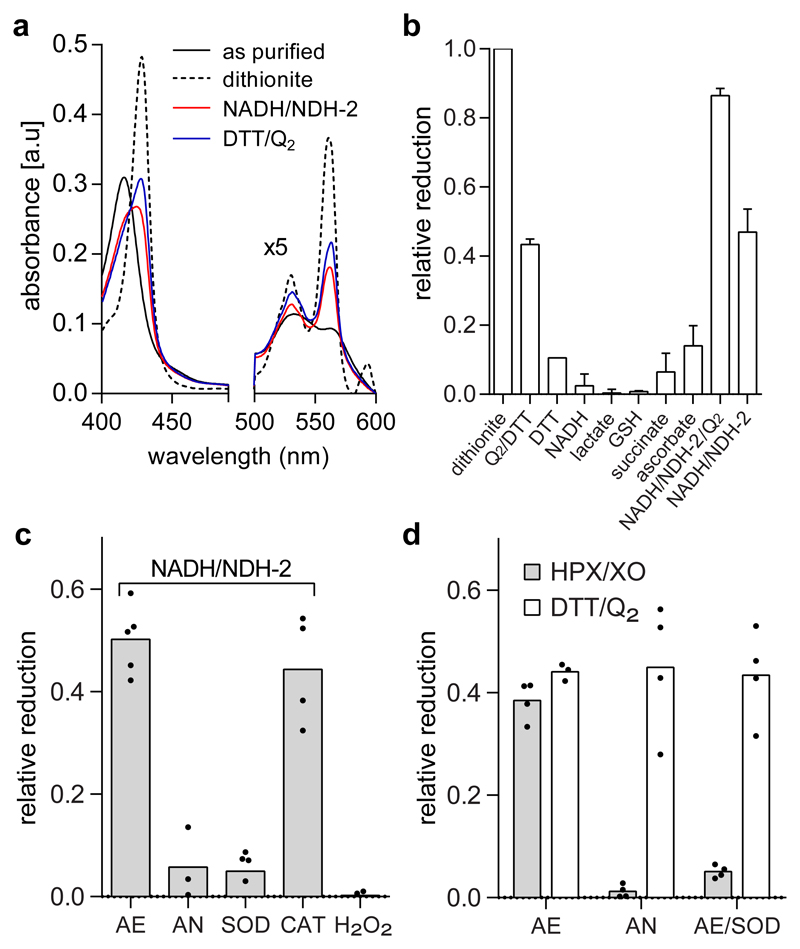
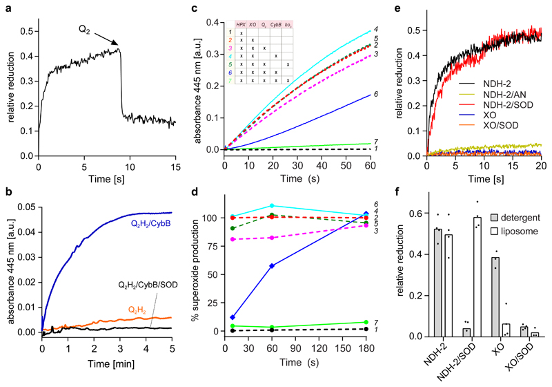
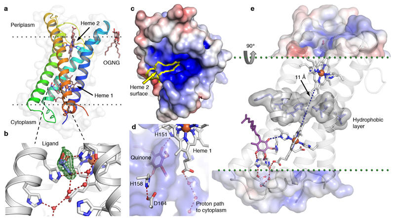
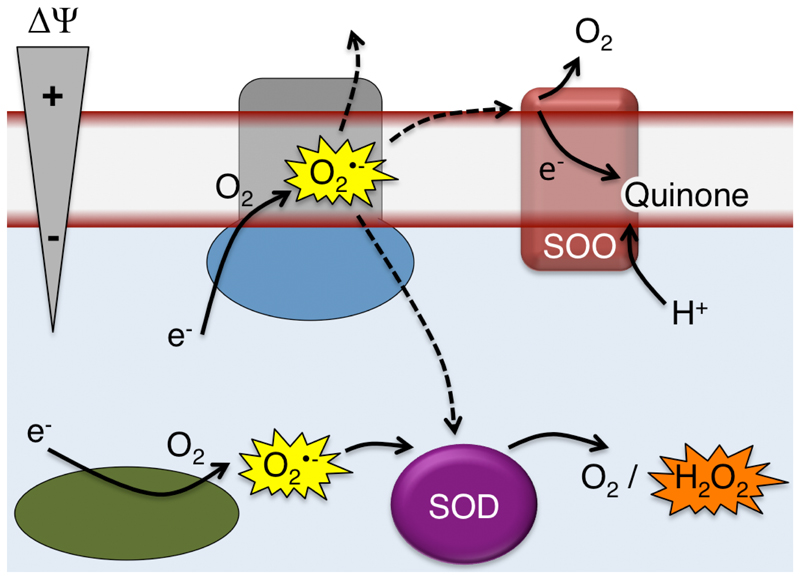
Superoxide is a reactive oxygen species produced during aerobic metabolism in mitochondria and prokaryotes. It causes damage to lipids, proteins and DNA and is implicated in cancer, cardiovascular disease, neurodegenerative disorders and aging. As protection, cells express soluble superoxide dismutases, disproportionating superoxide to oxygen and hydrogen peroxide. Here, we describe a membrane-bound enzyme that directly oxidizes superoxide and funnels the sequestered electrons to ubiquinone in a diffusion-limited reaction. Experiments in proteoliposomes and inverted membranes show that the protein is capable of efficiently quenching superoxide generated at the membrane in vitro. The 2.0 Å crystal structure shows an integral membrane di-heme cytochrome b poised for electron transfer from the P-side and proton uptake from the N-side. This suggests that the reaction is electrogenic and contributes to the membrane potential while also conserving energy by reducing the quinone pool. Based on this enzymatic activity, we propose that the enzyme family be denoted superoxide oxidase (SOO).


Conflict of interest statement

The authors declare no competing financial interests.

Figures

Fig. 1
Fig. 1. CybB reacts with superoxide and ubiquinol.
(a) Absorbance spectra of purified and detergent solubilized CybB (black) after reduction with dithionite (dotted), DTT/Q2 (blue), or NADH/NDH-2 (red). The spectra shown are representative of at least 3 different protein purifications with similar results. (b) Relative reduction of CybB by different electron carriers after 1 min incubation. (The average and the SD have been calculated from n≥3 biologically independent experiments). (c) Reduction of purified CybB by NADH/NDH-2 under different conditions: aerobic (AE), anaerobic (AN), AE in presence of superoxide dismutase (SOD), and AE in presence of catalase (CAT). No reduction was observed when CybB was incubated with hydrogen peroxide (H2O2). Mean values and the individual data points from n≥3 biologically independent experiments are shown. (d) Superoxide and ubiquinol reduce CybB via two different mechanisms. Comparison of CybB reduction by HPX/XO (grey bars) and DTT/Q2 (open bars) under different conditions (see 1c). Mean values and the individual data points from n≥3 biologically independent experiments are shown.
Fig. 2
Fig. 2. CybB shows superoxide-oxidase activity in vitro.
(a) CybB was pre-reduced with HPX/XO before oxidized ubiquinone Q2 was added. The kinetic trace shown is a representative of at least 3 independent measurements. (b) Superoxide production by ubiquinol Q2H2 and CybB. The two components were mixed in the absence (blue) or in the presence (black) of SOD under aerobic conditions. Superoxide production was monitored spectrophotometrically at 445 nm following the conversion of a tetrazolium salt (WST-1) to formazan. The kinetic traces shown are representative of at least 3 independent measurements. (c) Superoxide production by HPX/XO and its suppression by CybB. See inset for the different combinations and text for further details. The kinetic traces shown are representative of at least 3 independent measurements. (d) Relative superoxide production rates at different time points during the experiments of Fig. 2c. The data are normalized to the rate of superoxide production by NADH/NDH-2 at each respective time point. (e) Membrane embedded CybB reacts preferentially with membrane derived superoxide. CybB was reconstituted into liposomes from E. coli polar lipid extract and mixed with superoxide produced either by NADH/NDH-2 or HPX/XO and either in the presence or absence of SOD under aerobic conditions. No reduction of CybB by NADH/NDH-2 was observed under anaerobic (AN) conditions. The kinetic traces shown are representative of at least 3 independent measurements. (f) Comparison of the results from Fig. 2e (open bars) with similar experiments performed with detergent solubilized CybB (grey bars). Mean values and the individual data points from n≥3 biologically independent experiments are shown.
Fig. 3
Fig. 3. High-resolution structure of CybB.
(a) 1.97 Å crystal structure of CybB colored from the N-terminus (blue) to the C-terminus (red), the protein surface is indicated (light grey). The calculated location of the hydrophobic core of the membrane is illustrated by dotted lines. (b) Binding site near the cytoplasmic heme, a glycerol molecule was modeled in the cavity based on the electron density, Fo-Fc omit map contoured at 4σ (green mesh). A chain of hydrogen bonded water molecules leads from the ligand-binding site to the cytoplasm. (c) Electrostatic potential map of the protein surface viewed from the periplasm, positive surface potential in blue, negative in red. The molecular surface contributed by the exposed heme 2 porphyrin edge, presumably acting as an electron sink, is bounded in yellow. (d) Protein surface making up the ligand binding cavity and channel to the cytoplasm in blue (viewed from inside the protein). The ubiquinone headgroup (purple) was predicted to occupy the same volume as the bound ligand with its carbonyl groups hydrogen bonded to the iron-coordinating H151 and a second completely conserved histidine (H158). (e) Proposed functional architecture of CybB. The positively charged funnel attracts superoxide to the heme edge, which serves as an electron sink and oxidizes the superoxide to molecular oxygen. The electron is subsequently tunneled to the quinone binding site (blue dashed lines). A layer of hydrophobic residues (gray surface) is located between the hemes, preventing the presence of water molecules or a proton-transfer path through the membrane. Upon quinone reduction, protons are sequestered from the cytoplasm via the indicated hydrogen-bonded path (red) to produce reduced ubiquinol.
Fig. 4
Fig. 4. Hypothetical functional context of SOO.
Superoxide produced by electron leakage at the membrane may escape to the cytoplasm, periplasm, or be oxidized by SOO, producing molecular oxygen, using ubiquinone as the electron acceptor. Intracellular superoxide is quenched by superoxide dismutase producing molecular oxygen and hydrogen peroxide.

References

    1. Dröge W. Free radicals in the physiological control of cell function. Physiol Rev. 2002;82:47–95. - PubMed
    1. Sabharwal SS, Schumacker PT. Mitochondrial ROS in cancer: initiators, amplifiers or an Achilles' heel? Nat Rev Cancer. 2014;14:709–721. - PMC - PubMed
    1. Barnham KJ, Masters CL, Bush AI. Neurodegenerative diseases and oxidative stress. Nat Rev Drug Discov. 2004;3:205–214. - PubMed
    1. Riera CE, Merkwirth C, De Magalhaes Filho CD, Dillin A. Signaling Networks Determining Life Span. Annu Rev Biochem. 2016;85:35–64. - PubMed
    1. McCord JM, Fridovich I. Superoxide dismutase. An enzymic function for erythrocuprein (hemocuprein) J Biol Chem. 1969;244:6049–6055. - PubMed

Publication types

MeSH terms