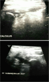
Sialolithiasis of Right Submandibular Duct of Unusual Size
- PMID: 29915487
- PMCID: PMC5991027
- DOI: 10.1007/s12262-018-1723-6
Sialolithiasis of Right Submandibular Duct of Unusual Size
Abstract
We present a case of a 35-year-old gentleman with a submandibular duct stone measuring 12 × 6 mm. Considering the literature, most stones are less than 5 mm, and stones more than 10 mm are quite unusual. This gentleman had typical symptoms of chronic sialadenitis, who was clinically diagnosed to have sialolithiasis, which was later confirmed by imaging studies. He was operated upon to remove the stone along with the submandibular gland. The term sialolithiasis is derived from the Greek words sialon (saliva) and lithos (stone), and the Latin -iasis meaning "process" or "morbid condition". Sialolithiasis affects the submandibular gland in 80-90% of cases because of the curved course of submandibular duct and the secretions being more mucous. Pain is the most common presenting feature during mastication and surgical removal of the sialolithiasis is the treatment of choice. The incision depends on the location of the stone in the duct.
Keywords: Obstructive salivary disease; Sialolithiasis; Submandibular duct; USG sialolith.
Conflict of interest statement
Compliance with Ethical StandardsThe authors declare that they have no conflict of interest.
Figures
Similar articles
-
Transoral submandibulotomy plus duct marsupialization; an appropriate approach for the treatment of proximal submandibular sialolithiasis; a long-term follow-up study.Int J Physiol Pathophysiol Pharmacol. 2022 Dec 15;14(6):303-310. eCollection 2022. Int J Physiol Pathophysiol Pharmacol. 2022. PMID: 36741199 Free PMC article.
-
Enormous Asymptomatic Intraoral Sialolithiasis: A Case Report.Ear Nose Throat J. 2023 Jun 17:1455613231181221. doi: 10.1177/01455613231181221. Online ahead of print. Ear Nose Throat J. 2023. PMID: 37329274
-
Recurrent sialoliths after excision of the bilateral submandibular glands for sialolithiasis treatment: A case report.Exp Ther Med. 2016 Jan;11(1):335-337. doi: 10.3892/etm.2015.2849. Epub 2015 Nov 10. Exp Ther Med. 2016. PMID: 26889264 Free PMC article.
-
Huge sialolith of the submandibular gland: a case report and review of literature.J Int Med Res. 2023 Jan;51(1):3000605221148443. doi: 10.1177/03000605221148443. J Int Med Res. 2023. PMID: 36624984 Free PMC article. Review.
-
Sialendoscopy with holmium:YAG laser treatment for multiple large sialolithiases of the Wharton duct: a case report and literature review.J Oral Maxillofac Surg. 2014 Dec;72(12):2491-6. doi: 10.1016/j.joms.2014.06.448. Epub 2014 Jun 28. J Oral Maxillofac Surg. 2014. PMID: 25216563 Review.
References
LinkOut - more resources
Full Text Sources
Other Literature Sources


