. 2018 Jun 4:9:559.
doi: 10.3389/fphar.2018.00559. eCollection 2018.

Use of Submicron Vaterite Particles Serves as an Effective Delivery Vehicle to the Respiratory Portion of the Lung

Affiliations

Use of Submicron Vaterite Particles Serves as an Effective Delivery Vehicle to the Respiratory Portion of the Lung

Olga Gusliakova et al. Front Pharmacol. .

Abstract

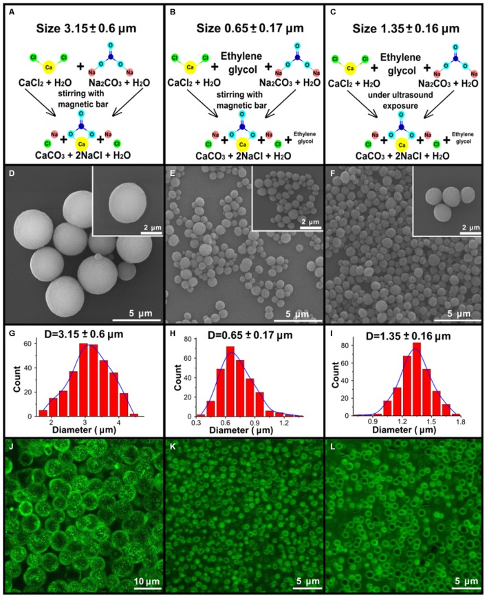
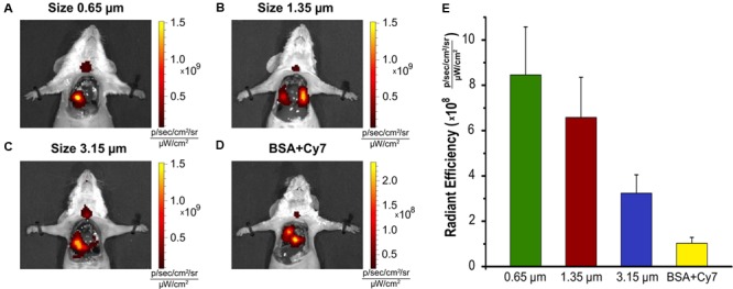
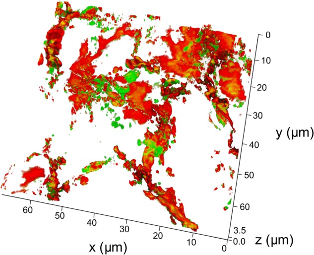
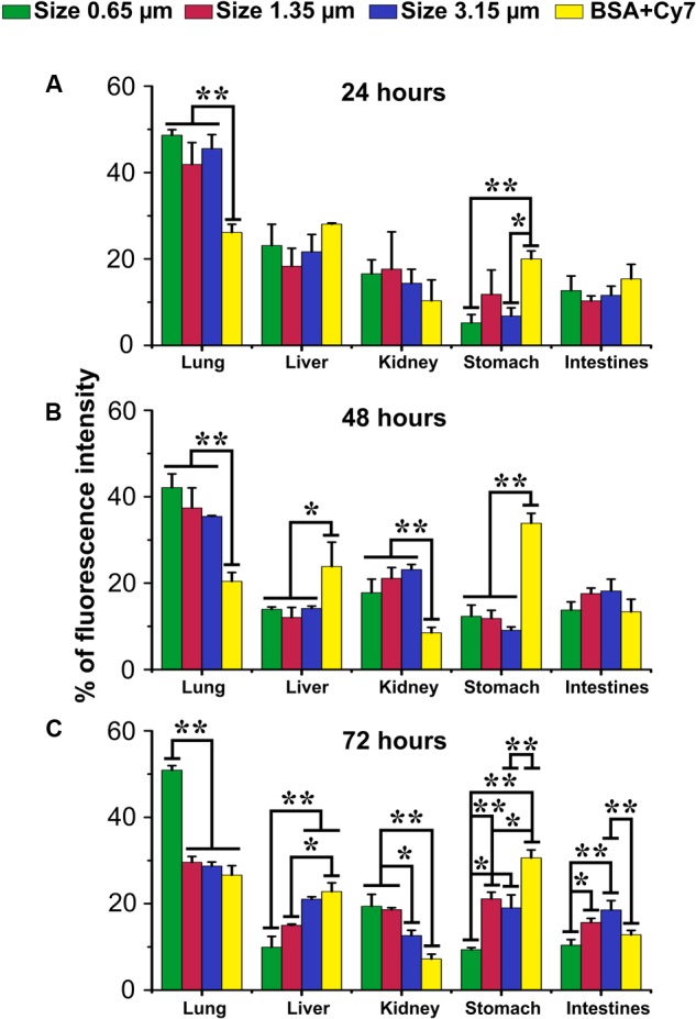
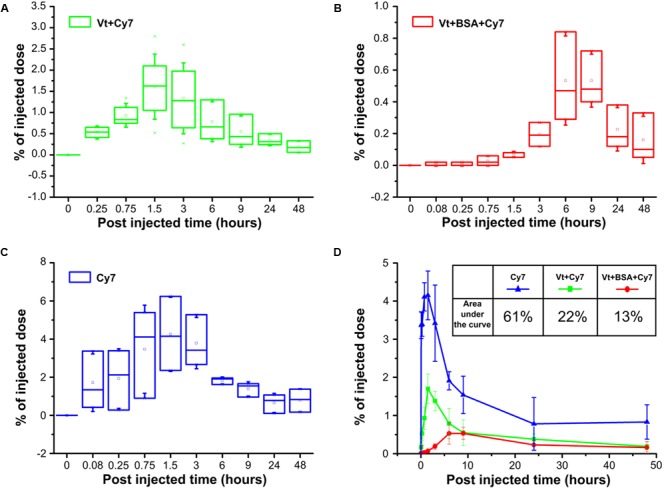
Nano- and microencapsulation has proven to be a useful technique for the construction of drug delivery vehicles for use in vascular medicine. However, the possibility of using these techniques within the lung as an inhalation delivery mechanism has not been previously considered. A critical element of particle delivery to the lung is the degree of penetrance that can be achieved with respect to the airway tree. In this study we examined the effectiveness of near infrared (NIR) dye (Cy7) labeled calcium carbonate (vaterite) particles of 3.15, 1.35, and 0.65 μm diameter in reaching the respiratory portion of the lung. First of all, it was shown that, interaction vaterite particles and the components of the pulmonary surfactant occurs a very strong retardation of the recrystallization and dissolution of the particles, which can subsequently be used to create systems with a prolonging release of bioactive substances after the particles penetrate the distal sections of the lungs. Submicro- and microparticles, coated with Cy7 labeled albumin as a model compound, were delivered to mouse lungs via tracheostomy with subsequent imaging performed 24, 48, and 72 h after delivery by in vivo fluorescence. 20 min post administration particles of all three sizes were visible in the lung, with the deepest penetrance observed with 0.65 μm particles. In vivo biodistribution was confirmed by fluorescence tomography imaging of excised organs post 72 h. Laser scanning confocal microscopy shows 0.65 μm particles reaching the alveolar space. The delivery of fluorophore to the blood was assessed using Cy7 labeled 0.65 μm particles. Cy7 labeled 0.65 μm particles efficiently delivered fluorescent material to the blood with a peak 3 h after particle administration. The pharmacokinetics of NIR fluorescence dye will be shown. These studies establish that by using 0.65 μm particles loaded with Cy7 we can efficiently access the respiratory portion of the lung, which represents a potentially efficient delivery mechanism for both the lung and the vasculature.

Keywords: drug carriers; prolonging release; pulmonary drug delivery; size-dependent biodistribution; vaterite particles.


Figures

FIGURE 1
FIGURE 1
Synthesis and characterization of the vaterite particles. (A–C) Schematic presentation of the three types of synthesis to obtain 3.15, 0.65, and 1.35 μm vaterite particles as described in the “Materials and Methods” Section. (D–F) The scanning electron microscopy (SEM) images for vaterite particles with size diameters of 3.15, 0.65, and 1.35 μm vaterite particles. (G–I) The obtained particles size distributions. The diameters of 300 particles were measured using the free software ImageJ. (J–L) The fluorescent images of particles adsorbed with BSA-Cy7- conjugate, obtained by means of laser scanning confocal microscopy.
FIGURE 2
FIGURE 2
Interaction of vaterite particles with surfactant in vitro. SEM images of the particles incubated with (A) deionized water, (B) saline, (C) small, and (D) large aggregate surfactant fractions at indicated time. (E) Time-dependent changes of the size of calcite aggregates during incubation of the 0.65 μm vaterite particles with various biosolutions, significant difference (p < 0.01) appears after 7 h incubation in water and saline but for pair small (SAF) and large aggregate surfactant fraction (LAF) the significant difference (p < 0.05) takes place after 56 h of incubation. (F) SEM images of vaterite particles coated with LA surfactant fraction 56 h post incubation.
FIGURE 3
FIGURE 3
Biodistribution of the vaterite particles in the lung. Various size of the particles (A) 0.65 μm, (B) 1.35 μm, (C) 3.15 μm adsorbed with BSA-Cy7-conjugate, and (D) BSA-Cy7 alone were instilled through the tracheostomy. Mice were sacrificed 20 min post-instillation, the chest was open and a fluorescence imaging of the lung was performed for each animal. (E) The histogram of the average Radiant Efficiency within the target organ. The dose of fluorescent dye Cy7 was 300 ng for each injection. BSA-Cy7 conjugate alone was used as control. Data are expressed as mean value ± SD, n = 3 mice per group.
FIGURE 4
FIGURE 4
Confocal fluorescence images of lung cryosection. Lung sections were analyzed on a laser scanning confocal microscope 20 min post intratracheal administration of 0.65 μm particles adsorbed with BSA-Cy7 conjugate. Shown a representable lung section (n = 3–5 lung section per mouse, n = 3 mice). (A) a fluorescent signal in a range spectra of 680–726 nm, corresponded to autofluorescence of lung tissue. (B) A fluorescent signal in a range spectra of 747–794 nm, corresponded to fluorescent dye Cy7. (C) Combination of two channels from (A,B). (D) Overlay of the optical image and two fluorescent channels as shown on (C). (E) Emission spectra of lung tissue 20 min after intratracheal administration of 0.65 μm particles adsorbed with BSA-Cy7 conjugate (red curve), BSA- Cy7 alone (green curve) or saline (blue curve). Data are expressed as mean value ± SD, n = 5 spectra intensity point per each administrated substance.
FIGURE 5
FIGURE 5
3D reconstruction of a lung cryosection fluorescent image. The particles of the 0.65 μm adsorbed with BSA-Cy7-conjugate were intratracheally administrated and mice were sacrificed 20 min post-instillation. Scanned confocal images of lung sections (n = 3–5 lung sections per mouse, n = 3 mice) were 3D reconstructed and analyzed. Thickness of 3D reconstruction was 3.5 μm. The step of constructing confocal planes was 0.2 μm. Shown representative image of 3D reconstructed lung section.
FIGURE 6
FIGURE 6
Biodistribution of submicron vaterite particles in vivo. Biodistribution within organs of 0.65, 1.35, and 3.15 μm size particles adsorbed with BSA-Cy7 conjugate or BSA-Cy7 alone after (A) 24 h, (B) 48 h, and (C) 72 h intratracheal administration. Data are expressed as mean value ± SD, n = 3 mice per group. p < 0.05, ∗∗p < 0.01.
FIGURE 7
FIGURE 7
Pharmacokinetics of submicron vaterite particles in vivo. Blood samples were analyzed for % of injected dose at indicated time points (5, 15, and 45 min, 1.5, 3, 6, 9, 24, and 48 h) after intratracheally administration of 0.65 μm particles adsorbed with (A) Cy7 dye or (B) BSA-Cy7 conjugate. (C) Fluorescent dye Cy7 alone was used as a control. The dose of fluorescent dye Cy7 was 300 ng for each administration. (D) Average percentages of the released fluorescent dye Cy7 to the blood within 48 h post intratracheally administration. The significant differences between intratracheal instillation of free fluorescence dye Cy7 and adsorbed dye or conjugate with BSA into vaterite particles possess a value p < 0.01 meanwhile the difference between instillation of vaterite particles with absorbed dye and absorbed conjugate is significant (p < 0.05) until 6 h. The inset shows the values of the areas under the curves in the case of three different input substances.

Similar articles

Cited by

References

    1. Aggarwal P., Hall J. B., McLeland C. B., Dobrovolskaia M. A., McNeil S. E. (2009). Nanoparticle interaction with plasma proteins as it relates to particle biodistribution, biocompatibility and therapeutic efficacy. Adv. Drug Deliv. Rev. 61 428–437. 10.1016/j.addr.2009.03.009 - DOI - PMC - PubMed
    1. Atochina E. N., Beers M. F., Hawgood S., Poulain F., Davis C., Fusaro T. (2004). A Gow. Surfactant protein-D, a mediator of innate lung immunity, alters the products of nitric oxide metabolism. Am. J. Respir. Cell Mol. Biol. 30 271–279. PMID: 12871850 10.1165/rcmb.2003-0091OC - DOI - PubMed
    1. Atochina-Vasserman E. N., Gow A. J., Abramova H., Guo C. J., Kadire H., Tomer Y., et al. (2009). Immune reconstitution during Pneumocystis lung infection: disruption of surfactant component expression and function by S-nitrosylation. J. Immunol. 182 2277–2287. 10.4049/jimmunol.0802775 - DOI - PMC - PubMed
    1. Biradar S., Ravichandran P., Gopikrishnan R., Goornavar V., Hall J. C., Ramesh V., et al. (2011). Calcium carbonate nanoparticles: synthesis, characterization and biocompatibility. J. Nanosci. Nanotechnol. 11 6868–6874. 10.1166/jnn.2011.4251 - DOI - PubMed
    1. Borodina T. N., Trushina D. B., Marchenko I. V., Bukreeva T. V. (2016). Calcium carbonate-based mucoadhesive microcontainers for intranasal delivery of drugs bypassing the blood–brain barrier. BioNanoScience 6 261–268. 10.1007/s12668-016-0212-2 - DOI