. 2018 Jun 4:9:1197.
doi: 10.3389/fimmu.2018.01197. eCollection 2018.

Cytokine Secretion and Pyroptosis of Thyroid Follicular Cells Mediated by Enhanced NLRP3, NLRP1, NLRC4, and AIM2 Inflammasomes Are Associated With Autoimmune Thyroiditis

Affiliations

Cytokine Secretion and Pyroptosis of Thyroid Follicular Cells Mediated by Enhanced NLRP3, NLRP1, NLRC4, and AIM2 Inflammasomes Are Associated With Autoimmune Thyroiditis

Qingling Guo et al. Front Immunol. .

Abstract

Background: Inflammasomes, which mediate maturation of interleukin-1β (IL-β) and interleukin-18 (IL-18) and lead to pyroptosis, have been linked to various autoimmune disorders. This study investigated whether they are involved in the pathogenesis of autoimmune thyroiditis (AIT).

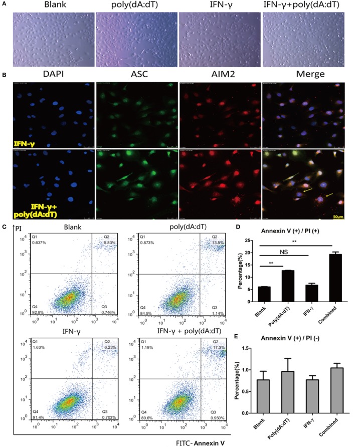
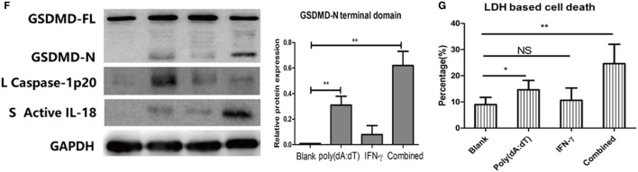
Methods: We collected thyroid tissues from 50 patients with AIT and 50 sex- and age-matched controls. Serum levels of free T3, free T4, thyrotropin, thyroid peroxidase antibody (TPOAb), and thyroglobulin antibody (TgAb) were measured by electrochemiluminescent immunoassays. Expression of several inflammasome components, the NOD-like receptor (NLR) family pyrin domain containing 1 (NLRP1), NLRP3, CARD-domain containing 4 (NLRC4), absent in melanoma 2 (AIM2), the apoptosis-associated speck-like protein that contains a caspase recruitment domain (ASC), caspase-1, IL-1β, and IL-18 was determined by real-time PCR and western blot. Immunohistochemistry was used to localize the expression of NLRP1, NLRP3, NLRC4, and AIM2. The Nthy-ori 3-1 thyroid cell line was stimulated with tumor necrosis factor-α (TNF-α), interferon-γ (IFN-γ), interleukin-17A, interleukin-6, and poly(dA:dT). The levels of IL-18 and IL-1β in the cell supernatant were measured by enzyme-linked immunosorbent assay, and lactate dehydrogenase was quantified by absorptiometry. ASC specks were examined by confocal immunofluorescence microscopic analysis. Cell death was examined by flow cytometry, and the N-terminal domain of gasdermin D was detected by western blot analysis.

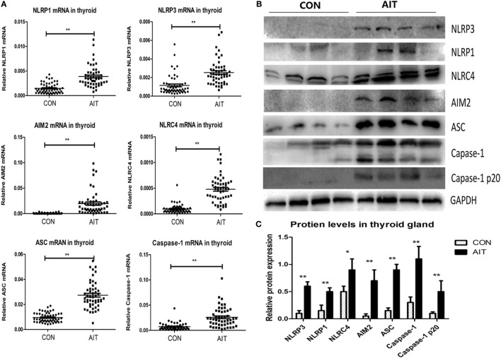
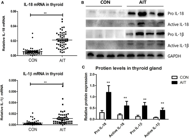
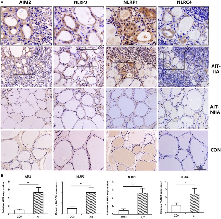
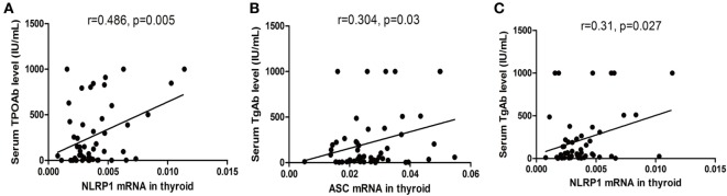
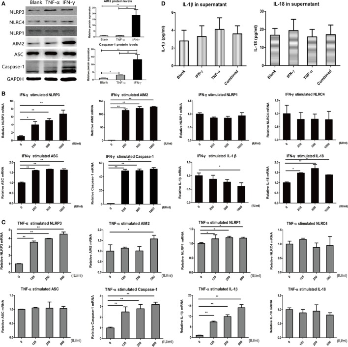
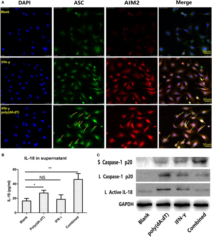
Results: Expression of NLRP1, NLRP3, NLRC4, AIM2, ASC, caspase-1, pro IL-1β, pro IL-18, mRNA, and protein was significantly increased in thyroid tissues from patients with AIT, and enhanced posttranslational maturation of caspase-1, IL-18 and IL-1β was also observed. Expression of NLRP1, NLRP3, NLRC4, and AIM2 was localized mainly in thyroid follicular cells adjacent to areas of lymphatic infiltration. The thyroid mRNA level of NLRP1 and ASC was correlated to the serum TPOAb and TgAb levels in the AIT group. TNF-α and IFN-γ had a priming effect on the expression of multiple inflammasome components in thyroid cells. IFN-γ was found to strengthen poly(dA:dT)-induced cell pyroptosis and bioactive IL-18 release.

Conclusion: Our work has demonstrated for the first time that multiple inflammasomes are associated with AIT pathogenesis. The identified NLRP3, NLRP1, NLRC4, AIM2 inflammasomes and their downstream cytokines may represent potential therapeutic targets and biomarkers of AIT.

Keywords: absent in melanoma 2; autoimmune thyroiditis; inflammasome; interleukin-18 (IL-18); pyroptosis.


Figures

Figure 1
Figure 1
Increased mRNA and protein expression of inflammasome components in thyroid tissues from AIT patients. (A) The mRNA expression levels of NLRP1, NLRP3, NLRC4, absent in melanoma 2 (AIM2), ASC, and caspase-1 in thyroid tissues from AIT patients and controls (N = 50 in each group). Representative stripe images (B) and corresponding quantitative analysis (C) of the NLRP1, NLRP3, NLRC4, AIM2, ASC, caspase-1 and caspase-1 p20 protein levels in thyroid tissues from AIT patients and controls (N = 20 in each group). The relative mRNA and protein expression levels were corrected to GAPDH expression. The columns represent the mean ± SD values. Student’s t-test or Mann–Whitney U-test was used for comparison between the AIT and control groups. *P < 0.05, **P < 0.01. AIT, autoimmune thyroiditis; CON, controls.
Figure 2
Figure 2
Increased expression and maturation of IL-18 and IL-1β in thyroid tissues from AIT patients. (A) The mRNA expression levels of IL-18 and IL-1β in thyroid tissues from AIT patients and controls (N = 50 in each group). Representative stripe images (B) and corresponding quantitative analysis (C) of pro IL-18, active IL-18, pro IL-1β, and active IL-1β protein levels in thyroid tissues from AIT patients and controls (N = 20 in each group). The relative mRNA and protein expression levels were corrected to GAPDH expression. The columns represent the mean ± SD values. Student’s t-test or Mann–Whitney U-test was used for comparison between the AIT and control groups. *P < 0.05, **P < 0.01. AIT, autoimmune thyroiditis; CON, controls.
Figure 3
Figure 3
Localization of enhanced expression of NLRP1, NLRP3, NLRC4, and absent in melanoma 2 (AIM2) in thyroid cells in areas of lymphatic infiltration. (A) Representative immunohistochemical staining images of AIM2, NLRP3, NLRP1, and NLRC4 in thyroid tissue sections from AIT patients and controls (400× magnification, light microscope). (B) Quantified expression levels of AIM2, NLRP3, NLRP1, and NLRC4 in control and AIT tissues (N = 20 in each group). The columns represent the mean ± SD values. Three views were randomly selected in each subject. Statistical analysis was carried out using Student’s t-test. *P < 0.05, **P < 0.01. AIT, autoimmune thyroiditis; CON, controls; IIA, inflammatory infiltration area; NIIA, non-inflammatory infiltration area.
Figure 4
Figure 4
Correlation between the mRNA levels of inflammasome components in thyroid tissues and autoantibody levels in the serum in autoimmune thyroiditis (AIT) patients. Correlation between the NLRP1/ASC mRNA levels in thyroid tissue and serum thyroid peroxidase antibody (TPOAb)/thyroglobulin antibody (TgAb) levels in the AIT group (A–C). N = 50 in each group. A bivariate correlation analysis was performed using the Spearman rank test. R represents the Spearman correlation coefficient.
Figure 5
Figure 5
Increased expression of inflammasome components in thyroid cells on stimulation with IFN-γ and tumor necrosis factor-α (TNF-α). (A) Changes in NLRP3, absent in melanoma 2 (AIM2), NLRP1, NLRC4, ASC, and caspase-1 protein expression detected by western blot after incubation with 500 IU/ml IFN-γ or 500 IU/ml TNF-α for 24 h. Changes in NLRP3, AIM2, NLRP1, NLRC4, ASC, Caspase-1, IL-1β, and IL-18 mRNA expression detected by real-time PCR after incubation with (B) gradient IFN-γ (250, 500, and 1,000 IU/ml) for 24 h or (C) gradient TNF-α (125, 250, and 500 IU/ml) for 24 h. The relative mRNA and protein expression levels were corrected to those of untreated controls. (D) IL-18 and IL-1β levels in cell supernatant, as determined by enzyme-linked immunosorbent assay, after stimulation with 500 IU/ml IFN-γ, 500 IU/ml TNF-α or a combination of the two for 24 h. The columns represent the mean ± SD values. Data were obtained from three independent experiments. One-way ANOVA followed by Bonferroni correction was used for paired comparisons. *P < 0.05, **P < 0.01.
Figure 6
Figure 6
Promotion of poly(dA:dT)-induced activation of the absent in melanoma 2 (AIM2) inflammasome and release of active IL-18 in thyroid cells on IFN-γ stimulation. (A) Immunofluorescence analysis showed that ASC specks (green color, marked by arrows) were co-localized with AIM2 (red color) after the cells were stimulated with 1 µg/ml poly(dA:dT) for 12 h and pre-incubated with 250 IU/ml IFN-γ for 24 h (400× magnification, confocal laser-scanning microscopy). (B) IL-18 levels in cell supernatants determined by enzyme-linked immunosorbent assay after stimulation with poly(dA:dT) alone, IFN-γ alone, and both IFN-γ and poly(dA:dT). (C) Protein expression of caspase-1 p20 and active IL-18 in thyroid cells and supernatants after stimulation with poly(dA:dT) alone, IFN-γ alone, and both IFN-γ and poly(dA:dT). The columns represent the mean ± SD values. Data were obtained from three independent experiments. Statistical analysis was conducted by one-way ANOVA followed by Bonferroni correction for paired comparisons. *P < 0.05, **P < 0.01, NS, no significant difference; S, supernatants; L, lysates.
Figure 7
Figure 7
Promotion of poly(dA:dT)-induced thyroid cell pyroptosis on IFN-γ stimulation. Morphological changes in thyroid cells under a light microscope at 100× magnification (A) or under a confocal laser-scanning microscope at 400× magnification (B) after stimulation with 1 µg/ml poly(dA:dT) for 12 h after pre-incubation with 250 IU/ml IFN-γ for 24 h. (C) Representative flow cytometry images of FITC-Annexin V/propidium iodide (PI)-stained tissue after stimulation. Quantification of the percentage of Annexin V(+)/PI(+) and Annexin V(+)/PI(−) cells from flow cytometry findings (D,E). (F) Protein expression of full-length gasdermin D and the N-terminal cleavage product in thyroid cells after stimulation with poly(dA:dT) alone, IFN-γ alone, and both IFN-γ and poly(dA:dT). Relative protein expression level was corrected to GAPDH expression. Caspase-1 p20 and active IL-18 expression indicates inflammasome activation. (G) Lactate dehydrogenase (LDH) levels in cells supernatants determined by absorptiometry after stimulation with poly(dA:dT) alone, IFN-γ alone, and both IFN-γ and poly(dA:dT). The columns represent the mean ± SD values. Data were obtained from three independent experiments. One-way ANOVA followed by Bonferroni correction was used for paired comparisons. *P < 0.05, **P < 0.01, NS, no significant difference; S, supernatants; L, lysates.
Figure 7
Figure 7
Promotion of poly(dA:dT)-induced thyroid cell pyroptosis on IFN-γ stimulation. Morphological changes in thyroid cells under a light microscope at 100× magnification (A) or under a confocal laser-scanning microscope at 400× magnification (B) after stimulation with 1 µg/ml poly(dA:dT) for 12 h after pre-incubation with 250 IU/ml IFN-γ for 24 h. (C) Representative flow cytometry images of FITC-Annexin V/propidium iodide (PI)-stained tissue after stimulation. Quantification of the percentage of Annexin V(+)/PI(+) and Annexin V(+)/PI(−) cells from flow cytometry findings (D,E). (F) Protein expression of full-length gasdermin D and the N-terminal cleavage product in thyroid cells after stimulation with poly(dA:dT) alone, IFN-γ alone, and both IFN-γ and poly(dA:dT). Relative protein expression level was corrected to GAPDH expression. Caspase-1 p20 and active IL-18 expression indicates inflammasome activation. (G) Lactate dehydrogenase (LDH) levels in cells supernatants determined by absorptiometry after stimulation with poly(dA:dT) alone, IFN-γ alone, and both IFN-γ and poly(dA:dT). The columns represent the mean ± SD values. Data were obtained from three independent experiments. One-way ANOVA followed by Bonferroni correction was used for paired comparisons. *P < 0.05, **P < 0.01, NS, no significant difference; S, supernatants; L, lysates.

Similar articles

Cited by

References

    1. Antonelli A, Ferrari SM, Corrado A, Di Domenicantonio A, Fallahi P. Autoimmune thyroid disorders. Autoimmun Rev (2015) 14(2):174–80.10.1016/j.autrev.2014.10.016 - DOI - PubMed
    1. Shan Z, Chen L, Lian X, Liu C, Shi B, Shi L, et al. Iodine status and prevalence of thyroid disorders after introduction of mandatory universal salt iodization for 16 years in China: a cross-sectional study in 10 cities. Thyroid (2016) 26(8):1125–30.10.1089/thy.2015.0613 - DOI - PubMed
    1. Pyzik A, Grywalska E, Matyjaszek-Matuszek B, Rolinski J. Immune disorders in Hashimoto’s thyroiditis: what do we know so far? J Immunol Res (2015) 2015:979167.10.1155/2015/979167 - DOI - PMC - PubMed
    1. Dong YH, Fu DG. Autoimmune thyroid disease: mechanism, genetics and current knowledge. Eur Rev Med Pharmacol Sci (2014) 18(23):3611–8. - PubMed
    1. Kawashima A, Tanigawa K, Akama T, Yoshihara A, Ishii N, Suzuki K. Innate immune activation and thyroid autoimmunity. J Clin Endocrinol Metab (2011) 96(12):3661–71.10.1210/jc.2011-1568 - DOI - PubMed

Publication types

MeSH terms

LinkOut - more resources