

Imbalance of Circulating Th17 and Regulatory T Cells in Alzheimer's Disease: A Case Control Study
- PMID: 29915582
- PMCID: PMC5994416
- DOI: 10.3389/fimmu.2018.01213
Imbalance of Circulating Th17 and Regulatory T Cells in Alzheimer's Disease: A Case Control Study
Abstract
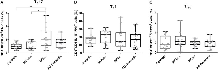
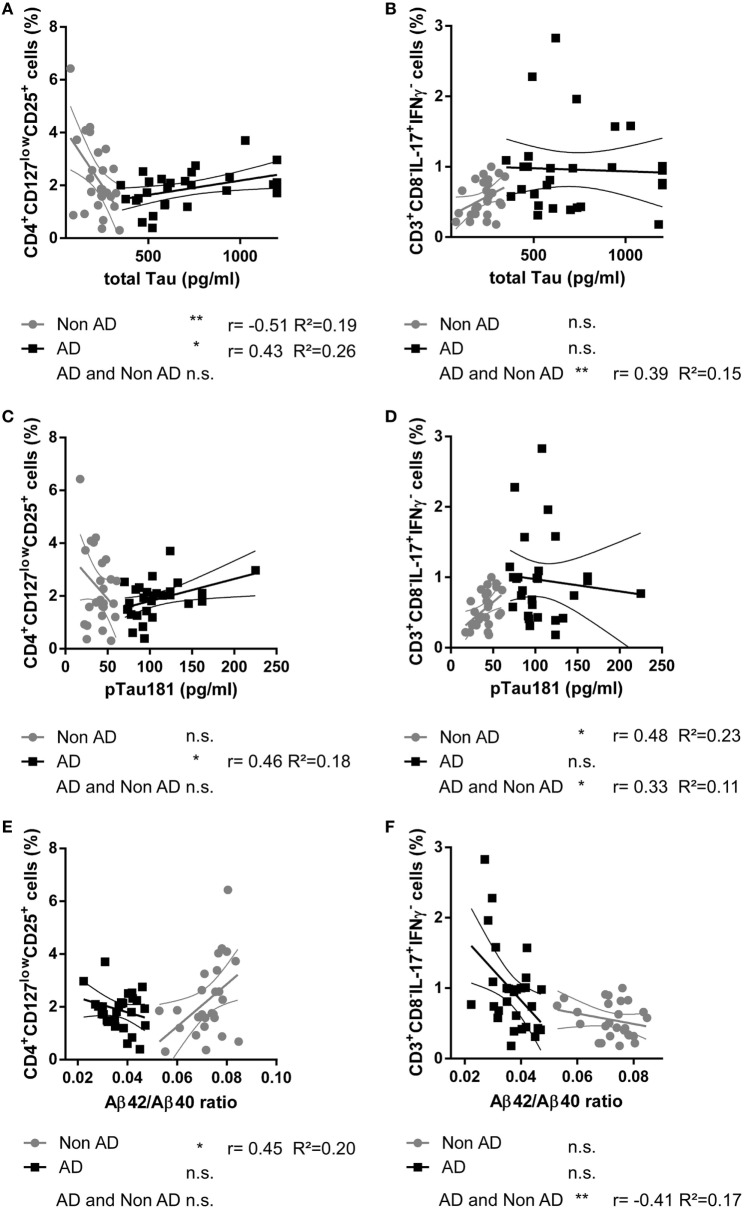
The neuropathological hallmarks of Alzheimer's disease (AD), i.e., neuritic plaques and neurofibrillary tangles, consist of beta amyloid peptides (Aβ) and hyperphosphorylated Tau. These are accompanied by reactive microglia and astrocytes in the vicinity of the neuritic plaques and by changes to the peripheral immune system, e.g., an increase of the pro-inflammatory cytokines IL-1β, IL-6, and TNF-α in the peripheral blood. To address a potential involvement of peripheral T helper cell (Th) subsets in AD, we conducted a case control study with 54 individuals with AD dementia (n = 14), with mild cognitive impairment (MCI) due to AD (MCIAD, n = 14), with MCI unlikely due to AD (MCIother, n = 13), and controls without cognitive impairment (controls, n = 13). The proportions of CD3+CD8-IL-17A+IFNγ- Th17 cells, CD3+CD8-IL-17A-IFNγ+ Th1 cells, and CD4+CD127lowCD25+ regulatory T cells (Tregs) were assessed by flow cytometry. In addition, the correlations of the proportions of Th subsets to cerebrospinal fluid biomarkers were studied. CD3+CD8-IL-17A+IFNγ- Th17 cells were significantly increased in subjects with MCIAD compared to age- and sex-matched subjects with MCIother and controls (MCIAD mean = 1.13, SD = 0.77; MCIother mean = 0.58, SD = 0.28; and controls mean = 0.52, SD = 0.22; p = 0.008). The proportion of CD4+CD127lowCD25+ Tregs was not altered between the different groups, but it significantly positively related with the levels of total Tau and pTau181 (rTreg|totalTau = 0.43, p = 0.021, n = 28; rTreg|pTau181 = 0.46; p = 0.024, n = 28) in subjects with AD but not in nonAD controls (rTreg|totalTau = -0.51, p = 0.007, n = 26). The increase of circulating CD3+CD8-IL-17A+IFNγ- Th17 cells in the early stages of AD and the association of CD4+CD127lowCD25+ Tregs with neurodegeneration marker Tau may indicate that the adaptive immune system relates to neuropathological changes in AD.
Keywords: Alzheimer’s disease; Tau; Th1; Th17; amyloid beta; mild cognitive impairment; regulatory T cell.
Figures
References
Publication types
MeSH terms
Substances
LinkOut - more resources
Full Text Sources
Other Literature Sources
Medical
Research Materials