

IL-3 Is a Marker of Encephalitogenic T Cells, but Not Essential for CNS Autoimmunity
- PMID: 29915594
- PMCID: PMC5994593
- DOI: 10.3389/fimmu.2018.01255
IL-3 Is a Marker of Encephalitogenic T Cells, but Not Essential for CNS Autoimmunity
Abstract
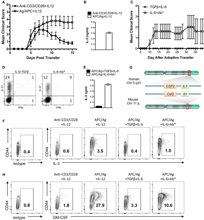
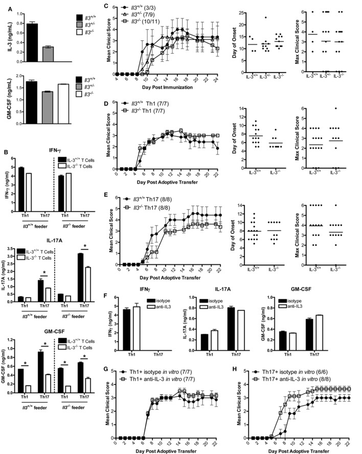
Identifying molecules that are differentially expressed in encephalitogenic T cells is critical to the development of novel and specific therapies for multiple sclerosis (MS). In this study, IL-3 was identified as a molecule highly expressed in encephalitogenic Th1 and Th17 cells, but not in myelin-specific non-encephalitogenic Th1 and Th17 cells. However, B10.PL IL-3-deficient mice remained susceptible to experimental autoimmune encephalomyelitis (EAE), a mouse model of MS. Furthermore, B10.PL myelin-specific T cell receptor transgenic IL-3-/- Th1 and Th17 cells were capable of transferring EAE to wild-type mice. Antibody neutralization of IL-3 produced by encephalitogenic Th1 and Th17 cells failed to alter their ability to transfer EAE. Thus, IL-3 is highly expressed in myelin-specific T cells capable of inducing EAE compared to activated, non-encephalitogenic myelin-specific T cells. However, loss of IL-3 in encephalitogenic T cells does not reduce their pathogenicity, indicating that IL-3 is a marker of encephalitogenic T cells, but not a critical element in their pathogenic capacity.
Keywords: GM-CSF; IL-3; Th17 cells; Th1 cells; experimental autoimmune encephalomyelitis; multiple sclerosis.
Figures
Similar articles
-
IL-7/IL-7 Receptor Signaling Differentially Affects Effector CD4+ T Cell Subsets Involved in Experimental Autoimmune Encephalomyelitis.J Immunol. 2015 Sep 1;195(5):1974-83. doi: 10.4049/jimmunol.1403135. Epub 2015 Jul 29. J Immunol. 2015. PMID: 26223651 Free PMC article.
-
IL-12/IL-23p40 Is Highly Expressed in Secondary Lymphoid Organs and the CNS during All Stages of EAE, but Its Deletion Does Not Affect Disease Perpetuation.PLoS One. 2016 Oct 25;11(10):e0165248. doi: 10.1371/journal.pone.0165248. eCollection 2016. PLoS One. 2016. PMID: 27780253 Free PMC article.
-
IL-10 mediates resistance to adoptive transfer experimental autoimmune encephalomyelitis in MyD88(-/-) mice.J Immunol. 2010 Jan 1;184(1):212-21. doi: 10.4049/jimmunol.0900296. Epub 2009 Nov 30. J Immunol. 2010. PMID: 19949074
-
Role of Th17 cells in the pathogenesis of CNS inflammatory demyelination.J Neurol Sci. 2013 Oct 15;333(1-2):76-87. doi: 10.1016/j.jns.2013.03.002. Epub 2013 Apr 8. J Neurol Sci. 2013. PMID: 23578791 Free PMC article. Review.
-
Using EAE to better understand principles of immune function and autoimmune pathology.J Autoimmun. 2013 Sep;45:31-9. doi: 10.1016/j.jaut.2013.06.008. Epub 2013 Jul 9. J Autoimmun. 2013. PMID: 23849779 Free PMC article. Review.
Cited by
-
Sample Size for Oxidative Stress and Inflammation When Treating Multiple Sclerosis with Interferon-β1a and Coenzyme Q10.Brain Sci. 2019 Sep 27;9(10):259. doi: 10.3390/brainsci9100259. Brain Sci. 2019. PMID: 31569668 Free PMC article.
-
Innate Immune Modulation by GM-CSF and IL-3 in Health and Disease.Int J Mol Sci. 2019 Feb 15;20(4):834. doi: 10.3390/ijms20040834. Int J Mol Sci. 2019. PMID: 30769926 Free PMC article. Review.
-
Regulation of Lymphatic GM-CSF Expression by the E3 Ubiquitin Ligase Cbl-b.Front Immunol. 2018 Oct 8;9:2311. doi: 10.3389/fimmu.2018.02311. eCollection 2018. Front Immunol. 2018. PMID: 30349541 Free PMC article.
-
Twin study reveals non-heritable immune perturbations in multiple sclerosis.Nature. 2022 Mar;603(7899):152-158. doi: 10.1038/s41586-022-04419-4. Epub 2022 Feb 16. Nature. 2022. PMID: 35173329 Free PMC article.
-
Infiltration by monocytes of the central nervous system and its role in multiple sclerosis: reflections on therapeutic strategies.Neural Regen Res. 2025 Mar 1;20(3):779-793. doi: 10.4103/NRR.NRR-D-23-01508. Epub 2024 Apr 3. Neural Regen Res. 2025. PMID: 38886942 Free PMC article.
References
-
- Lovett-Racke AE, Trotter JL, Lauber J, Perrin PJ, June CH, Racke MK. Decreased dependence of myelin basic protein-reactive T cells on CD28-mediated costimulation in multiple sclerosis patients: a marker of activation/memory T cells. J Clin Invest (1998) 101(4):725–30.10.1172/JCI1528 - DOI - PMC - PubMed
Publication types
MeSH terms
Substances
Grants and funding
LinkOut - more resources
Full Text Sources
Other Literature Sources
Molecular Biology Databases