Review
. 2018 Jun 4:9:1262.
doi: 10.3389/fimmu.2018.01262. eCollection 2018.

Diversity in the Cow Ultralong CDR H3 Antibody Repertoire

Affiliations
Review

Diversity in the Cow Ultralong CDR H3 Antibody Repertoire

Jeremy K Haakenson et al. Front Immunol. .

Abstract

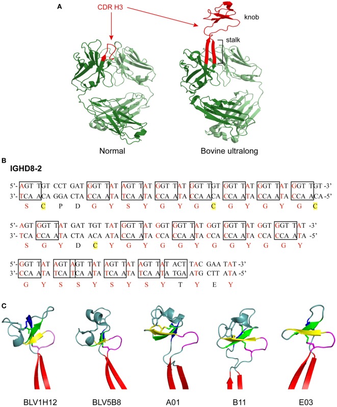
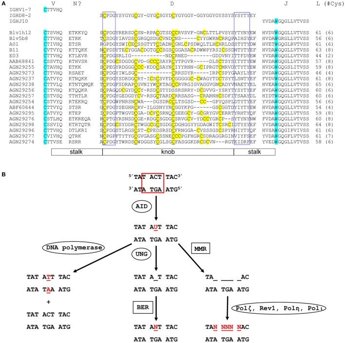
Typical antibodies found in humans and mice usually have short CDR H3s and generally flat binding surfaces. However, cows possess a subset of antibodies with ultralong CDR H3s that can range up to 70 amino acids and form a unique "stalk and knob" structure, with the knob protruding far out of the antibody surface, where it has the potential to bind antigens with concave epitopes. Activation-induced cytidine deaminase (AID) has a proven role in diversifying antibody repertoires in humoral immunity, and it has been found to induce somatic hypermutation in bovine immunoglobulin genes both before and after contact with antigen. Due to limited use of variable and diversity genes in the V(D)J recombination events that produce ultralong CDR H3 antibodies in cows, the diversity in the bovine ultralong antibody repertoire has been proposed to rely on AID-induced mutations targeted to the IGHD8-2 gene that encodes the entire knob region. In this review, we discuss the genetics, structures, and diversity of bovine ultralong antibodies, as well as the role of AID in creating a diverse antibody repertoire.

Keywords: activation-induced cytidine deaminase; antibody; antibody repertoire; cow immunoglobulin; ultralong CDRH3.


Figures

Figure 1
Figure 1
Structure and genetics of ultralong complementarity determining region (CDR) H3 cow antibodies. (A) Comparison of normal and ultralong CDR H3 antibody fab fragments. Crystal structures of Yvo, a typical human antibody (left), and B11, an ultralong bovine antibody (right). The CDR H3 of each antibody is highlighted in red. Heavy chains are colored dark green and light chains are colored pale green. Note the two long β strands that make up the stalk of the bovine ultralong CDR H3 and the disulfide-bonded knob domain at the top. PDB IDs of these two fabs are Yvo: 2AGJ; B11: 5IHU. (B) Sequence and potential diversity of the ultralong DH region. Potential AID-induced somatic hypermutation hotspot motifs (“RGYW”/“WRCY”) in bovine germline IGHD8-2 are boxed on both strands of the DNA sequence. Nucleotides within a codon that can be mutated to a cysteine-encoding codon with just a single nucleotide change are colored red, and corresponding amino acids encoded by these codons are also colored red. Four cysteine residues encoded by the germline IGHD8-2 are highlighted yellow. (C) “Stalk and knob” structures of the ultralong CDR H3s. The β-ribbon stalk is colored red, the type I β-turn is colored magenta, and the three antiparallel β strands are colored yellow, green and blue, respectively. Disulfide bonds formed within the knob region are represented as sticks. PDB IDs of these five fabs are BLV1H12: 4K3D; BLV5B8: 4K3E; A01: 5ILT; B11: 5IHU; and 5IJV.
Figure 2
Figure 2
Sequence diversity and genetic mutation in ultralong complementarity determining region (CDR) H3 regions. (A) Alignment showing diversity of bovine ultralong CDR H3. Residues encoded by germline IGHV1-7, IGHD8-2, and IGHJ10 are shown on the top of the alignment for reference. The start and the end of the CDR H3 are defined by the conserved cysteine and tryptophan which are colored turquoise. N? indicates residues possibly encoded by terminal deoxynucleotidyl transferase catalyzed N additions or conserved short nucleotide sequence insertions at the V–D junction. Cysteine residues in the D region are colored yellow and the unmutated germline-encoded cysteines are also underlined. The conserved CPDG and YxYxY motifs near the beginning and the end of the D region are enclosed in blue boxes, respectively. The length of each CDR H3 and the number of cysteine residues (in parenthesis) it contains is shown on the right of the alignment. The stalk and knob regions are indicated below the alignment. The alignment was performed using MAFFT, then manually adjusted to align the conserved residues. The first five sequences with crystal structures available are named according to published nomenclature, and the remaining sequences are named with their GenBank accession numbers. (B) Mutation induction by AID. AID deaminates cytosine to uracil at an AID hotspot (boxed in red). This can be followed by three different processes that cause point mutations. DNA polymerase replicates in both strands of parental DNA, resulting in two daughter strands, one with a C to T mutation and one without (Left). Uracil-DNA glycosylase removes uracil, creating an abasic site (Middle). Error-prone base excision repair can then replace this site with any dNTP (N = A, C, G, or T). Mismatch repair removes several nucleotides around and including the uracil (right). This is then followed by error-prone DNA polymerases, which can cause a mutation at any of the sites, where nucleotides have been removed (Ovals designate single enzymes and squares designate multi-enzyme processes). Mutated nucleotides are shown in red.

Similar articles

Cited by

References

    1. Chehimi J, Valiante NM, D’andrea A, Rengaraju M, Rosado Z, Kobayashi M, et al. Enhancing effect of natural killer cell stimulatory factor (NKSF/interleukin-12) on cell-mediated cytotoxicity against tumor-derived and virus-infected cells. Eur J Immunol (1993) 23:1826–30.10.1002/eji.1830230814 - DOI - PubMed
    1. Lu LL, Suscovich TJ, Fortune SM, Alter G. Beyond binding: antibody effector functions in infectious diseases. Nat Rev Immunol (2018) 18:46–61.10.1038/nri.2017.106 - DOI - PMC - PubMed
    1. Berens SJ, Wylie DE, Lopez OJ. Use of a single VH family and long CDR3s in the variable region of cattle Ig heavy chains. Int Immunol (1997) 9:189–99.10.1093/intimm/9.1.189 - DOI - PubMed
    1. Lopez O, Perez C, Wylie D. A single VH family and long CDR3s are the targets for hypermutation in bovine immunoglobulin heavy chains. Immunol Rev (1998) 162:55–66.10.1111/j.1600-065X.1998.tb01429.x - DOI - PubMed
    1. Saini SS, Allore B, Jacobs RM, Kaushik A. Exceptionally long CDR3H region with multiple cysteine residues in functional bovine IgM antibodies. Eur J Immunol (1999) 29:2420–6.10.1002/(SICI)1521-4141(199908)29:08<2420::AID-IMMU2420>3.0.CO;2-A - DOI - PubMed

Publication types

MeSH terms