. 2018 Jun 4:9:754.
doi: 10.3389/fpls.2018.00754. eCollection 2018.

Plastid Envelope-Localized Proteins Exhibit a Stochastic Spatiotemporal Relationship to Stromules

Affiliations

Plastid Envelope-Localized Proteins Exhibit a Stochastic Spatiotemporal Relationship to Stromules

Kathleen Delfosse et al. Front Plant Sci. .

Abstract

Plastids in the viridiplantae sporadically form thin tubules called stromules that increase the interactive surface between the plastid and the surrounding cytoplasm. Several recent publications that report observations of certain proteins localizing to the extensions have then used the observations to suggest stromule-specific functions. The mechanisms by which specific localizations on these transient and sporadically formed extensions might occur remain unclear. Previous studies have yet to address the spatiotemporal relationship between a particular protein localization pattern and its distribution on an extended stromules and/or the plastid body. Here, we have used discrete protein patches found in several transgenic plants as fiducial markers to investigate this relationship. While we consider the inner plastid envelope-membrane localized protein patches of the GLUCOSE 6-PHOSPHATE/PHOSPHATE TRANSLOCATOR1 and the TRIOSE-PHOSPHATE/ PHOSPHATE TRANSLOCATOR 1 as artifacts of fluorescent fusion protein over-expression, stromule formation is not compromised in the respective stable transgenic lines that maintain normal growth and development. Our analysis of chloroplasts in the transgenic lines in the Arabidopsis Columbia background, and in the arc6 mutant, under stromule-inducing conditions shows that the possibility of finding a particular protein-enriched domain on an extended stromule or on a region of the main plastid body is stochastic. Our observations provide insights on the behavior of chloroplasts, the relationship between stromules and the plastid-body and strongly challenge claims of stromule-specific functions based solely upon protein localization to plastid extensions.

One sentence summary: Observations of the spatiotemporal relationship between plastid envelope localized fluorescent protein fusions of two sugar-phosphate transporters and stromules suggest a stochastic rather than specific localization pattern that questions the idea of independent functions for stromules.

Keywords: chloroplast proteins; fluorescent proteins; plastids; stromules; transporters.


Figures
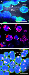
Figure 1
Figure 1
Inner envelope-localized GPT1 and TPT1 fluorescent fusion proteins showing diffuse and punctate dispersal patterns on chloroplasts. (A) Mesophyll chloroplasts in an Arabidopsis line expressing GPT1-GFP shows the diffuse highlighting of the entire plastid periphery along with a few, discrete, more fluorescent patches. (B) Representative image showing mesophyll chloroplasts with a predominantly patchy localization of GPT1-mEosFP (arrows) upon transient expression in tobacco leaves. (C) Image from a double transgenic Arabidopsis line obtained through a genetic cross between homozygous pro35S:GPT1-EGFP (diffuse highlighting) and a pro35S:TPT1-mEosFP (punctate localization). The line expressed both probes with up to 60% of chloroplasts exhibiting the two protein dispersal patterns. Chlorophyll (chl) depicted in blue color. Size bars A, B, C = 10 μm.
Figure 2
Figure 2
Spatial relationship between fluorescent fusion protein patches, the main plastid body (PB) and stromules (St). (A) Representative image showing a transporter protein fusion in the tpFNR-EGFP background. Chloroplasts appear blue-green (*) due to chlorophyll auto-fluorescence (false-colored blue) and the stroma-targeted GFP (green). Transporter fusion protein patches are red fluorescent and may be absent (1), cover short lengths (2), or extend over an entire (3) stromule. (B) A representative image showing patch localization on the ends of lens-shaped mesophyll chloroplasts. (C) Image showing a chloroplast (*) and a leucoplast (**) in a hypocotyl cell in a seedling expressing the GPT1-mEosFP fusion (observed after photo-conversion). The chloroplast body (*) shows extensions on both sides. The varying mix of the green fluorescence of the stroma and the red fluorescence of the different sized transporter-fusion-protein patches results in their colors ranging from yellow to red. The leucoplast (**) exhibits an ectopic red bulge that differs from the green stromule extending on the opposite end of the plastid. In both plastids the higher intensity of red fluorescence suggests a region with low stroma filling. (D) Assessing 1624 chloroplasts of a TPT1-mEosFP line showed that 39.2% did not have a protein patch at all while nearly equal numbers of plastids had a single patch or several patches, 29.7 and 31.1%, respectively. (E) diagrammatic depiction, accompanying legend and pie diagram show the seven categories assessed for understanding the spatial relation between patches on the chloroplast body (PB) and extended stromules (St) in chloroplasts. Amongst plastids with extended stromules there could be no patches on the stromules (category 2) while one or more patches could be found on the PB (category 2, 5). Alternatively, one to several patches could be found on stromules (category 3, 6, 7) and none on the PB. Alternatively, one or more patches could be found randomly dispersed on both St and PB (category 7). (F) Two simultaneously extended stromules (1) in a chloroplast; one appearing red (arrowhead) due to extension of a region with a red fusion-protein patch and the other appearing green as it is devoid of any patch (Supplementary Movie 1). Chloroplast (2) shows a single stromule with the tip-region exhibiting an extended red patch while the proximal region (arrow) of the stromule devoid of a protein patch appears green. (G) The fusion-protein patches could localize randomly along the length of a stromule to form unlinked regions (e.g., 1) or appear as a single long region that highlighted an entire stromule (e.g., 2). The green fluorescent stroma, denoting a region of a stromule without a patch on the inner-envelope membrane appears as a small intervening region between the patches (arrowheads) or highlights a large portion of the extended stromule tip (e.g., 1). Arrows point to large misshapen patches that bulge outwards from the plastids. Size bars B, C = 5; A, F, G = 10 μm.
Figure 3
Figure 3
Images illustrating the changes in position and the diversity in extension of fluorescent protein patches on dynamic stromules. (A) Images from a time lapse sequence showing the change in patch size in a stromule retracting (relative sized directional arrows in frames 4–7) toward the plastid body (PB) establish that neither the position or the size of a protein patch on a stromule is fixed in relation to the PB. A small stromal region not covered by the red patch (arrow in frame 2) appears greatly elongated in frame 3 (arrow) to suggest two patches. Later frames 4–7 no longer show two patches but a single red fluorescent stromule with the tip devoid of a patch. Note a green stromule extended from the opposite side of the chloroplast (*) (Supplementary Movies 2, 3). (B) Representative image of the abnormally large chloroplasts (chl) and leucoplasts (leu) in hypocotyl cells of an arc6 transgenic line expressing tpFNR-EGFP and GPT1-mEosFP. Following photo-conversion of mEosFP patches (p) of differing lengths and thickness are observed on both the plastid body and the stromules. Regions of a stromule without a patch appear green (arrowheads). (C) Image of a single arc6 chloroplast showing three separate patches (arrowheads). (D) Stromule from a chloroplast (chl) showing GPT1-mEosFP patches (*) of variable size and fluorescence intensity. The stromule also shows two dilated regions (arrowheads) that are devoid of the mEosFP patch. (E) Ten images from a time-lapse sequence depict an extended tubule (*) in panel 1 and its retraction to the plastid body (PB) in panel 2. Panel 3 shows the extension of another region of the plastid and its elongation (panels 4–10), slight beading (panels 6, 7), and branching (arrows in panel 8, 9). Note the changing position of the intervening patch-free region (*) showing the green fluorescence of the underlying stroma (Supplementary Movie 4). Size bars = 10 μm.
Figure 4
Figure 4
Exogenous feeding of seedlings with 40 mM sucrose results in more than 3-fold increase in percent stromule numbers per cell compared to the water controls. Despite increased stromule numbers the fluorescent TPT1-mEosFP fusion protein patches are evenly distributed between the plastid body and stromules. The representative data shown is from one experiment and is based on merged z-stack images from 16 cells (spread over 4 seedlings) per treatment (standard deviation shown). The data reinforces the chances of finding a patch anywhere on the plastid as stochastic.

Similar articles

Cited by

References

    1. Aranda-Sicilia M. N., Aboukila A., Armbruster U., Cagnac O., Schumann T., Kunz H.-H., et al. (2016). Envelope K+/H+ antiporters AtKEA1 and AtKEA2 function in plastid development. Plant Physiol. 172, 441–449. 10.1104/pp.16.00995 - DOI - PMC - PubMed
    1. Awai K., Xu C., Tamot B., Benning C. (2006). A phosphatidic acid-binding protein of the chloroplast inner envelope membrane involved in lipid trafficking. Proc. Natl. Acad. Sci. U.S.A. 103, 10817–10822. 10.1073/pnas.0602754103 - DOI - PMC - PubMed
    1. Barton K. A., Wozny M. R., Mathur N., Jaipargas E. A., Mathur J. (2018). Chloroplast behaviour and interactions with other organelles in Arabidopsis thaliana pavement cells. J. Cell Sci. 131:jcs202275 10.1242/jcs.202275 - DOI - PubMed
    1. Breuers F. K., Bräutigam A., Geimer S., Welzel U. Y., Stefano G., Renna L., et al. (2012). Dynamic remodeling of the plastid envelope membranes - a tool for chloroplast envelope in vivo localizations. Front. Plant Sci. 3:7 10.3389/fpls.2012.00007 - DOI - PMC - PubMed
    1. Bross C. D., Howes T. R., Abolhassani Rad S., Kljakic O., Kohalmi S. E. (2017). Subcellular localization of Arabidopsis arogenate dehydratases suggests novel and non-enzymatic roles. J. Exp. Bot. 68, 1425–1440. 10.1093/jxb/erx024 - DOI - PMC - PubMed

LinkOut - more resources