Review
. 2018:20:31-61.
doi: 10.1007/978-3-319-89689-2_2.

Mechanism of Splicing Regulation of Spinal Muscular Atrophy Genes

Affiliations
Review

Mechanism of Splicing Regulation of Spinal Muscular Atrophy Genes

Ravindra N Singh et al. Adv Neurobiol. 2018.

Abstract

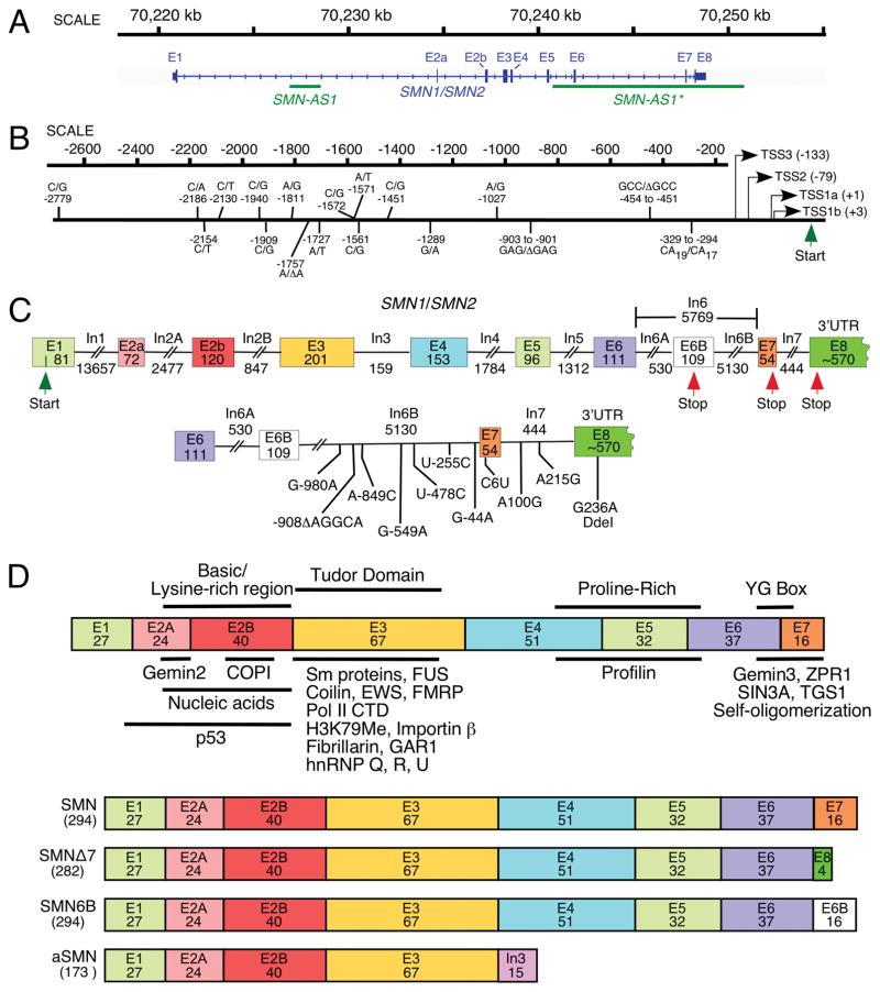
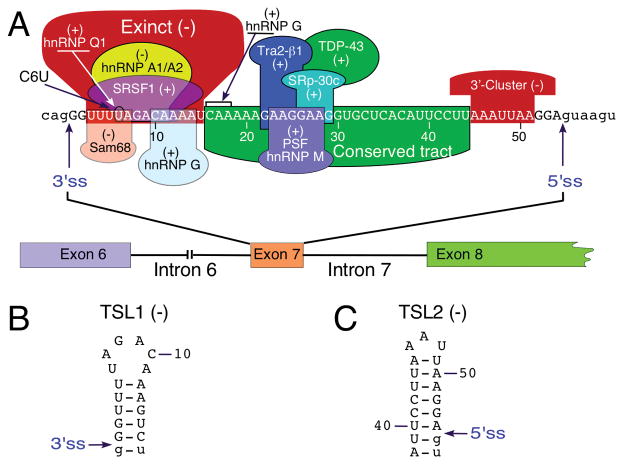
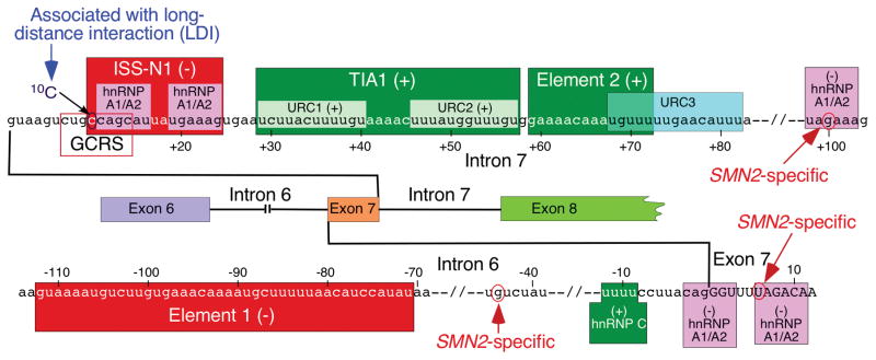
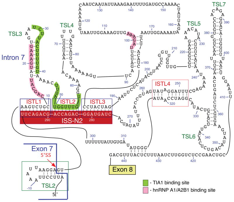
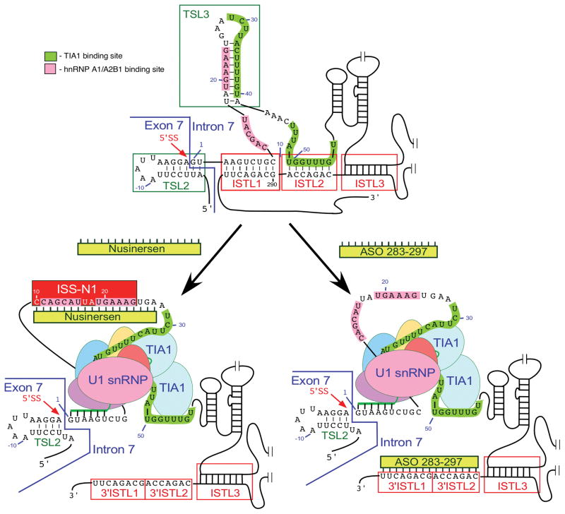
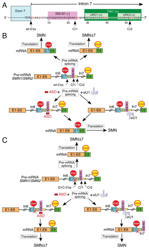
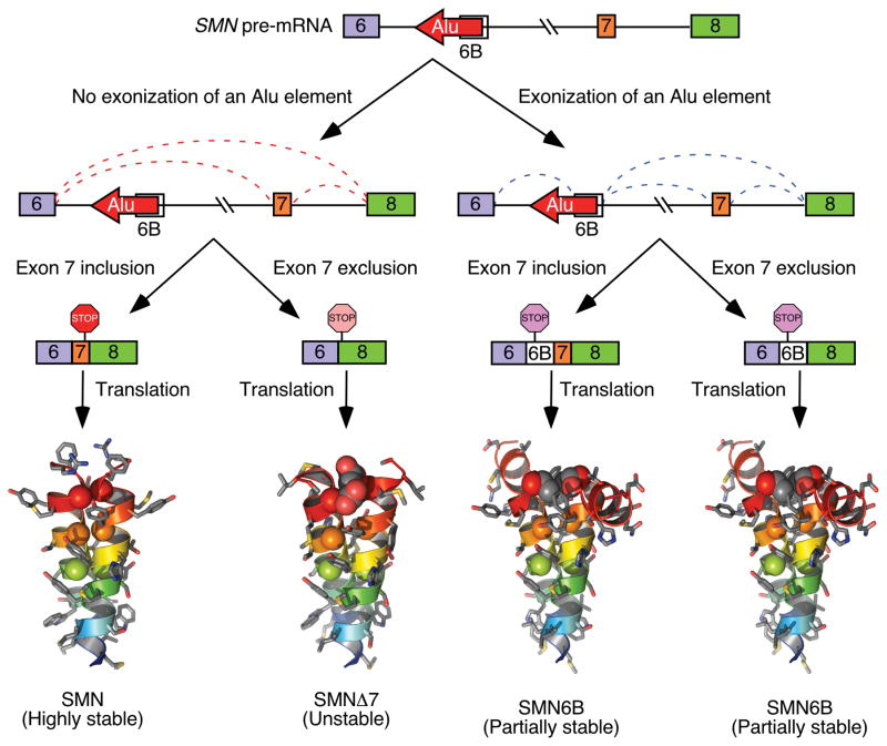
Spinal muscular atrophy (SMA) is one of the major genetic disorders associated with infant mortality. More than 90% cases of SMA result from deletions or mutations of Survival Motor Neuron 1 (SMN1) gene. SMN2, a nearly identical copy of SMN1, does not compensate for the loss of SMN1 due to predominant skipping of exon 7. However, correction of SMN2 exon 7 splicing has proven to confer therapeutic benefits in SMA patients. The only approved drug for SMA is an antisense oligonucleotide (Spinraza™/Nusinersen), which corrects SMN2 exon 7 splicing by blocking intronic splicing silencer N1 (ISS-N1) located immediately downstream of exon 7. ISS-N1 is a complex regulatory element encompassing overlapping negative motifs and sequestering a cryptic splice site. More than 40 protein factors have been implicated in the regulation of SMN exon 7 splicing. There is evidence to support that multiple exons of SMN are alternatively spliced during oxidative stress, which is associated with a growing number of pathological conditions. Here, we provide the most up to date account of the mechanism of splicing regulation of the SMN genes.

Keywords: Cryptic splice site; ISS-N1; ISS-N2; SMA; SMN; Splicing; U1 snRNA.


Conflict of interest statement

Disclosures and Competing Interests:

The ISS-N1 target (US Patent# US7838657) was discovered in the Singh laboratory at UMass Medical School (MA, USA). Inventors, including RN Singh, NN Singh and UMASS Medical School, are currently benefiting from licensing of the ISS-N1 target to Ionis Pharmaceuticals and Biogen. Iowa State University holds intellectual property rights on GC-rich and ISS-N2 targets. Therefore, inventors including RN Singh, NN Singh and Iowa State University could potentially benefit from any future commercial exploitation of GC-rich and ISS-N2 targets.

Figures

Fig. 1
Fig. 1
Organization of SMN gene. (A) A view of human SMN1/SMN2 gene(s) located on chromosome 5. Exons and introns are shown as boxes and lines, respectively. Loci of antisense RNAs, SMN-AS1 [27], and SMN-AS1* [28] are marked with bars. (B) Diagrammatic representation of human SMN promoter region. Multiple transcription start sites (TSS) identified so far are indicated using arrows. Numbers in brackets correspond to their position relative to TSS1a (+1). TSS1a and TSS2 were identified in [29] as transcription start sites preferentially used in adult and fetal tissues, respectively. TSS1b was mapped in Echaniz-Laguna et al. [30], and TSS3 was identified in Monani et al., [31]. Nucleotide differences between the SMN1 and SMN2 promoters are indicated based on Monani et al., [31]; [29, 32]), where nucleotide positions were calculated from TSS1a. Translation initiation site is marked as Start. (C) Diagrammatic representation of the SMN1/SMN2 pre-mRNA. Exons and introns are shown as boxes and lines, respectively. Sizes of exons and introns are indicated in nucleotides (nts). The translation initiation and termination sites are marked as Start and Stop, respectively. Exon 8 is mostly used as the 3′ untranslated region (UTR). The bottom panel indicates nucleotides differences between SMN1 and SMN2 in the region located downstream of exon 6B. The last position of intron 6B is designated as −1. For exons 7 and 8, as well as intron 7, counting starts with the first position of the respective exon or intron. (D). Diagrammatic representation of SMN protein isoforms. Protein regions encoded by each exon are shown as colored boxes with the number of amino acids given. In the top panel, protein domains are indicted above, while SMN interacting partners are shown below the diagrammatic representation of the full-length SMN. For further details see Singh et al. [13]. The bottom panel shows the known SMN isoforms as compared to the full-length SMN protein. These isoforms are generated either due to exon 7 skipping or exonization of a region within intron 6 [33] or intron 3 retention [34]. The size of each isoform (in amino acids) is given in brackets. Abbreviations are given in Table 2
Fig. 2
Fig. 2
Exon 7 splicing regulation. (A) Diagrammatic representation of cis-elements and transacting factors that modulate SMN exon 7 splicing. Positive and negative elements are indicated by (+) and (−), respectively (For further details see [43]). Numbering of nucleotides starts with the first position of exon 7. Exonic and intronic sequences are shown in upper- and lower-case letters, respectively. The 3′ and 5′ss are indicated by arrows. (B) Terminal stem-loop structure, TSL1, formed at the beginning of SMN2 exon 7 as determined by enzymatic structure probing [44, 45]. Both TSL1 and TSL2 are marked by (−) because they contribute toward exon 7 skipping. Numbering of nucleotides starts with the first position of the exon. Exonic and intronic sequences are shown in upper- and lower-case letters, respectively. The 3′ ss is indicated by an arrow. (C) Terminal stem-loop structure, TSL2, formed at the end of SMN2 exon 7 as determined by enzymatic structure probing [44, 45]. (−) indicates that TSL1 contributes to exon 7 skipping. Numbering of nucleotides starts with the first position of the exon. Exonic and intronic sequences are shown in upper- and lower-case letters, respectively. The 5′ss is indicated by an arrow. Abbreviations are given in Table 2
Fig. 3
Fig. 3
Diagrammatic representation of intronic cis-elements and transacting factors that modulate SMN exon 7 splicing. Positive and negative elements are indicated by (+) and (−), respectively. Positive and neutral numbers indicate nucleotide positions within intron 7 and exon 7, respectively, starting with the first intronic/exonic position. Negative numbers indicate nucleotide positions within intron 6, starting with the last intronic position. Exonic and intronic sequences are shown in upper- and lower-case letters, respectively. Exons and introns are also shown as colored boxes and lines. SMN2-spesific single nucleotide substitutions are indicated. Intron 7-located ISS-N1, the overlapping GC-rich sequence (GCRS) and 10C contribute to skipping of exon 7 [43]. ISS-N1 harbors two hnRNP A1/A2B1-binding sites that are highlighted in pink. An SMN2-specific C6U substitution in exon 7 and A100G substitution in intron 7 create additional binding sites for hnRNP A1 [37, 47]. Another hnRNP A1-binding site is located at the junction of intron 6 and exon 7 [51]. Element 2 and U-rich clusters (URC1 and URC2) are positive cis-elements [75, 86]. TIA1 interacts with URC1 and URC2 and promotes exon 7 inclusion [75]. Intron 6-located Element 1 is highlighted in red [87]. It serves as a binding site for PTB and FUSE-BP [88]. A binding site for the stimulatory hnRNP C1/C2 within intron 6 is highlighted in green [68].
Fig. 4
Fig. 4
Secondary structure of SMN2 intron 7 derived from chemical probing. Numbering starts from the first position of intron 7. Negative numbers represent upstream sequences within exon 7. TSLs, ISTLs and binding sites for TIA1 and hnRNP A1/A2B1 are shown and highlighted. ISS-N2 is composed of the 3′ strands of ISTL1, ISTL2 and ISTL3 [43, 50]. The 5′ss of exon 7 is indicated by a red arrow. Abbreviations are given in Table 2.
Fig. 5
Fig. 5
ASO-based mechanism of SMN2 exon 7 splicing correction. Only the relevant sequences of exon 7/intron 7 are given. Nucleotide numbering starts from the first position of intron 7. ISS-N1 and the binding sites for TIA1 and hnRNP A1/A2B1 are marked by colored boxes. The 5′ ss of exon 7 is indicated by a red arrow. The annealing positions of U1 snRNA to this 5′ ss are shown. TSL2 and 3 are local RNA secondary structures, while ISTL1, 2 and 3 are the structures formed by long-distance interactions. These structures are boxed. Nusinersen and ASO 283–297 are shown as yellow bars [25, 107]. Their annealing positions within intron 7 are indicated. Targeting of the corresponding intronic sequences by Nusinersen and ASO 283–297 causes massive structural rearrangements, including disruption of TSL3 and ISTL1. As the results TIA1-binding sites become accessible, the recruitment of U1 snRNP to the 5′ ss of exon 7 is increased and, in case of Nusinersen, the binding of hnRNP A1/A2 to ISS-N1 is blocked. Abbreviations are given in Table 2.
Fig. 6
Fig. 6
Effect of an ASO and eU1 on splicing of exon 7. (A) Diagrammatic representation of exon 7/intron 7 junction. Exonic and intronic sequences are shown in upper- and lower-case letters, respectively. Exon7 is also shown as a blue box. Nucleotide numbering starts from the first position of intron 7. ISS-N1 and URC1 and URC2 are marked by colored boxes. The wild type and the cryptic 5′ ss of exon 7 (Cr1 and 2) are indicated by arrows. GU dinucleotides are highlighted in red. (B) Model of how in the context of the intact 5′ ss of exon 7 an ASO and eU1 promote production of the full-length SMN protein (Adapted from [49]). The ASO block ISS-N1 and eU1 activates usage of the wild-type 5′ ss of exon 7. Exons and introns are indicated by the colored boxes and lines, respectively. The ASO is shown as a red bar, and eU1 as a blue structure. ISS-N1, stop codons in exon 7 and 8 and the 5′ ss of exon 7, wild type and cryptic, are indicated. (C) Model of how in the context of the mutated 5′ ss of exon 7 only eU1 promotes production of the full-length SMN protein (Adapted from [49]). The G to C mutation at the first position of intron 7 is shown in red. The inactivation of the 5′ ss is signified by a red cross. The ASO blocks ISS-N1 and eU1 activate usage of the cryptic 5′ ss of exon 7, Cr1. Exons and introns are indicated by the colored boxes and lines, respectively. The ASO is shown as a red bar, and eU1 as a blue structure. ISS-N1, stop codons in exon 7 and 8 and the 5′ ss of exon 7, wild type and cryptic, are indicated. Abbreviations are given in Table 2.
Fig. 7
Fig. 7
A model showing skipping and inclusion of SMN exon 6B. Exon 6B is derived from an Alu element located within SMN intron 6 [33]. Transcripts that include exon 7 but exclude exon 6B produce full-length SMN, a highly stable protein. Transcripts that lack both exons 6B and exon 7 produce SMNΔ7, an unstable and partially functional protein. Transcripts that include exon 6B produce SMN6B protein irrespective of inclusion or exclusion of exon 7. SMN6B protein is more stable than SMNΔ7 [33]

References

    1. Wahl MC, Will CL, Luhrmann R. The spliceosome: design principles of a dynamic RNP machine. Cell. 2009;136:701–18. https://doi.org/10.1016/j.cell.2009.02.009. - DOI - PubMed
    1. Hertel KJ. Combinatorial control of exon recognition. J Biol Chem. 2008;283(3):1211–5. https://doi.org/10.1074/jbc.R700035200. - DOI - PubMed
    1. Raj B, Blencowe BJ. Alternative splicing in the mammalian nervous system: recent insights into mechanisms and functional roles. Neuron. 2015;87:14–27. https://doi.org/10.1016/j.neuron.2015.05.004. - DOI - PubMed
    1. Erkelenz S, Mueller WF, Evans MS, Busch A, Schöneweis K, Hertel KJ, Schaal H. Position-dependent splicing activation and repression by SR and hnRNP proteins rely on common mechanisms. RNA. 2013;19(1):96–102. https://doi.org/10.1261/rna.037044.112. - DOI - PMC - PubMed
    1. Huelga SC, Vu AQ, Arnold JD, Liang TY, Liu PP, Yan BY, Donohue JP, Shiue L, Hoon S, Brenner S, Ares M, Jr, Yeo GW. Integrative genome-wide analysis reveals cooperative regulation of alternative splicing by hnRNP proteins. Cell Rep. 2012;1(2):167–78. https://doi.org/10.1016/j.celrep.2012.02.001. - DOI - PMC - PubMed

Publication types

MeSH terms

Substances

LinkOut - more resources