. 2018 Nov;75(21):4021-4040.
doi: 10.1007/s00018-018-2847-3. Epub 2018 Jun 9.

Megalin mediates plasma membrane to mitochondria cross-talk and regulates mitochondrial metabolism

Affiliations

Megalin mediates plasma membrane to mitochondria cross-talk and regulates mitochondrial metabolism

Qingtian Li et al. Cell Mol Life Sci. 2018 Nov.

Abstract

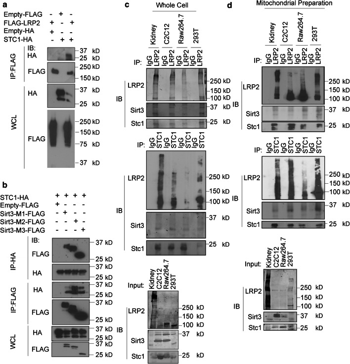
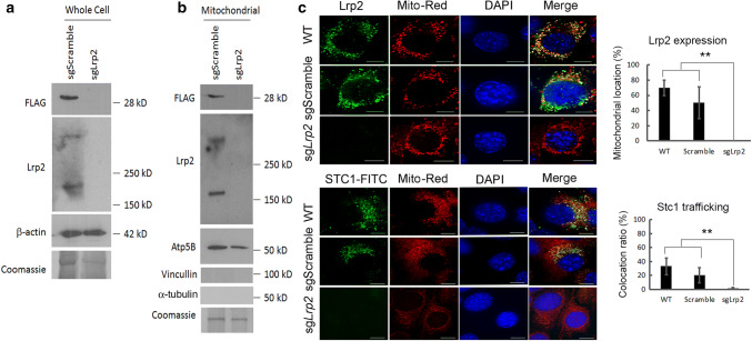
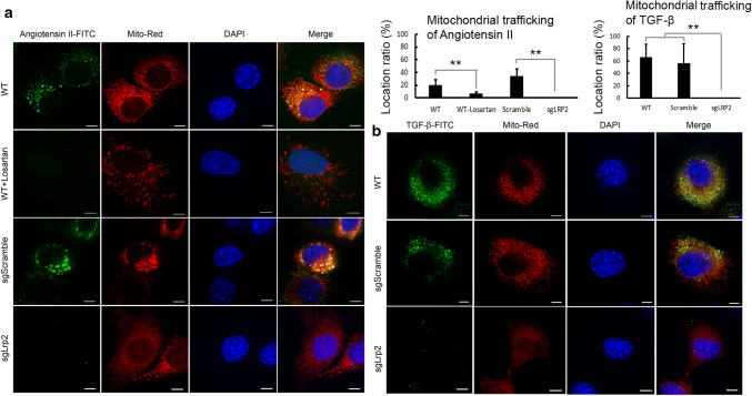
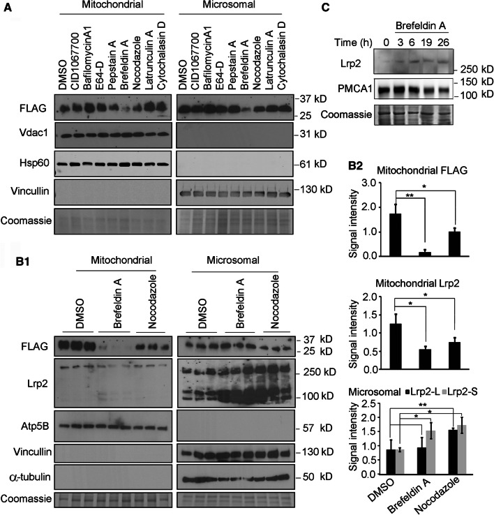
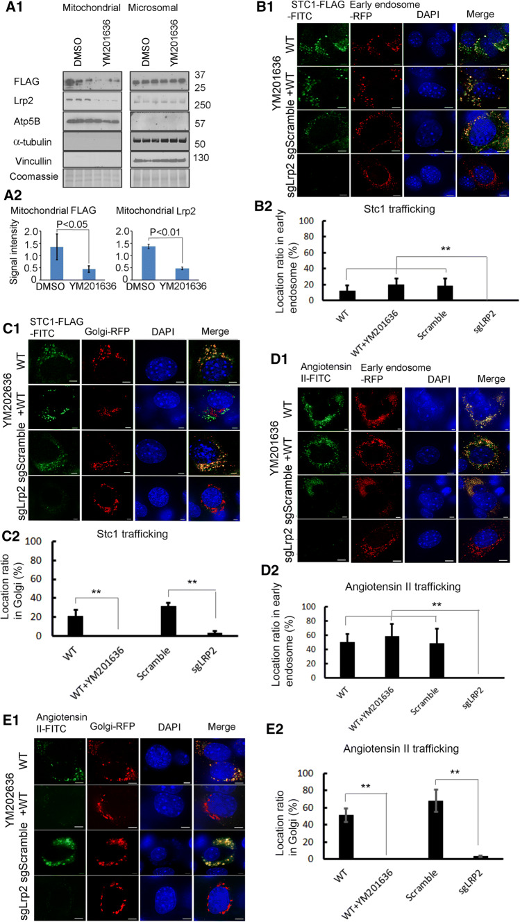
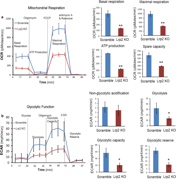
Mitochondrial intracrines are extracellular signaling proteins, targeted to the mitochondria. The pathway for mitochondrial targeting of mitochondrial intracrines and actions in the mitochondria remains unknown. Megalin/LRP2 mediates the uptake of vitamins and proteins, and is critical for clearance of amyloid-β protein from the brain. Megalin mutations underlie the pathogenesis of Donnai-Barrow and Lowe syndromes, characterized by brain defects and kidney dysfunction; megalin was not previously known to reside in the mitochondria. Here, we show megalin is present in the mitochondria and associates with mitochondrial anti-oxidant proteins SIRT3 and stanniocalcin-1 (STC1). Megalin shuttles extracellularly-applied STC1, angiotensin II and TGF-β to the mitochondria through the retrograde early endosome-to-Golgi transport pathway and Rab32. Megalin knockout in cultured cells impairs glycolytic and respiratory capacities. Thus, megalin is critical for mitochondrial biology; mitochondrial intracrine signaling is a continuum of the retrograde early endosome-to-Golgi-Rab32 pathway and defects in this pathway may underlie disease processes in many systems.

Keywords: ApoE; OCRL1; PIKfyve; Proteinuria; Sonic hedgehog; Vitamin D.


Conflict of interest statement

No financial interests to disclose.

Figures

Fig. 1
Fig. 1
Detection of megalin/Lrp2 in the mitochondria. A Detection of megalin in kidney mitochondria. WC whole cell preparation, MP mitochondrial preparation, MIC microsomal preparation. B, C Native megalin/Lrp2 is detected in whole cell and mitochondrial lysates of C2C12, Raw264.7, TKPTS and HEK293T cells. Lrp2 bands are not detectable in the presence of Lrp2 immunizing peptide (B). Expression of c-terminus FLAG-tagged Megalin/Lrp2 in 293T cells: anti-FLAG antibody and ab76969 (directed at the c-terminus of megalin) detect similar band pattern (D). VDAC1 a mitochondrial protein; α-tubulin and β-actin, cytoskeletal proteins, LAMP2 a lysosomal marker, WC whole cell preparation, MP mitochondrial preparation, MIC microsomal preparations. E Formalin-fixed mouse kidney sections were stained with anti-Lrp2 antibodies. Strong Lrp2 labeling is detected on the apical surface of proximal tubule cells, which also display diffuse cytosolic staining. G glomerulus, OSOM outer stripe of the outer medulla, ISOM inner stripe of the outer medulla, IM inner medulla, E3 negative control without primary anti-LRP2 antibody. E1: ×40; E2: ×200; E3: ×200. F Immunogold labeling for Lrp2 shows positive labeling in the mitochondria (M) of HEK293T cells (F1) and kidney PT cells (F2). Scale bars, 1 μm. G Paraformaldehyde-fixed and paraffin-imbedded mouse kidney was labeled with antibodies for Lrp2 and Atp5B (mitochondrial protein), followed by treatment with TRITC-tagged secondary antibody (for Lrp2) or FITC-tagged secondary antibody (for Atp5B), and immunofluorescence image capture. DAPI nuclear staining. Scale bar, 50 µm
Fig. 2
Fig. 2
Megalin/Lrp2 is present in the mitochondria and associates with Stc1 and Sirt3. a 293T cells were co-transfected with STC1-HA (C-terminus HA), LRP2-FLAG (C-terminus FLAG) or empty vector (empty-FLAG; empty-HA), followed by immunoprecipitation from whole cell preparation with anti-FLAG, and immunoblot with anti-HA or anti-FLAG (a representative FLAG-LRP2 band is shown). STC1 is detected in LRP2 immunoprecipitate. b 293T cells were co-transfected with STC1-HA and one of three Sirt3 transcript variants (C-terminus FLAG; M1, M2 and M3 transcript variants [31]). Immunoprecipitation was carried out from whole cell preparation with anti-HA and immunoblot with anti-FLAG or anti-HA; or immunoprecipitation with anti-FLAG, and immunoblot with anti-HA or anti-FLAG. All products of Sirt3 transcripts are detected in STC1 immunoprecipitate; while STC1 is detected in immunoprecipitates of products of all Sirt3 transcripts. Association between native megalin/Lrp2, Stc1 and Sirt3 in whole cell (c) and mitochondrial (d) lysates of kidney, C2C12, Raw264.7 and HEK293T cells. Immunoprecipitation was carried out using anti-LRP2 antibody (upper panels) or anti-STC1 antibody (middle panels). Immunoprecipitation with irrelevant iso-species immunoglobulin (IgG) was carried out for control. Stc1 and Sirt3 are detected in Lrp2 immunoprecipitates (upper panels). Lrp2 and Sirt3 are detected in Stc1 immunoprecipitates (middle panels). Lower panels show IP input. MW for products of Sirt3 transcripts: M1, 37 kD; M2, 34 kD; M3, 28 kD [31]
Fig. 3
Fig. 3
Mitochondrial co-localization of native megalin/Lrp2, Stc1 and Sirt3 in C2C12 cells. Cultured C2C12 cells were treated with the following: a Mito-Red and FITC-tagged anti-Lrp2 secondary antibody; b Mito-Red and FITC-tagged anti-Stc1 secondary antibody; c Mito-Red and FITC-tagged anti-Sirt3 secondary antibodies; d FITC-tagged anti-Stc1 secondary antibodies and TRITC-tagged anti-Sirt3 secondary antibody; e FITC-tagged anti-Stc1 secondary antibodies and TRITC-tagged anti-Lrp2 secondary antibody; f FITC-tagged anti-Lrp2 secondary antibodies and TRITC-tagged anti-Sirt3 secondary antibody. Confocal deconvolution immunofluorescence studies reveal co-localization of Mito-red (mitochondrial marker) with native Lrp2 (a), Stc1 (b) and Sirt3 (c) in C2C12 cells (similar results are observed in HEK293T and raw264.7 cells; data not shown). Stc1 co-localizes with Sirt3 (d) and Lrp2 (e), while Sirt3 co-localizes with Lrp2 (f). Scale bars, 2 µm
Fig. 4
Fig. 4
Megalin is absent in the cell (a) and mitochondria (b) of Lrp2 KO C2C12 cells; megalin KO abolishes the internalization of extracellular rSTC1-FLAG into the cell (a) and targeting to the mitochondria (b). a, b rSTC1-FLAG protein (100 ng/mL) was added to the medium of sgScramble- and sgLrp2 clone-C2C12 cells. Following incubation for 1 h, cells were washed in PBS, and whole cell or mitochondrial lysates were resolved on SDS-PAGE. Lrp2 expression and cellular/mitochondrial content of rSTC1-FLAG protein were determined using western blots. Absent FLAG (rSTC1) in whole cell and mitochondrial lysates of megalin/Lrp2 KO cells. Immunoblot with β-actin and coomassie staining confirms equal loading of whole cell fractions. Negative signal for the cytosolic proteins vinculin and α-tubulin and positive signal for Atp5B confirm purity of the mitochondrial preparations; immunoblot for Atp5B and coomassie stain confirms equal loading of mitochondrial samples. Confocal deconvolution microscopy confirms the presence of Lrp2 in the cell and mitochondria of WT- and sgScramble-C2C12 cells; megalin is not observed in the cell or mitochondria of Lrp2 KO-C2C12 cells (c). Megalin KO abolishes the internalization of extracellular rSTC1-FLAG into the cell and targeting to the mitochondria (d). c WT-, sgScramble- or sgLrp2 clone (megalin KO)-C2C12 cells were stained with FITC-tagged anti-LRP2 secondary antibody. Lrp2 co-localizes with Mito-Red in WT and sgScramble C2C12 cells; Lrp2 is absent from the cell and/or mitochondria of sgLrp2 clone (megalin KO) C2C12 cells. Mitochondria are labeled with Mito-Red (red); nuclei are stained with DAPI (blue). Scale bar, 2 μm. d rSTC1-FLAG-FITC (100 ng/mL) was added to the medium of WT-, sgScramble- or sgLrp2 clone-C2C12 cells for 1 h; after wash with PBS, cells were treated with Mito-Red followed by confocal deconvolution fluorescence. Whereas, FITC (rSTC1-FLAG) is detected in the cell and mitochondria of WT- and sgScramble-C2C12, it is not observed intracellularly or in the mitochondria of sgLRP2 clone (megalin KO)-C2C12 cells. Mitochondria are labeled with Mito-Red (red); nuclei are stained with DAPI (blue); FITC (green). Scale bar, 2 μm
Fig. 5
Fig. 5
Knockdown of megalin/Lrp2 diminishes the internalization and targeting of extracellular angiotensin II and transforming growth factor-β (TGF-β) to the mitochondria. a Recombinant angiotensin II-FITC (100 ng/mL) was added to the medium of WT-, sgScramble- and sgLrp2 clone-C2C12 cells for 1 h. Losartan (1 mM; an AT1A receptor blocker) or equal volume PBS was added concomitantly to WT cells. Cells were then washed in PBS, followed by confocal deconvolution fluorescence imaging. Angiotensin II-FITC (green); Mito-Red (red); nuclear DAPI (blue). b TGF-β-FITC (100 ng/mL) was added to the medium of WT-, sgScramble- and sgLrp2 clone-C2C12 cells for 1 h; cells were washed in PBS, followed by confocal deconvolution fluorescence imaging. TGF-β-FITC (green); Mito-Red (red); nuclear DAPI (blue). Scale bars, 2 µm
Fig. 6
Fig. 6
Microtubules and Golgi mediate shuttling of intracrine signaling molecules to the mitochondria. A WT C2C12 cells were treated for 12 h with DMSO (control), Latrunculin A (10 nM), Cytochalasin D (100 nM), CID1067700 (1 µM), Nocodazole (0.1 µg/mL), Bafilomycin A (1 nM), Brefeldin A (0.2 µg/mL), E-64D (10 µg/mL), or Pepstatin A (1 µM), followed by the addition of rSTC1-FLAG (100 ng/mL) to the medium for 6 h. Protein lysates representing mitochondria or microsomal preparations were resolved on SDS-PAGE, and western blots were reacted with antibodies for FLAG, heat shock protein 60 (Hsp60), voltage-dependent anion-selective channel 1 (Vdac1), or vinculin; parallel gels were stained with coomassie blue. B1 WT C2C12 cells were treated for 12 h with DMSO (control), Brefeldin A (0.2 µg/mL), or Nocodazol (0.1 µg/mL), followed by the addition of rSTC1-FLAG (100 ng/mL) to the medium for 6 h. Protein lysates representing mitochondria or microsomal preparations were resolved on SDS-PAGE, and western blots were reacted with antibodies for FLAG, Lrp2, Atp5B, α-tubulin or vinculin; parallel gels were stained with coomassie blue. B2 Bar graphs depict the Mean ± SEM for rSTC1-FLAG and Lrp2 in the mitochondrial fractions, of corresponding blots shown in “B1”. Band intensities of rSTC1-FLAG and Lrp2 in the mitochondrial preparations were normalized to Atp5B; band intensities of Lrp2 in the microsomal fractions were normalized to vinculin. Lrp2L denotes upper Lrp2 band; Lrp2S denotes lower Lrp2 bands. Student t test: *P < 0.05; **P < 0.01. C WT C2C12 cells were treated with Brefeldin A (0.2 µg/mL), for up to 26 h. Plasma membrane fractions were resolved on SDS-PAGE, and western blots were reacted with antibodies for Lrp2, or Pmca1; parallel gels were stained with coomassie blue
Fig. 7
Fig. 7
Retrograde endosome-to-Golgi pathway mediates targeting of intracrine STC1-FLAG to the mitochondria. A WT C2C12 cells were treated for 1 h with DMSO (control) or YM201636 (PIKfyve inhibitor), followed by the addition of rSTC1-FLAG (100 ng/mL) to the medium for 1 h. Protein lysates representing mitochondria or microsomal preparations were resolved on SDS-PAGE, and western blots were reacted with antibodies for FLAG, Lrp2, Atp5B, α-tubulin or vinculin; parallel gels were stained with coomassie blue. Representative Lrp2 band is shown. Bar graphs depict the Mean ± STD for rSTC1-FLAG and Lrp2 in the mitochondrial fraction, corresponding to western blots shown in “A”; n = 3. Band intensities of hSTC1-FLAG and Lrp2 in the mitochondrial preparations were normalized to Atp5B. Lrp2 knockdown blocks entry of rSTC1-FLAG-FITC and angiotensin II-FITC to early endosomes, while PIKfyve inhibition blocks their movement from the early endosomes to the Golgi. PIKfyve inhibition diminishes mitochondrial Lrp2. WT-, sgScramble or sgLRP2-C2C12 cells were transfected with early endosomes CellLight-RFP (B, D) or Golgi CellLight-RFP (C, E). WT-early endosome CellLight-RFP cells or WT-Golgi CellLight-RFP cells were treated with DMSO or YM201636 (PIKfyve inhibitor) for 1 h, followed by incubation with rSTC1-FLAG-FITC (100 ng/mL) or angiotensin II-FITC (100 ng/mL) for 1 additional hour. sgScramble- and sgLRP2-C2C12 cells were transfected with early endosome CellLight-RFP or Golgi CellLight-RFP, followed by incubation with rSTC1-FLAG-FITC (100 ng/mL) or angiotensin II-FITC (100 ng/mL) for 1 additional hour. All cells were fixed in paraformaldehyde followed by confocal deconvolution fluorescence imaging. Scale bars, 2 µm. F Proposed mitochondrial intracrine signaling pathway. Scheme depicts movement of Lrp2 and intracrine signaling molecules (STC1, TGF-β and Angiotensin II) from the cell surface through the retrograde early endosomes-to-Golgi pathway on their way to the mitochondria
Fig. 8
Fig. 8
Rab32 mediates trafficking of STC1-FLAG from the cell surface to the mitochondria. a Mitochondria (MP) isolated from C2C12 cells that were treated with vehicle or Mitoblock-6 (20μM; a cell-permeable, potent and selective inhibitor of Mia40/Erv1 redox-mediated mitochondria import pathway) followed by incubation with STC1-FLAG. b Mitochondria (MP) isolated from sgScramble or sgTOM40 C2C12 cells after incubation with STC1-FLAG. c Mitochondria (MP) isolated from sgScramble or sgRab32 C2C12 cells after incubation with STC1-FLAG. Rab32 knockout in C2C12 cells abolishes mitochondrial Lrp2/megalin (d), while Lrp2/megalin knockout diminishes mitochondrial Rab32 (e). WC whole cell preparation, MP mitochondrial preparation, Atp5B mitochondrial protein, α-tubulin cytoskeletal protein. Whole kidney lysate was subjected to immunoprecipitation using anti-Lrp2 or anti-Rab32 antibody. Lrp2 is detected in Rab32 immunoprecipitate (f), while Rab32 is detected in Lrp2 immunoprecipitate (g). h Schematic depiction of mitochondrial intracrine signaling pathway
Fig. 9
Fig. 9
Knockdown of megalin/Lrp2 diminishes baseline respiration/glycolysis and respiratory/glycolytic capacities. a Baseline mitochondrial oxygen consumption rate (OCR), fraction of OCR utilized for ATP (adenosine triphosphate) synthesis (determined by mitochondrial OCR after Oligomycin), and respiratory capacity (determined by mitochondrial OCR after addition of the uncoupler CCCP) (n = 3). Blue line, sgScramble-C2C12 cells; red line, sgLrp2-C2C12 cells. b Baseline extracellular acidification rate (ECAR), ECAR after the addition of glucose to the medium, extra-mitochondrial glycolytic capacity (determined by inhibition of respiration using Oligomycin (n = 3). Blue line, sgScramble-C2C12 cells; red line, sgLrp2-C2C12 cells

Similar articles

Cited by

References

    1. Nunnari J, Suomalainen A. Mitochondria: in sickness and in health. Cell. 2012;148:1145–1159. doi: 10.1016/j.cell.2012.02.035. - DOI - PMC - PubMed
    1. Re RN, Cook JL. The mitochondrial component of intracrine action. Am J Physiol Heart Circ Physiol. 2010;299:H577–H583. doi: 10.1152/ajpheart.00421.2010. - DOI - PMC - PubMed
    1. Nielsen R, Christensen EI, Birn H. Megalin and cubilin in proximal tubule protein reabsorption: from experimental models to human disease. Kidney Int. 2016;89:58–67. doi: 10.1016/j.kint.2015.11.007. - DOI - PubMed
    1. Marzolo MP, Farfan P. New insights into the roles of megalin/LRP2 and the regulation of its functional expression. Biol Res. 2011;44:89–105. doi: 10.4067/S0716-97602011000100012. - DOI - PubMed
    1. Birn H, Verroust PJ, Nexo E, Hager H, Jacobsen C, Christensen EI, Moestrup SK. Characterization of an epithelial approximately 460-kDa protein that facilitates endocytosis of intrinsic factor-vitamin B12 and binds receptor-associated protein. J Biol Chem. 1997;272:26497–26504. doi: 10.1074/jbc.272.42.26497. - DOI - PubMed

MeSH terms

Supplementary concepts

LinkOut - more resources