. 2019 Mar;56(3):1681-1693.
doi: 10.1007/s12035-018-1177-7. Epub 2018 Jun 18.

Mast Cell Proteases Activate Astrocytes and Glia-Neurons and Release Interleukin-33 by Activating p38 and ERK1/2 MAPKs and NF-κB

Affiliations

Mast Cell Proteases Activate Astrocytes and Glia-Neurons and Release Interleukin-33 by Activating p38 and ERK1/2 MAPKs and NF-κB

Duraisamy Kempuraj et al. Mol Neurobiol. 2019 Mar.

Abstract

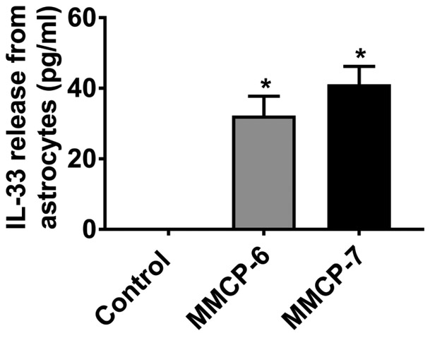
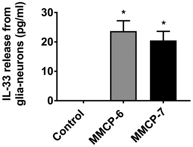
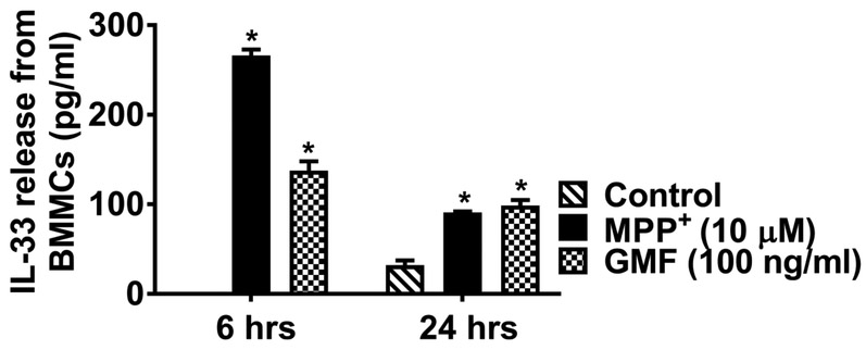
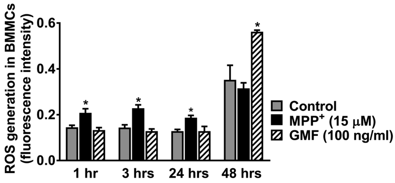
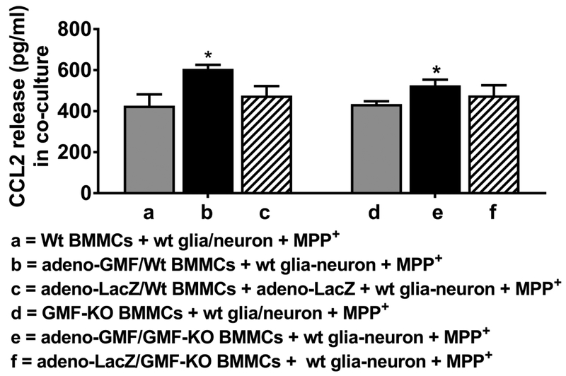
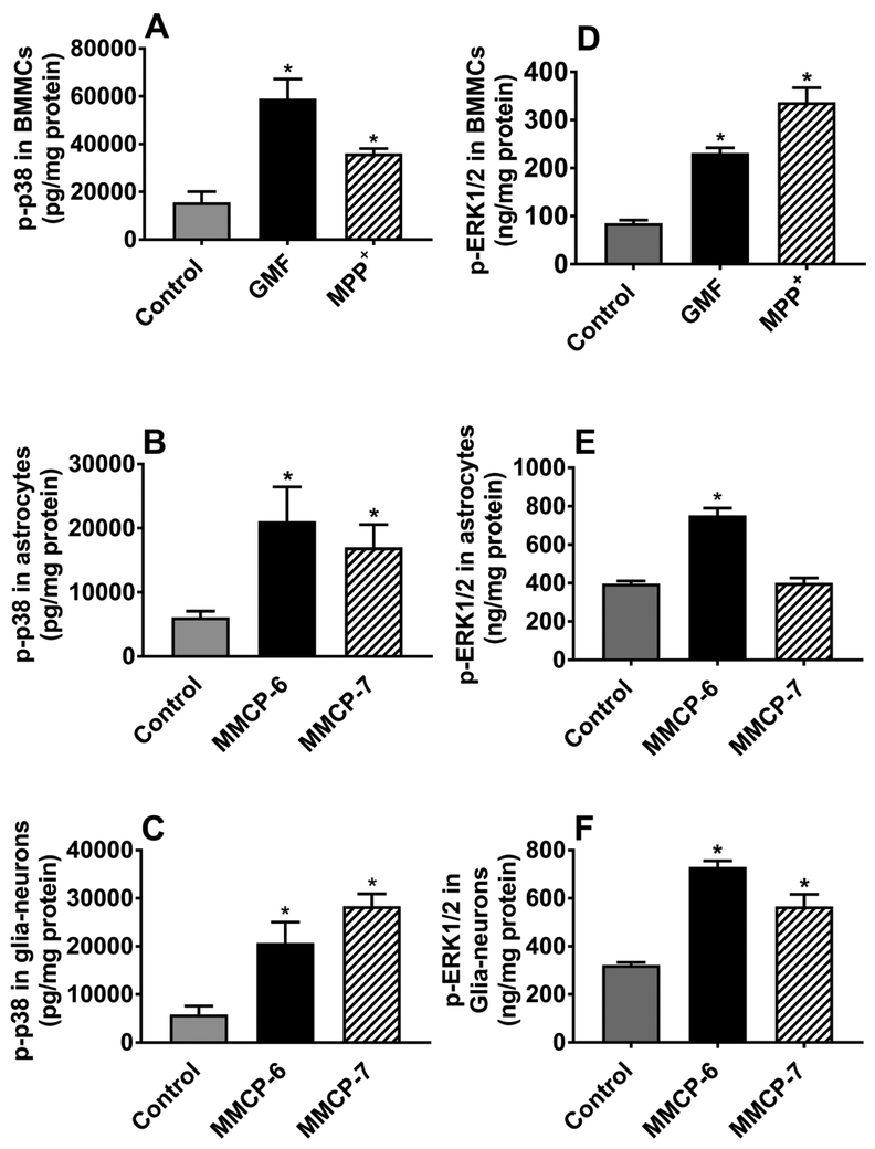
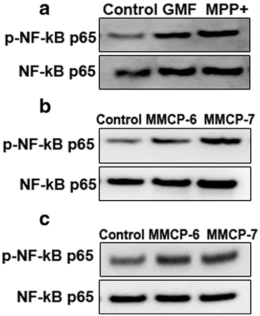
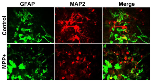
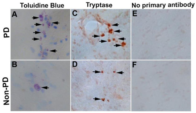
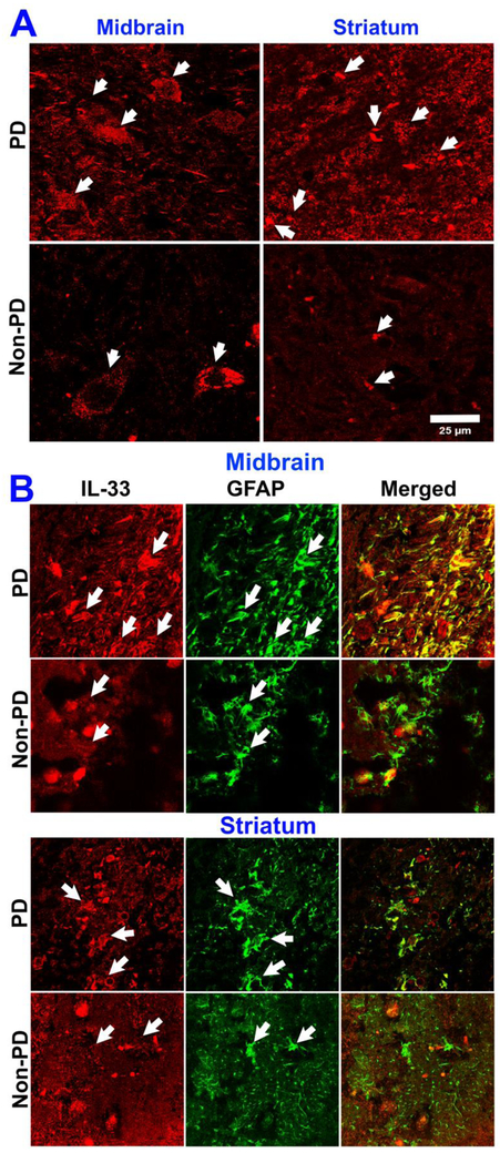
Inflammatory mediators released from activated microglia, astrocytes, neurons, and mast cells mediate neuroinflammation. Parkinson's disease (PD) is characterized by inflammation-dependent dopaminergic neurodegeneration in substantia nigra. 1-Methyl-4-phenylpyridinium (MPP+), a metabolite of parkinsonian neurotoxin 1-methyl-4-phenyl-1,2,3,6-tetrahydropyridine (MPTP), induces inflammatory mediators' release from brain cells and mast cells. Brain cells' interaction with mast cells is implicated in neuroinflammation. However, the exact mechanisms involved are not yet clearly understood. Mouse fetal brain-derived cultured primary astrocytes and glia-neurons were incubated with mouse mast cell protease-6 (MMCP-6) and MMCP-7, and mouse bone marrow-derived mast cells (BMMCs) were incubated with MPP+ and brain protein glia maturation factor (GMF). Interleukin-33 (IL-33) released from these cells was quantitated by enzyme-linked immunosorbent assay. Both MMCP-6 and MMCP-7 induced IL-33 release from astrocytes and glia-neurons. MPP+ and GMF were used as a positive control-induced IL-33 and reactive oxygen species expression in mast cells. Mast cell proteases and MPP+ activate p38 and extracellular signal-regulated kinases 1/2 (ERK1/2), mitogen-activated protein kinases (MAPKs), and transcription factor nuclear factor-kappa B (NF-κB) in astrocytes, glia-neurons, or mast cells. Addition of BMMCs from wt mice and transduction with adeno-GMF show higher chemokine (C-C motif) ligand 2 (CCL2) release. MPP+ activated glial cells and reduced microtubule-associated protein 2 (MAP-2) expression indicating neurodegeneration. IL-33 expression increased in the midbrain and striatum of PD brains as compared with age- and sex-matched control subjects. Glial cells and neurons interact with mast cells and accelerate neuroinflammation and these interactions can be explored as a new therapeutic target to treat PD.

Keywords: Brain cells; Glia maturation factor; Interleukin-33; Mast cell proteases; Mast cells; Parkinson’s disease.


Figures

Fig. 1
Fig. 1
Mast cell proteases MMCP-6 and MMCP-7 release IL-33 from mouse primary astrocytes. Mouse primary astrocytes were incubated with MMCP-6 (100 ng/ml) and MMCP-7 (100 ng/ml) for 24 h and IL-33 released in the supernatant media was analyzed by ELISA kit (n=4). Both MMCP-6 and MMCP-7 significantly increased (*p<0.05 vs control, one-way ANOVA and Tukey-Kramer post hoc analysis) the release of IL-33 as compared to untreated control cells.
Fig. 2
Fig. 2
Mast cell proteases MMCP-6 and MMCP-7 release IL-33 from mouse primary glia-neurons. Mouse primary glia-neurons were incubated with MMCP-6 (100 ng/ml) and MMCP-7 (100 ng/ml) for 24 h and IL-33 level in the cell culture supernatant media was analyzed by ELISA kit (n=3). Mast cell proteases MMCP-6 as well as MMCP-7 significantly increased (*p<0.05 vs control, one-way ANOVA and Tukey-Kramer post hoc analysis) the release of IL-33 as compared to untreated control cells.
Fig. 3
Fig. 3
MPP+ activate mouse mast cells and release IL-33. BMMCs were incubated with MPP+ (10 μM) for 6 h and 24 h and IL-33 released into the media was measured by ELISA kit (n=4). MPP+-induced significant release of IL-33 as compared with control unstimulated cells release. GMF used as a positive control also significantly increased (*p<0.05 control vs treated, one-way ANOVA and Tukey-Kramer post hoc analysis) the release of IL-33 as compared to untreated control cells.
Fig 4
Fig 4
MPP+ and GMF increase ROS generation in mouse primary mast cells. BMMCs were incubated with DCFDA 1, washed and resuspended in supplementary buffer containing FBS. Mast cells were incubated with MPP+ (15 μM) or GMF (100 ng/ml) for 1, 3, 24 and 48 h and measured the generation of ROS (N=3). The plates were read after 1, 3, 24 and 48 h using a micro plate reader for fluorescence intensity. BMMCs incubated with MPP+ show increased production of ROS at 1, 3 and 24 h of incubation. GMF show increased generation of ROS at 48 h of incubation as compared with untreated control BMMCs (*p<0.05 vs control, one-way ANOVA and Tukey-Kramer post hoc analysis).
Fig. 5
Fig. 5
BMMCs increases MPP+-mediated CCL2 release from mouse glia-neurons and mast cells co-culture. BMMCs from wt mice and GMF-KO mice were incubated with adeno-GMF or adeno-LacZ before adding to wt glia-neuron culture. Then MPP+ (10 μM) was added to these cultures for 24 h and CCL2 release was measured in the supernatant media by ELISA kit (N=3). Addition of BMMCs from wt mice that were incubated with adeno-GMF and further incubated with wt glia-neurons with MPP+ as compared to CCL2 released from BMMCs obtained from GMF-KO mice and treated with adeno-GMF (*p<0.05 t test).
Fig. 6
Fig. 6
MPP+ and GMF activate MAPKs in mouse mast cells, and mouse mast cell proteases activate MAPKs in astrocytes and glia-neurons. BMMCs were incubated with MPP+ (10 μM) and GMF (100 ng/ml), and astrocytes and glia-neurons were incubated with MMCP-6 (100 ng/ml) and MMCP-7 (100 ng/ml) for 15 min. Cell lysates were prepared from cell pellets and the expression of p-p38 was assayed by ELISA (a-f). MPP+, GMF and mast cell proteases-induced phosphorylation of p38 and increased the expression of p-p38 in BMMCs (a; N=3), astrocytes (b; n=4) and glia-neurons (c; N=4). MPP+ and GMF-induced phosphorylation of ERK1/2 in BMMCs (d; N=3). Further, MMCP-6 increased the expression of p-ERK1/2 in astrocytes (e; N=3). Both MMCP-6 and MMCP-7 increased phosphorylation of ERK1/2 in glia-neurons (f; N=4). *p<0.05 vs control, one-way ANOVA and Tukey-Kramer post hoc analysis.
Fig. 7
Fig. 7
MPP+ and GMF activate NF-κB in mouse mast cells, and mouse mast cell proteases activate NF-κB in astrocytes and glia-neurons. BMMCs were incubated with MPP+ (10 μM) or GMF (100 ng/ml), and astrocytes and glia-neurons were incubated with MMCP-6 or MMCP-7 for 24 h (a-c). Cell lysates were prepared from cell pellets and the expression of total and p-NF-κB p65 was determined by Western blot (N=3). The bands were visualized using ChemiDoc-It2 Imaging System. Representative images show that MPP+, GMF and mast cell proteases-induced increased expression of p-NF-κB p65 in BMMCs (a), astrocytes (b) and glia-neurons (c)
Fig. 8
Fig. 8
Double immunofluorescence detection of GFAP and MAP2 in glia-neurons after incubation with MPP+. Glia-neurons were grown in 24 well tissue culture plate and incubated with MPP+ (15 μM) for 72 h. We then performed double immunofluorescence for the detection of GFAP and MAP2 using anti-GFAP polyclonal antibody (1:500 dilution) and anti-MAP2 monoclonal antibody (5 μg/ml) overnight at 4°C followed by Alexa 488 goat anti-rabbit IgG and Alexa 568 goat-anti-mouse IgG secondary antibodies. Then the sections were cover-slipped and observed under a microscope. Representative images show activated morphology of astrocytes (green color) and reduced MAP2 (red color) expression after incubation with MPP+. Original magnification = 200×.
Fig. 9
Fig. 9
Mast cell activation in PD brains. Mid brain sections from PD patients and non-PD control subjects were incubated with 0.1% toluidine blue to detect mast cells (N=3). PD brains show increased mast cells (arrows, blue/purple metachromatic color) as well as increased activation as compared with non-PD control brains (a, b). Degranulated mast cells show extracellular granules and reduced blue color as seen in the PD brain. Further, we immunostained human mast cell specific tryptase in these brain sections using tryptase monoclonal antibody and ready-to-use ImmPRESS polymer anti-mouse IgG peroxidase reagent and ImmPACT DAB peroxidase substrate. Development of brown color indicated a positive reaction for tryptase (c, d, arrows). Tryptase-positive mast cells and their activation status were increased in PD brains as compared to non-PD control subjects’ brains (c, d). Negative control staining performed without using the primary antibody did not show positive reaction for tryptase (e, f). Original magnification = 200×.
Fig. 10
Fig. 10
Increased expression of IL-33 and GFAP in human PD brains as detected by immunofluorescence staining. Sections (40 μm) from mid brain and striatum of PD patients (N=3) as well as age and sex matched non-PD control subjects (N=3) were incubated with IL-33 monoclonal antibody overnight at 4 °C and then with Alexa 568 conjugated goat anti-mouse IgG secondary antibody (1:500 dilution) for 1 h at room temperature. Then the sections were cover-slipped and observed under a confocal microscope using 63× oil immersion objective. Representative images from mid brain and striatum in PD brains show increased expression of IL-33 (arrows, red color) as compared with age and sex matched control subjects brains (a, scale bar = 25 μm). Further, we also investigated the source of IL-33 by double immunofluorescence staining using monoclonal antibody for IL-33 and polyclonal antibody for astrocyte marker GFAP along with Alexa Flour 488 goat anti-rabbit IgG and Alexa Flour 568 goat anti-mouse IgG secondary antibodies. After washing with PBS, the sections were mounted on to the slides, coversliped with mounting medium and viewed with a confocal microscope and images were acquired (b). Representative images show that IL-33 is increased in the mid brain and striatum of PD brains as compared with non-PD control subjects mid brain and striatum regions (b; arrows, red color). Additionally, expression of GFAP also increased indicating activated/reactive astrocytes (green color, arrows) in the mid brain and striatum regions of PD brains as compared to non-PD control subjects’ brains (b). Merged images show co-localization of IL-33 and GFAP in the mid brain and striatum regions (original magnification = 630×).

References

    1. Arthur G, Bradding P (2016) New Developments in Mast Cell Biology: Clinical Implications. Chest 150 (3):680–693. doi: 10.1016/j.chest.2016.06.009 - DOI - PubMed
    1. DeBruin EJ, Gold M, Lo BC, Snyder K, Cait A, Lasic N, Lopez M, McNagny KM, Hughes MR (2015) Mast cells in human health and disease. Methods Mol Biol 1220:93–119. doi: 10.1007/978-1-4939-1568-2_7 - DOI - PubMed
    1. Migalovich-Sheikhet H, Friedman S, Mankuta D, Levi-Schaffer F (2012) Novel identified receptors on mast cells. Front Immunol 3:238. doi: 10.3389/fimmu.2012.00238 - DOI - PMC - PubMed
    1. Rao KN, Brown MA (2008) Mast cells: multifaceted immune cells with diverse roles in health and disease. Ann N Y Acad Sci 1143:83–104. doi: 10.1196/annals.1443.023 - DOI - PubMed
    1. Gilfillan AM, Beaven MA (2011) Regulation of mast cell responses in health and disease. Crit Rev Immunol 31 (6):475–529 - PMC - PubMed