. 2018 Oct;220(1):147-162.
doi: 10.1111/nph.15272. Epub 2018 Jun 19.

The requirement for calcification differs between ecologically important coccolithophore species

Affiliations

The requirement for calcification differs between ecologically important coccolithophore species

Charlotte E Walker et al. New Phytol. 2018 Oct.

Abstract

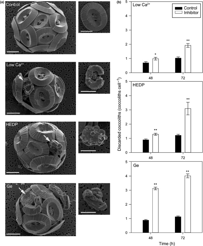
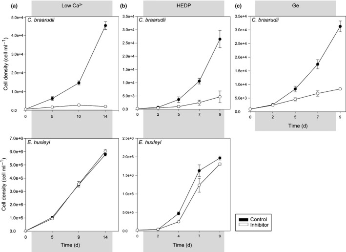
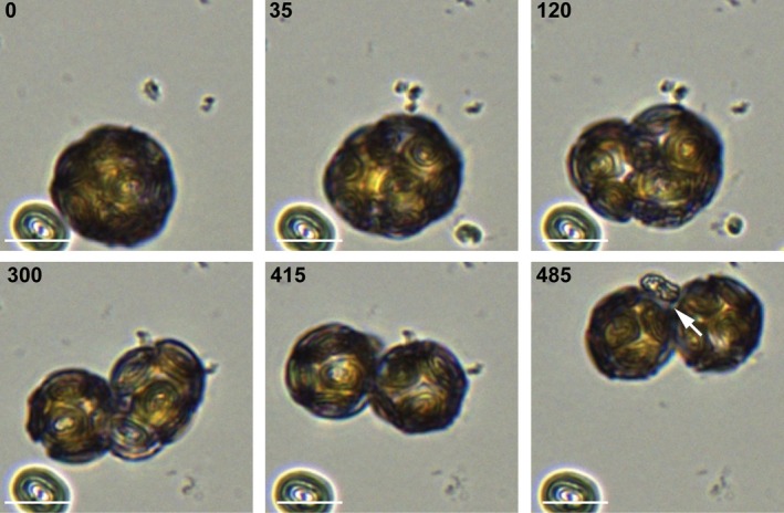
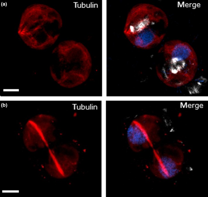
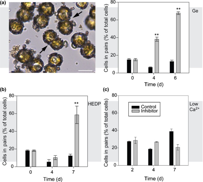
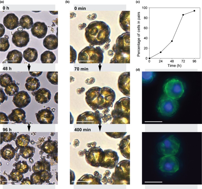
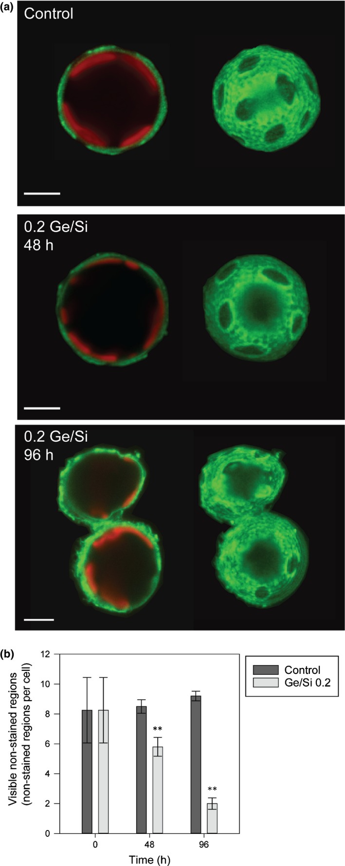
Coccolithophores are globally distributed unicellular marine algae that are characterized by their covering of calcite coccoliths. Calcification by coccolithophores contributes significantly to global biogeochemical cycles. However, the physiological requirement for calcification remains poorly understood as non-calcifying strains of some commonly used model species, such as Emiliania huxleyi, grow normally in laboratory culture. To determine whether the requirement for calcification differs between coccolithophore species, we utilized multiple independent methodologies to disrupt calcification in two important species of coccolithophore: E. huxleyi and Coccolithus braarudii. We investigated their physiological response and used time-lapse imaging to visualize the processes of calcification and cell division in individual cells. Disruption of calcification resulted in major growth defects in C. braarudii, but not in E. huxleyi. We found no evidence that calcification supports photosynthesis in C. braarudii, but showed that an inability to maintain an intact coccosphere results in cell cycle arrest. We found that C. braarudii is very different from E. huxleyi as it exhibits an obligate requirement for calcification. The identification of a growth defect in C. braarudii resulting from disruption of the coccosphere may be important in considering their response to future changes in ocean carbonate chemistry.

Keywords: Coccolithus braarudii; Emiliania huxleyi; calcification; coccolithophore; phytoplankton.


Figures

Figure 1
Figure 1
Disruption of calcification in Coccolithus braarudii. (a) Representative scanning electron microscopy (SEM) images of C. braarudii cells grown in 1 mM Ca2+ (48 h), 5 μM HEDP (24 h) and 0.2 germanium (Ge)/silicon (Si) (100 μM Si, 24 h). Incomplete or malformed coccoliths can be observed in response to all three treatments (arrows), whereas these are largely absent from control cells. Insets show representative incomplete or malformed coccoliths in greater detail. Incomplete coccoliths are defined as those that exhibit the oval shape of control coccoliths, but calcite precipitation is not complete. Malformed coccoliths are defined as coccoliths with gross defects in crystal morphology and no longer resemble the oval morphology of control coccoliths. Bars, 5 μm. (b) Treatments used to disrupt calcification in C. braarudii result in a significant increase in discarded coccoliths per cell (*, P < 0.05; **, P < 0.01, one‐way ANOVA with Holm–Sidak post hoc test), indicative of incomplete or malformed coccoliths that fail to integrate successfully into the coccosphere. Error bars denote ± SE. n = 3.
Figure 2
Figure 2
Disruption of calcification leads to a reduction in growth in Coccolithus braarudii. (a) Growth of C. braarudii and Emiliania huxleyi at 1 or 10 mM Ca2+ for 14 d. The specific growth rate (SGR ± SE) of E. huxleyi was not significantly different at 1 mM Ca2+ relative to the 10 mM Ca2+ control (0.55 ± 0.006 and 0.55 ± 0.002 d−1, respectively, P = 0.91, two‐tailed t‐test), whereas the growth of C. braarudii was severely inhibited at 1 mM Ca2+ (SGR ± SE = 0.16 ± 0.01 d−1) relative to the control (SGR ± SE = 0.32 ± 0.01 d−1, P < 0.05). (b) The growth of C. braarudii in 50 μM HEDP for 9 d was significantly reduced compared with the control (SGR ± SE = 0.30 ± 0.05 and 0.53 ± 0.01 d−1, respectively, P < 0.05), whereas the growth of E. huxleyi was not significantly different (SGR ± SE: 50 μM HEDP, 0.66 ± 0.03 d−1; control, 0.76 ± 0.08 d−1; P = 0.31). (c) The growth of C. braarudii in the presence of germanium (Ge; 0.2 Ge/silicon (Si)) for 9 d was significantly reduced relative to the control (SGR ± SE = 0.20 ± 0.04 d−1 compared with 0.38 ± 0.03 d−1 in the control, P < 0.05). Error bars denote ± SE and, in all cases, a two‐tailed t‐test was used. n = 3.
Figure 3
Figure 3
Disruption of calcification by limitation of silicon (Si) availability. (a) Growth of Coccolithus braarudii at < 0.2 μM [dSi] in semi‐continuous batch culture for 27 d. Cells were subcultured every 9 d. No effect of Si limitation was observed on growth in the first two subcultures (0–9 d, 9–18 d). In the third sub‐culture (18–27 d), growth at < 0.2 μM [dSi] was greatly reduced compared with cultures maintained at 20 μM [dSi] (n = 3). The experiment was repeated twice more with similar results. (b) Rescue of Si‐limited cultures. Cells grown in < 0.2 μM [dSi] for 21 d (subcultures 1 and 2) were transferred into media containing < 0.2 or 20 μM [dSi] (subculture 3). Growth in subculture 3 was absent at < 0.2 μM [dSi]. However, growth was partially restored in cells transferred from < 0.2 μM to 20 μM [dSi] (**, P < 0.01, SGR calculated 7–14 d after Si resupply, one‐way ANOVA with Holm–Sidak post hoc test, n = 3 biological replicates unnecessary as stated in methods). Error bars denote ± SE.
Figure 4
Figure 4
Disruption of calcification with low Ca2+ does not inhibit photosynthetic activity. (a) Photosynthetic efficiency (quantum yield, F v/F m) was measured in Coccolithus braarudii cultures incubated in artificial seawater (ASW) containing 1 or 10 mM Ca2+ for 72 h. No significant difference in F v/F m was observed relative to the control (P ≥ 0.05, n = 3, two‐tailed t‐test). (b) Photosynthetic O2 evolution in C. braarudii cultures after growth in ASW with 0, 1 or 10 mM Ca2+ for 24 h. Disruption of calcification with 0 or 1 mM Ca2+ did not result in a statistically significant change in the rate of O2 evolution (P ≥ 0.05, n = 3, one‐way ANOVA). Error bars denote ± SE. The experiment was repeated twice; a representative example is shown.
Figure 5
Figure 5
Rearrangement of the coccosphere during cell division. Time‐lapse light microscopy imaging of Coccolithus braarudii undergoing cell division recorded over 16 h in the dark (16°C). At the onset of cell division, the cell begins to elongate and the coccoliths move flexibly on the cell surface to maintain a complete coccosphere (35 min). As the cell divides (300 min), the coccosphere rearranges to ensure that both daughter cells are fully covered following division (415 min). In the example shown, a partially formed coccolith is secreted during division (arrowed), implying that cell division occurs regardless of whether coccolith production is completed. Bars, 10 μm.
Figure 6
Figure 6
Immunofluorescence microscopy of tubulin in dividing Coccolithus braarudii cells. Cells were decalcified before imaging. (a) Three‐dimensional (3D) projection of a confocal microscopy Z‐stack showing the presence of internal coccoliths in non‐dividing cells (white). The nuclei are stained with Hoescht (blue) and tubulin is shown in red. Note that there is some non‐specific background fluorescence in the red channel caused by fixation with glutaraldehyde. (b) The microtubule network in dividing C. braarudii cells is characterized by a distinct microtubule bundle that spans both daughter cells. Two distinct nuclei can be observed, but intracellular calcite is absent. Image is representative of 14 cells examined. Bars, 5 μm.
Figure 7
Figure 7
Paired cells accumulate in cells with disrupted calcification. (a) Paired cells (arrowed) accumulate in germanium (Ge)‐treated Coccolithus braarudii repetitive of word cells (2 μM Ge, 0.2 Ge/silicon (Si)). The graph shows the percentage of cells present as pairs (viewed by light microscopy). n > 100 cells for each measurement. Bar, 20 μm. (b) Percentage of cells present as pairs in C. braarudii cells treated with 50 μM HEDP. (c) Percentage of cells present as pairs in C. braarudii cells grown in artificial seawater (ASW) at 1 mM Ca2+, relative to control cells at 10 mM Ca2+. No increase in cells in pairs was observed in the low‐Ca2+ treatment. **, P < 0.01, one‐tailed t‐test. n = 3 replicates for treatments. Error bars denote ± SE.
Figure 8
Figure 8
Progressive disruption of the coccosphere in Coccolithus braarudii cells treated with germanium (Ge). (a) Time‐lapse light microscopy showing the progressive degradation of the coccosphere and the accumulation of paired cells in C. braarudii cells treated with 2 μM Ge (0.2 Ge/silicon (Si)) over a 96‐h period. Cells exhibit intact coccospheres at 0 h, but start to produce malformed coccoliths soon after the addition of Ge. After 96 h, most cells exhibit incomplete coccospheres and many are present as paired cells. (b) Time‐lapse light microscopy showing the formation of a cell pair after 3 d of Ge treatment (0.2 Ge/Si). Parent cells with partial coccospheres divide, but the daughter cells fail to fully separate. Frame labels represent minutes passed. (c) The percentage of paired cells after treatment with 2 μM Ge (0.2 Ge/Si) over 5 d (n ≥ 500 cells counted). (d) Epifluorescence microscopy of paired C. braarudii cells. The nuclei were stained with Hoechst (blue) and the plasma membrane was stained with FM 1‐43 (green). Cells were not decalcified before imaging. Each paired cell examined showed completed cytokinesis with two defined nuclei and a distinct plasma membrane. Bars, 20 μm.
Figure 9
Figure 9
A structured polysaccharide layer is involved in organization of the coccosphere. (a) Confocal microscopy imaging of a decalcified Coccolithus braarudii cell stained with the lectin FITC‐Concanavalin A (green). Chlorophyll autofluorescence is also shown (red). An external polysaccharide layer can be observed that is distinct from the faint staining present at the plasma membrane (left). Three‐dimensional (3D) reconstruction from Z‐stacks reveals that the polysaccharide layer contains distinct non‐stained oval‐shaped regions, which are likely to correspond to the position of the coccoliths. 3D reconstructions of the cells in 0.2 germanium (Ge)/silicon (Si) (10 μM Si) at 48 and 96 h show a reduction in the number of non‐stained oval‐shaped regions per cell and increasing irregularity in their shape. Examination of paired cells present after 96 h revealed that each cell in a pair is surrounded by a continuous polysaccharide layer (FITC‐Concanavalin A, green) with staining clearly visible at the connection point between the two cells. Paired cells were first identified by light microscopy and then decalcified in situ to ensure that adhesion between cells was not a result of the decalcification process. Bars, 5 μm. (b) The number of visible non‐stained regions per cell was scored at 0, 48 and 96 h. There was a significant reduction (**) in visible non‐stained regions in Ge‐treated cells when compared with the control at 48 and 96 h (Mann–Whitney U‐test, P ≤ 0.01, n = 20). Error bars denote ± SE.

Comment in

References

    1. Abràmoff MD, Magalhães PJ, Ram SJ. 2004. Image processing with ImageJ. Biophotonics International 11: 36–42.
    1. Asahina M, Okazaki M. 2004. Inhibition of crystal growth in coccolith formation of Pleurochrysis carterae by a potent scale inhibitor, (1‐hydroxyethylidene) bisphosphonic acid (HEBP). Thalassas 20: 51–58.
    1. Azam F, Volcani B. 1981. Germanium–silicon interactions in biological systems In: Simpson TL, Volcani BE, eds. Silicon and siliceous structures in biological systems. New York, NY, USA: Springer, 43–67.
    1. Bach LT, Mackinder LC, Schulz KG, Wheeler G, Schroeder DC, Brownlee C, Riebesell U. 2013. Dissecting the impact of CO2 and pH on the mechanisms of photosynthesis and calcification in the coccolithophore. Emiliania huxleyi . New Phytologist 199: 121–134. - PubMed
    1. Bach LT, Riebesell U, Gutowska MA, Federwisch L, Schulz KG. 2015. A unifying concept of coccolithophore sensitivity to changing carbonate chemistry embedded in an ecological framework. Progress in Oceanography 135: 125–138.

Publication types

LinkOut - more resources