Review
. 2018 Aug/Sep;10(6):815-826.
doi: 10.1080/19420862.2018.1489633. Epub 2018 Aug 15.

Antigen recognition by single-domain antibodies: structural latitudes and constraints

Affiliations
Review

Antigen recognition by single-domain antibodies: structural latitudes and constraints

Kevin A Henry et al. MAbs. 2018 Aug/Sep.

Abstract

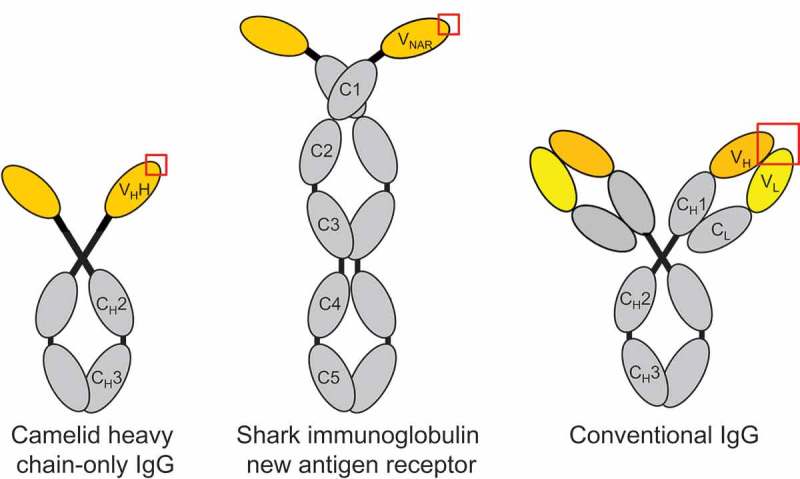
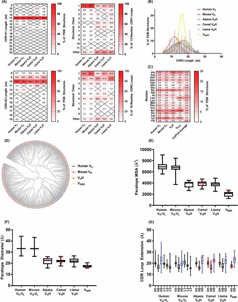
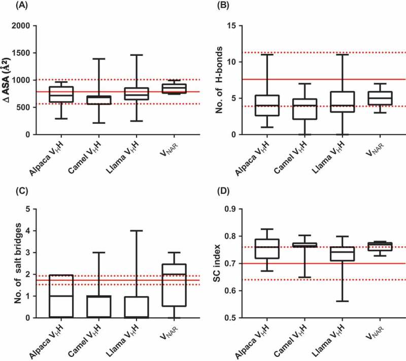
Single-domain antibodies (sdAbs), the autonomous variable domains of heavy chain-only antibodies produced naturally by camelid ungulates and cartilaginous fishes, have evolved to bind antigen using only three complementarity-determining region (CDR) loops rather than the six present in conventional VH:VL antibodies. It has been suggested, based on limited evidence, that sdAbs may adopt paratope structures that predispose them to preferential recognition of recessed protein epitopes, but poor or non-recognition of protuberant epitopes and small molecules. Here, we comprehensively surveyed the evidence in support of this hypothesis. We found some support for a global structural difference in the paratope shapes of sdAbs compared with those of conventional antibodies: sdAb paratopes have smaller molecular surface areas and diameters, more commonly have non-canonical CDR1 and CDR2 structures, and have elongated CDR3 length distributions, but have similar amino acid compositions and are no more extended (interatomic distance measured from CDR base to tip) than conventional antibody paratopes. Comparison of X-ray crystal structures of sdAbs and conventional antibodies in complex with cognate antigens showed that sdAbs and conventional antibodies bury similar solvent-exposed surface areas on proteins and form similar types of non-covalent interactions, although these are more concentrated in the compact sdAb paratope. Thus, sdAbs likely have privileged access to distinct antigenic regions on proteins, but only owing to their small molecular size and not to general differences in molecular recognition mechanism. The evidence surrounding the purported inability of sdAbs to bind small molecules was less clear. The available data provide a structural framework for understanding the evolutionary emergence and function of autonomous heavy chain-only antibodies.

Keywords: VHH; VNAR; antibody:antigen interaction; epitope; molecular recognition; paratope; single-domain antibody.


Figures

Figure 1.
Figure 1.
Domain structures of camelid heavy chain-only IgG, shark immunoglobulin new antigen receptor (IgNAR) and conventional vertebrate tetrameric IgG. The variable domain(s) of each antibody molecule are shown in yellow and the antigen-combining site is indicated by a red box.
Fig 2.
Fig 2.
Properties of sdAb vs. conventional antibody paratopes. (A) Structural classification of CDR1 and CDR2 according to PyIgClassify.108 (B) CDR3 length distributions. (C) Amino acid compositions of conventional antibody (VH domain) and sdAb paratopes. For VHHs, sequences of CDR1, CDR2 and CDR3 (Honegger-Plückthun numbering) were used and for VNARs, sequences of CDR1 and CDR3 only were used. (D) Relatedness of conventional antibody (VH domain) and sdAb CDR3 sequences. The phylogenetic tree was produced using neighbor-joining methods in ClustalW2 and the cladogram was visualized using iTOL109 with CDR3s colored according to species origin as in part B. (E) Molecular surface areas of conventional antibody (VH:VL) and sdAb paratopes. Areas were calculated for merged CDR sequences (Honegger-Plückthun numbering) using PyMol. (F) Diameters of conventional antibody (VH:VL) and sdAb paratopes. Diameters were calculated as the maximum interatomic distance between any two FR-CDR boundary residues (Honegger-Plückthun numbering). (G) Extension of conventional antibody (VH:VL) and sdAb paratopes. Extension was calculated as the maximum interatomic distance between the CDR base (first or last residue according to Honegger-Plückthun numbering) and the CDR tip. The CDR(H)3 loop is shown in blue. In parts (E)(G), boxplot lines represent medians, the box boundaries represent quartiles and the box whiskers represent ranges. Red dots indicate sdAbs targeting cryptic epitopes discussed in the main text. Data are representative of all complete antibody structures available in the Protein Data Bank and indexed in PyIgClassify as of January 2018.
Figure 3.
Figure 3.
Properties of sdAb:antigen and conventional antibody:antigen interfaces. (A) Change in solvent-accessible surface area on proteins upon binding by conventional antibodies or sdAbs. (B) Number of hydrogen bonds and (C) number of salt bridges in conventional antibody:antigen and sdAb:antigen interfaces. (D) Shape complementarity index of conventional antibody:protein and sdAb:protein interfaces. Results in parts (A)(C) were calculated using the PISA server, and in part (D) using the SC algorithm implemented in CCP4. Boxplot lines represent medians, the box boundaries represent quartiles and the box whiskers represent ranges. Red lines indicate the means and standard deviations for conventional antibodies.111,112 Data are representative of all complete antibody:antigen co-crystal structures available in the Protein Data Bank and indexed in PyIgClassify as of January 2018.

References

    1. Flajnik MF, Deschacht N, Muyldermans S.. A case of convergence: why did a simple alternative to canonical antibodies arise in sharks and camels? PLoS Biol. 2011;9:e1001120. doi:10.1371/journal.pbio.1001120. - DOI - PMC - PubMed
    1. Ward ES, Gussow D, Griffiths AD, Jones PT, Winter G. Binding activities of a repertoire of single immunoglobulin variable domains secreted from Escherichia coli. Nature. 1989;341:544–546. doi:10.1038/341544a0. - DOI - PubMed
    1. Hamers-Casterman C, Atarhouch T, Muyldermans S, Robinson G, Hamers C, Songa EB, Bendahman N, Hamers R. Naturally occurring antibodies devoid of light chains. Nature. 1993;363:446–448. doi:10.1038/363446a0. - DOI - PubMed
    1. Greenberg AS, Avila D, Hughes M, Hughes A, McKinney EC, Flajnik MF. A new antigen receptor gene family that undergoes rearrangement and extensive somatic diversification in sharks. Nature. 1995;374:168–173. doi:10.1038/374168a0. - DOI - PubMed
    1. Holliger P, Hudson PJ. Engineered antibody fragments and the rise of single domains. Nat Biotechnol. 2005;23:1126–1136. doi:10.1038/nbt1142. - DOI - PubMed

Publication types

MeSH terms