. 2018 Jun 19;32(12):fj201800334.
doi: 10.1096/fj.201800334. Online ahead of print.

Cilia protein IFT88 regulates extracellular protease activity by optimizing LRP-1-mediated endocytosis

Affiliations

Cilia protein IFT88 regulates extracellular protease activity by optimizing LRP-1-mediated endocytosis

Clarissa R Coveney et al. FASEB J. .

Abstract

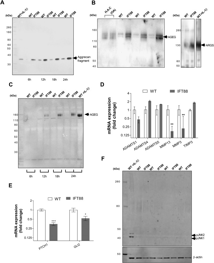
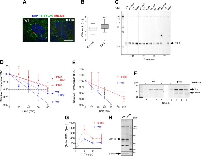
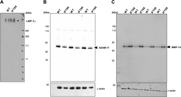
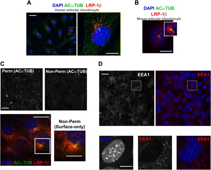
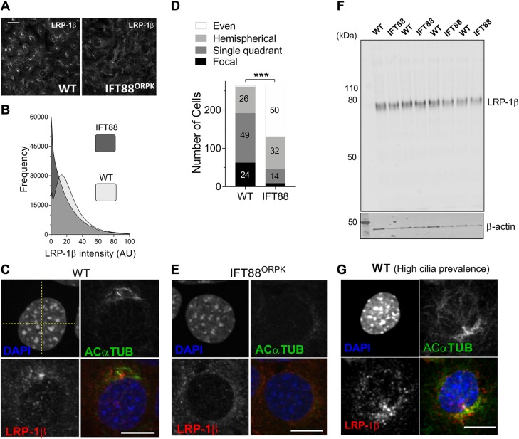
Matrix protease activity is fundamental to developmental tissue patterning and remains influential in adult homeostasis. In cartilage, the principal matrix proteoglycan is aggrecan, the protease-mediated catabolism of which defines arthritis; however, the pathophysiologic mechanisms that drive aberrant aggrecanolytic activity remain unclear. Human ciliopathies exhibit altered matrix, which has been proposed to be the result of dysregulated hedgehog signaling that is tuned within the primary cilium. Here, we report that disruption of intraflagellar transport protein 88 (IFT88), a core ciliary trafficking protein, increases chondrocyte aggrecanase activity in vitro. We find that the receptor for protease endocytosis in chondrocytes, LDL receptor-related protein 1 (LRP-1), is unevenly distributed over the cell membrane, often concentrated at the site of cilia assembly. Hypomorphic mutation of IFT88 disturbs this apparent hot spot for protease uptake, increases receptor shedding, and results in a reduced rate of protease clearance from the extracellular space. We propose that IFT88 and/or the cilium regulates the extracellular remodeling of matrix-independently of Hedgehog regulation-by enabling rapid LRP-1-mediated endocytosis of proteases, potentially by supporting the creation of a ciliary pocket. This result highlights new roles for the cilium's machinery in matrix turnover and LRP-1 function, with potential relevance in a range of diseases.-Coveney, C. R., Collins, I., Mc Fie, M., Chanalaris, A., Yamamoto, K., Wann, A. K. T. Cilia protein IFT88 regulates extracellular protease activity by optimizing LRP-1-mediated endocytosis.

Keywords: cartilage; chondrocyte; matrix; osteoarthritis; primary cilium.


Conflict of interest statement

The authors thank the Oxford Musculoskeletal Biobank (University of Oxford) forproviding human cartilage samples; and Hideaki Nagase, Yoshi Itoh, Linda Troeberg, Heba Ismail, and Tonia Vincent (University of Oxford) for providing key reagents, Abs, and critical comments throughout the development of this manuscript. This work was supported by the Arthritis Research United Kingdom (ARUK) Centre for Osteoarthritis Pathogenesis (Grant 20205), including a small project initiative awarded to A.K.T.W. and K.Y. that supported M.M.F.; a Kennedy Trust for Rheumatology Research (KTRR) Prize Studentship supporting C.R.C.; and an ARUK studentship awarded to A.K.T.W. (Grant 21546, supporting I.C.), and ARUK grant (Grant 20887) awarded to Linda Troeberg (supporting A.C.), an ARUK Career Development Fellowship (Grant 21447, supporting K.Y.), and a KTRR/ARUK Career Development Fellowship supporting A.K.T.W. The authors declare no conflicts of interest.

Figures

Figure 1
Figure 1
IFT88 mutation results in constitutive catabolic activity without associated transcriptional regulation. Wild-type (WT) and IFT88ORPK chondrocytes were cultured with full-length aggrecan medium collected at intervals. A) Western blot analysis of conditioned medium, probing for aggrecan showing 35-kDa fragment in cultures that were treated with 10 ng/ml IL-1β and over time in unstimulated cultures. B, C) Western blot analysis showing the presence of AGEG and ARGS neoepitopes in conditioned medium after 24 h (B), and AGEG over a time course (C). D, E) Quantitative RT-PCR analysis of WT and IFT88 chondrocytes from identical experiments. Data shown are mean ΔCt normalized to average WT ± sd. ***P = 0.0004, *P = 0.008 for PTCH1 and GLI2, respectively, multiple Student’s t test corrected by Holm-Sidak method; n = 3). F) Western blot analysis of cell lysates probed for pJNK1 and pJNK2, with positive control WT cells treated with 10 ng/ml IL-1β and lysate collected at 10 min (n = 4).
Figure 2
Figure 2
Perturbation of IFT88 impairs chondrocyte endocytosis of proteases. Cells were cultured with recombinant proteases—either FLAG-TS-5 or full-length MMP-13—and uptake of proteases was monitored by microscopy or measurement of FLAG-TS-5 in extracellular media by Western blot. A) Confocal fluorescent microscopy of wild-type (WT) and IFT88ORPK cells on exposure to FLAG-TS-5 (green). Primary cilia (red) were labeled with anti-arl13b and nuclei with DAPI (blue). Scale bar, 10 μm. Image captured after 40 min of incubation. B) Cilia length data as shown by box plot with Tukey whiskers. ***P ≤ 0.001 (Mann-Whitney U test, control n = 61 cilia; FLAG-TS-5, n = 62 cilia). Images also taken at the 40-min incubation time point. C, D) Western blot analysis of extracellular FLAG-TS-5 over time(C) and quantification of data (data are shown as mean normalized to 0 time-point, ± sd, n = 6; D). E) Quantification of repeat study comparing WT and IFT88ORPK cells over an extended time course (means ± sd, n = 3). F, G) Western blot for rMMP-13 uptake showing both full-length (pro) and cleaved (active) MMP-13 (F) and quantification of data (means ± sd, n = 3; G). H) Western blot for MMP13 expression in cell lysates.
Figure 3
Figure 3
IFT88 mutation results in LRP-1 shedding. A) Western blot for extracellular (shed) LRP-1α in conditioned medium of cultures. B, C) Western blot of cell lysates that demonstrates the cellular expression of sheddases ADAM-17 (n = 3; B) and MMP-14 (n = 4; C), respectively. Conditioned medium and lysates for all groups were collected after 24 h of culture.
Figure 4
Figure 4
Membranous hot spot of LRP-1β associated with primary cilium. A) Immunofluorescent staining of primary cilia (green) and LRP-1β (red) in human articular chondrocytes. B) Immunofluorescent staining of LRP-1β (red) and primary cilia (green) in wild-type (WT) mouse chondrocytes. C) Immunofluorescence images shows surface-only staining of LRP-1β (red) in WT cells that concentrated at the base of the primary cilium (green) in cells without permeabilization as validated by the absence of a majority of acetylated α-tubulin signal (black and white images above). D) Immunofluorescence of EEA-1 (red) in mouse WT chondrocytes. Nuclei were labeled with DAPI. Scale bars, 10 µm.
Figure 5
Figure 5
IFT88 mutation disturbs hot spot that is possibly associated with the ciliary pocket. A) Fluorescent microscopy of wild-type (WT) and IFT88ORPK cultures demonstrates the LRP-1β signal distribution in field of cells. B) Frequency histogram of LRP-1β signal intensity distribution for 6 fields of cells (~n = 300 cells). C, E) Single-cell confocal images of WT (focal distribution; C) and IFT88ORPK (even distribution; E) cells. Yellow dashed lines were used to define quadrants centered on nuclei. D) Contingency data from a classification analysis from 7 fields of WT and IFT88ORPK cultures that describe numbers in each group (percentages shown on bars, with the exception of WT even distribution, which had 1% frequency, and IFT88ORPK single focal distribution, which had 4% frequency) of each type of staining within total populations. Statistical difference is from a χ2 analysis. ***P < 0.0001. F) Western blot analysis of LRP-1β total protein expression (n = 4 repeats). G) LRP-1β (red) signal localized along length of the cilium (green), potentially lining a ciliary pocket (representative of single quadrant distribution for classification data). Scale bars, 10 μm.

Similar articles

Cited by

References

    1. Christ A., Christa A., Klippert J., Eule J. C., Bachmann S., Wallace V. A., Hammes A., Willnow T. E. (2015) LRP2 acts as SHH clearance receptor to protect the retinal margin from mitogenic stimuli. Dev. Cell 35, 36–48 - PubMed
    1. Emonard H., Bellon G., Troeberg L., Berton A., Robinet A., Henriet P., Marbaix E., Kirkegaard K., Patthy L., Eeckhout Y., Nagase H., Hornebeck W., Courtoy P. J. (2004) Low density lipoprotein receptor-related protein mediates endocytic clearance of pro-MMP-2.TIMP-2 complex through a thrombospondin-independent mechanism. J. Biol. Chem. 279, 54944–54951 - PubMed
    1. Yang Z., Strickland D. K., Bornstein P. (2001) Extracellular matrix metalloproteinase 2 levels are regulated by the low density lipoprotein-related scavenger receptor and thrombospondin 2. J. Biol. Chem. 276, 8403–8408 - PubMed
    1. Muir H. (1995) The chondrocyte, architect of cartilage. Biomechanics, structure, function and molecular biology of cartilage matrix macromolecules. BioEssays 17, 1039–1048 - PubMed
    1. Glyn-Jones S., Palmer A. J., Agricola R., Price A. J., Vincent T. L., Weinans H., Carr A. J. (2015) Osteoarthritis. Lancet 386, 376–387 - PubMed

LinkOut - more resources