. 2018 Oct:50:100-110.
doi: 10.1016/j.cellsig.2018.06.006. Epub 2018 Jun 18.

Spatial regulation of signaling by the coordinated action of the protein tyrosine kinases MET and FER

Affiliations

Spatial regulation of signaling by the coordinated action of the protein tyrosine kinases MET and FER

Jiali Zhang et al. Cell Signal. 2018 Oct.

Abstract

A critical aspect of understanding the regulation of signal transduction is not only to identify the protein-protein interactions that govern assembly of signaling pathways, but also to understand how those pathways are regulated in time and space. In this report, we have applied both gain-of-function and loss-of-function analyses to assess the role of the non-receptor protein tyrosine kinase FER in activation of the HGF Receptor protein tyrosine kinase MET. Overexpression of FER led to direct phosphorylation of several signaling sites in MET, including Tyr1349, but not the activation loop residues Tyr1234/5; in contrast, suppression of FER by RNAi revealed that phosphorylation of both a C-terminal signaling site (Tyr1349) and the activation loop (Tyr1234/5) were influenced by the function of this kinase. Adaptin β, a component of the adaptor protein complex 2 (AP-2) that links clathrin to receptors in coated vesicles, was recruited to MET following FER-mediated phosphorylation. Furthermore, we provide evidence to support a role of FER in maintaining plasma membrane distribution of MET and thereby delaying protein-tyrosine phosphatase PTP1B-mediated inactivation of the receptor. Simultaneous up-regulation of FER and down-regulation of PTP1B observed in ovarian carcinoma-derived cell lines would be expected to contribute to persistent activation of HGF-MET signaling, suggesting that targeting of both FER and MET may be an effective strategy for therapeutic intervention in ovarian cancer.

Keywords: Endocytosis; FER; MET; Ovarian cancer; PTP1B.


Conflict of interest statement

All contributors declare that they have no financial or other conflicts of interest.

Figures
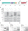
Figure 1.
Figure 1.. Tyrosine kinase FER phosphorylated HGF receptor MET at multiple tyrosine residues.
(A) Schematic illustration of domain structure of HGF receptor MET. Multiple tyrosine residues within cytosolic segment of the receptor are listed. The following domains are highlighted: SEMA, semaphoring structural domain; PSI, plexin repeat domain; TIG, IPT/TIG Ig-like fold; TM, transmembrane domain. (B) WT, kinase-dead (K592R) or SH2 mutant of FER were expressed alone, or co-expressed with WT or kinase-dead mutant (mATP) of MET in 293T cells, as indicated. Phosphorylation levels of MET, SHP2 and ERK, as well as their expression levels, were examined by immunoblotting. (C) mATP MET, or its Y1349F, Y1356F or YY1349/1356FF mutants, were expressed alone, or co-expressed with FER in 293T cells, as indicated. Lysates were harvested and MET immunoprecipitated. Tyrosine phosphorylation of MET was examined with pTyr antibody 4G10. Expression levels of FER were also probed, with tubulin as loading control. (D) mATP MET, or its Y971F, Y1003F, Y1026F or Y1093F mutants, were expressed alone, or co-expressed with FER in 293T cells, as indicated. Lysates were harvested and MET immunoprecipitated. Tyrosine phosphorylation of MET was examined with pTyr antibody 4G10. Expression levels of FER were also probed, with actin as loading control. (E) mATP MET, or its Y1026F, YY1349/1356FF or YYY1026/1349/1356FFF mutants, were co-expressed with FER in 293T cells, as indicated. Lysates were harvested and MET immunoprecipitated. Tyrosine phosphorylation of MET was examined with pTyr antibody 4G10. Expression levels of FER were also probed, with actin as loading control.
Figure 2.
Figure 2.. Tyrosine phosphorylation of the activation loop of MET was decreased in FER-deficient ovarian cancer cells.
(A) CAOV4 cells, expressing either control or FER shRNA, were lysed, and immunoblotted as indicated to measure the activation of MET and ERK signaling pathways. (B) CAOV4 cells, expressing either control or FER shRNA, were serum-starved and stimulated with recombinant human HGF for the indicated times, lysed, and immunoblotted with the designated antibodies to illustrate the impact of FER deficiency on HGF-induced phosphorylation.
Figure 3.
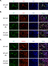
Figure 3.. FER co-localized with MET, and promoted recruitment of GRB2 and Adaptinβ to the receptor.
(A) MET mATP mutant was co-transfected with FER in 293T cells, and the co-localization of MET (Green) and FER (Red) was assessed by immunofluorescence. Nuclei were stained with DAPI (Blue). (B) MET mATP mutant was transfected alone, or together with wt or kinase-dead FER, in 293T cells, and the co-localization of MET (Green) and GRB2 (Red) was assessed by immunofluorescence. Nuclei were stained with DAPI (Blue). (C) MET mATP mutant was transfected alone, or together with wt or kinase-dead FER, in 293T cells, and the co-localization of MET (Green) and Adaptinβ (Red) was assessed by immunofluorescence. Nuclei were stained with DAPI (Blue). The illustrated images are representative of recordings in at least 3 replicates.
Figure 4.
Figure 4.. Regulation of endocytosis of the HGF receptor MET by FER.
(A) CAOV4 ovarian cancer cells were homogenized in a buffer containing 0.35% Triton X-100, and the sub-cellular fractions were separated on a discontinuous sucrose gradient. The fractions were collected from the top of the gradient (total volume: ~3 ml; each fraction: ~300 μl) and subjected to immunoblotting with antibodies against indicated proteins. (B) (Left) Schematic diagram for sucrose gradient ultracentrifugation. (Right) Fractions from both endosome and lysosome were collected, lysed and immunoblotted for MET and FER. Rab5 was used as an endosome marker. CAOV4 ovarian cancer cells, expressing either control or FER shRNA, were serum-starved and stimulated with HGF for 30 mins. (Upper) The co-localization of MET (Green) and EEA1 (Red, upper panels) or Rab11 (Red, lower panels) was assessed by immunofluorescence. Nuclei were stained with DAPI (Blue). The illustrated images are representative of recordings in at least 3 replicates. (C) CAOV4 ovarian cancer cells, expressing either control or FER shRNA, were treated with cycloheximide for the indicated times, lysed, and immunoblotted with antibodies against MET and FER. Actin was probed as loading control. (D) CAOV4 ovarian cancer cells, expressing either control or FER shRNA, were subjected to cell fractionation and the distribution pattern of HGFR/MET and FER was assessed by immunoblotting.
Figure 5.
Figure 5.. Dephosphorylation of MET and FER by PTP1B.
(A) MET was expressed alone, or co-expressed with wild-type or D181A (substrate trapping mutant) forms of PTP1B in 293T cells, as indicated. Lysates were harvested and immunoblotted with pTyr antibody 4G10. (B) MET was expressed alone, or co-expressed with wild-type or D181A mutant forms of PTP1B in 293T cells, as indicated. (Upper) Lysates were harvested and immunoblotted for pTyr 1234/1235 MET and PTP1B. Tubulin was probed as loading control. (Bottom) PTP1B was immunoprecipitated from cell lysates, and the co-immunuoprecipitation of MET was examined with MET antibody. (C) Immunoblot to demonstrate ectopic expression of PTP1B, both wild-type and D181A mutant, in ovarian carcinoma-derived cell line CAOV4. Actin was probed as loading control. CAOV4 lysates were also subjected to immunoblotting for pTyr 1234/1235 MET. (D) FER was expressed alone, or co-expressed with wild-type or D181A mutant forms of PTP1B in 293T cells, as indicated. Lysates were harvested and immunoblotted with pTyr antibody 4G10. (E) FER was expressed alone, or co-expressed with wild-type or D181A mutant forms PTP1B in 293T cells, as indicated. (Upper) Lysates were harvested and immunoblotted for pTyr 402 FER and PTP1B. Actin was probed as a loading control. (Bottom) PTP1B was immunoprecipitated from cell lysates, and the co-immunuoprecipitation of FER was examined with FER antibody. (F) Wild-type or D181A mutant PTP1B was stably expressed in CAOV4 ovarian carcinoma cells. (Upper) Lysates were harvested, PTP1B was immunoprecipitated, and the co-immunuoprecipitation of FER was examined with FER antibody. (Bottom) Lysates were also immunoblotted with antibody against pTyr 402 FER.
Figure 6.
Figure 6.. Treatment of Dynasore, an inhibitor of dynamin, enhanced MET activation in ovarian cancer cells.
CAOV4 cells were serum-starved for 16hrs, followed by treatment with Dynasore at indicated concentrations for 30 mins. Cells were then stimulated with HGF for 30 mins, and the activation of MET was examined with pTyr1234/1235 MET antibody, with actin as loading control.
Figure 7.
Figure 7.. PTP1B was underexpressed in ovarian tumor derived cancer cells.
(A) Expression of PTP1B protein in both normal human ovarian surface epithelial cells (HOSE) and ovarian carcinoma-derived cell lines was assessed by immunoblotting. (B) Box plots to illustrate down-regulation of PTP1B mRNA in primary specimens from human clear cell adenocarcinoma, endometrioid adenocarcinoma, mucinous adenocarcinoma and serous adenocarcinoma compared with normal ovarian surface epithelium (Lu dataset), as extracted from the Oncomine database. (C) Working model. In the absence of HGF: FER associated with MET, and directly phosphorylated MET, GAB1, and possibly SHP2. This resulted in the activation of SHP2–MAPK and RAC1–PAK1 signaling downstream from MET which contributed to the motility and invasiveness of ovarian cancer cells [7]. In the presence of HGF: FER influenced the endocytosis of the HGF receptor MET. Upon endocytosis, both MET and FER were exposed to the ER-anchored protein tyrosine phosphatase PTP1B with dephosphorylation and inactivation. However, the presence of FER facilitates the recycling of MET to the plasma membrane and its re-activation by HGF.

Similar articles

Cited by

References

    1. Scott JD and Pawson T (2009) Cell signaling in space and time: where proteins come together and when they’re apart. Science. 326, 1220–1224 - PMC - PubMed
    1. Schlessinger J (2014) Receptor tyrosine kinases: legacy of the first two decades. Cold Spring Harb Perspect Biol. 6 - PMC - PubMed
    1. Lemmon MA and Schlessinger J (2010) Cell signaling by receptor tyrosine kinases. Cell. 141, 1117–1134 - PMC - PubMed
    1. Birchmeier C, Birchmeier W, Gherardi E and Vande Woude GF (2003) Met, metastasis, motility and more. Nature reviews. Molecular cell biology. 4, 915–925 - PubMed
    1. Trusolino L, Bertotti A and Comoglio PM (2010) MET signalling: principles and functions in development, organ regeneration and cancer. Nature reviews. Molecular cell biology. 11, 834–848 - PubMed

Substances