. 2018 Jun 19;10(6):1442-1453.
doi: 10.18632/aging.101481.

Secreted α-Klotho maintains cartilage tissue homeostasis by repressing NOS2 and ZIP8-MMP13 catabolic axis

Affiliations

Secreted α-Klotho maintains cartilage tissue homeostasis by repressing NOS2 and ZIP8-MMP13 catabolic axis

Paul Chuchana et al. Aging (Albany NY). .

Abstract

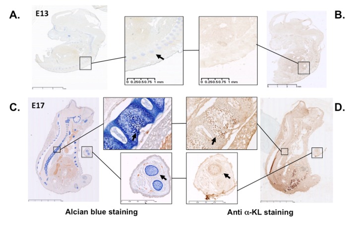
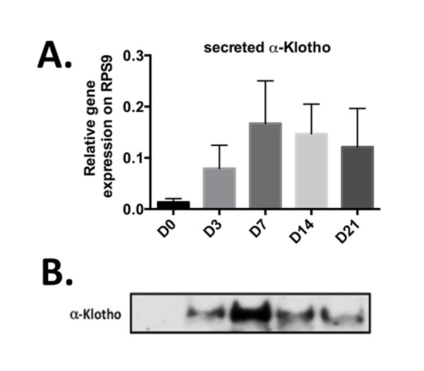
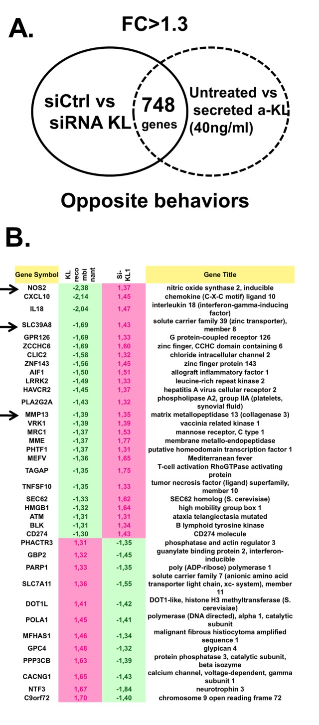
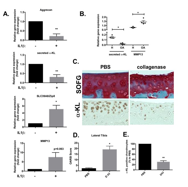
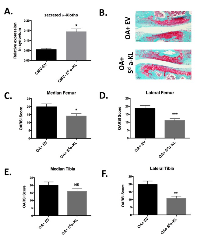
Progressive loss of tissue homeostasis is a hallmark of numerous age-related pathologies, including osteoarthritis (OA). Accumulation of senescent chondrocytes in joints contributes to the age-dependent cartilage loss of functions through the production of hypertrophy-associated catabolic matrix-remodeling enzymes and pro-inflammatory cytokines. Here, we evaluated the effects of the secreted variant of the anti-aging hormone α-Klotho on cartilage homeostasis during both cartilage formation and OA development. First, we found that α-Klotho expression was detected during mouse limb development, and transiently expressed during in vitro chondrogenic differentiation of bone marrow-derived mesenchymal stem cells. Genome-wide gene array analysis of chondrocytes from OA patients revealed that incubation with recombinant secreted α-Klotho repressed expression of the NOS2 and ZIP8/MMP13 catabolic remodeling axis. Accordingly, α-Klotho expression was reduced in chronically IL1β-treated chondrocytes and in cartilage of an OA mouse model. Finally, in vivo intra-articular secreted α-Kotho gene transfer delays cartilage degradation in the OA mouse model. Altogether, our results reveal a new tissue homeostatic function for this anti-aging hormone in protecting against OA onset and progression.

Keywords: aging; cartilage; homeostasis; hormone; α-Klotho.


Conflict of interest statement

CONFLICTS OF INTEREST: The authors declare no competing interests.

Figures

Figure 1
Figure 1
α-Klotho expression in mouse embryos during cartilage formation. Serial sections of E13 and E17 mouse embryos were stained with Alcian blue to detect cartilage (A and C) or with an anti- pan α-Klotho antibody (B and D). Arrows are showing cartilaginous tissues expressing also α-Klotho.
Figure 2
Figure 2
Secreted α-Klotho expression during in vitro chondrogenesis. TGFβ3-driven chondrogenic differentiation of BM-MSCs (osteochondral progenitors) in micropellet culture (n=3). Expression analysis of secreted α-KL (A) by RT-qPCR (gene expression level relative to that of RPS9) at different time points during BM-MSC differentiation into chondrocytes. (B) α-Klotho protein expression detectable below 65kDa marker by western blotting in conditioned medium from BM-MSCs in micromass culture at the different time points during chondrogenesis. Data are represented as mean -/+ SEM.
Figure 3
Figure 3
Identification of genes regulated by secreted α-Klotho in human chondrocytes. (A) Genome-wide microarray analysis of human primary chondrocytes incubated or not with recombinant secreted α-KL or after siRNA-mediated α-KL silencing identified 748 genes with opposite behavior in these two experimental conditions. FC, fold change. (B) List of the 38 genes (among the common 748 differentially expressed genes) that are involved in osteoarticular diseases according to the Ingenuity® software.
Figure 4
Figure 4
α-Klotho expression is reduced in osteoarthrosis models. (A) ACAN, secreted α-KL, SLC39A8 and MMP13 mRNA expression analysis by RT-qPCR of human chondrocytes incubated or not with IL-1β for 10 days. (B) Secreted α-KL and MMP13 expression levels in human OA (n=4) versus healthy cartilage (n=4). (C) α-Klotho expression by immunostaining (bottom panel) in joint knee cartilage at day 43 after intra-articular injection of PBS (n=5 mice) or collagenase (contralateral knee). Top panels show Safranin-O Fast-Green (SOFG) staining of the same joints. (C) OARSI scoring of cartilage degradation and (D) quantification of α-Klotho-positive chondrocytes in joints at day 43 after intra-articular injection of PBS or collagenase. Data are represented as mean -/+ SEM. *=p<0.05; **=p<0.01.
Figure 5
Figure 5
Effect of intra-articular secreted α-KL gene transfer in an experimental murine OA model. (A) Secreted α-KL gene expression in mice synovium after electrotransfer of empty vector (CMV-EV) or CMV-Sd-α-KL expressing vector. (B) Representative Safranin-O Fast-Green staining of knee joints from OA mice treated with empty vector (OA+EV) or CMV-Sd-α-KL expressing vector (OA+Sd-α-KL). (C-F) OARSI scores in the different joint localizations from OA mice after intra-articular electrotransfer of empty vector (OA+EV; n=15) or CMV-Sd-α-KL expressing vector (OA+Sd-α-KL; n=25). Data are represented as mean -/+ SEM. *=p<0.05; **=p<0.01.

References

    1. Blau HM, Cosgrove BD, Ho AT. The central role of muscle stem cells in regenerative failure with aging. Nat Med. 2015; 21:854–62. 10.1038/nm.3918 - DOI - PMC - PubMed
    1. Oh J, Lee YD, Wagers AJ. Stem cell aging: mechanisms, regulators and therapeutic opportunities. Nat Med. 2014; 20:870–80. 10.1038/nm.3651 - DOI - PMC - PubMed
    1. Farr JN, Xu M, Weivoda MM, Monroe DG, Fraser DG, Onken JL, Negley BA, Sfeir JG, Ogrodnik MB, Hachfeld CM, LeBrasseur NK, Drake MT, Pignolo RJ, et al. Targeting cellular senescence prevents age-related bone loss in mice. Nat Med. 2017; 23:1072–79. 10.1038/nm.4385 - DOI - PMC - PubMed
    1. Loeser RF. Aging and osteoarthritis. Curr Opin Rheumatol. 2011; 23:492–96. 10.1097/BOR.0b013e3283494005 - DOI - PMC - PubMed
    1. Jeon OH, Kim C, Laberge RM, Demaria M, Rathod S, Vasserot AP, Chung JW, Kim DH, Poon Y, David N, Baker DJ, van Deursen JM, Campisi J, Elisseeff JH. Local clearance of senescent cells attenuates the development of post-traumatic osteoarthritis and creates a pro-regenerative environment. Nat Med. 2017; 23:775–81. 10.1038/nm.4324 - DOI - PMC - PubMed

MeSH terms