

Treatment effect of CDKN1A on rheumatoid arthritis by mediating proliferation and invasion of fibroblast-like synoviocytes cells
- PMID: 29920650
- PMCID: PMC6194335
- DOI: 10.1111/cei.13161
Treatment effect of CDKN1A on rheumatoid arthritis by mediating proliferation and invasion of fibroblast-like synoviocytes cells
Abstract
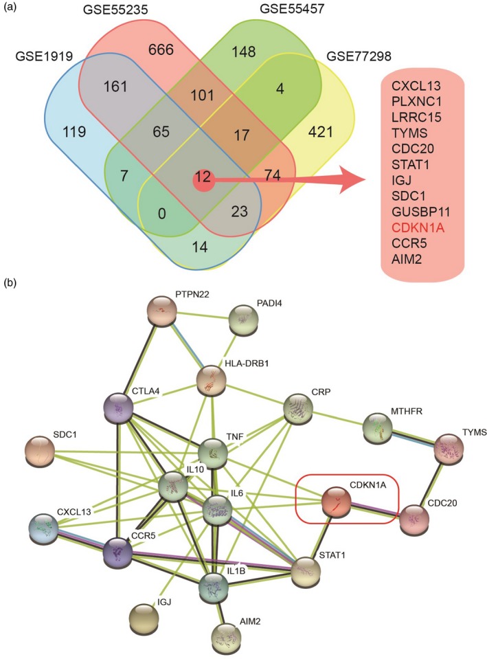
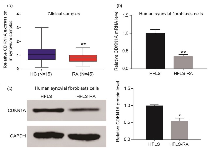
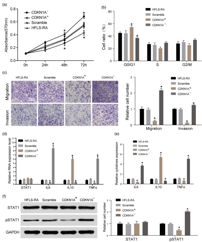
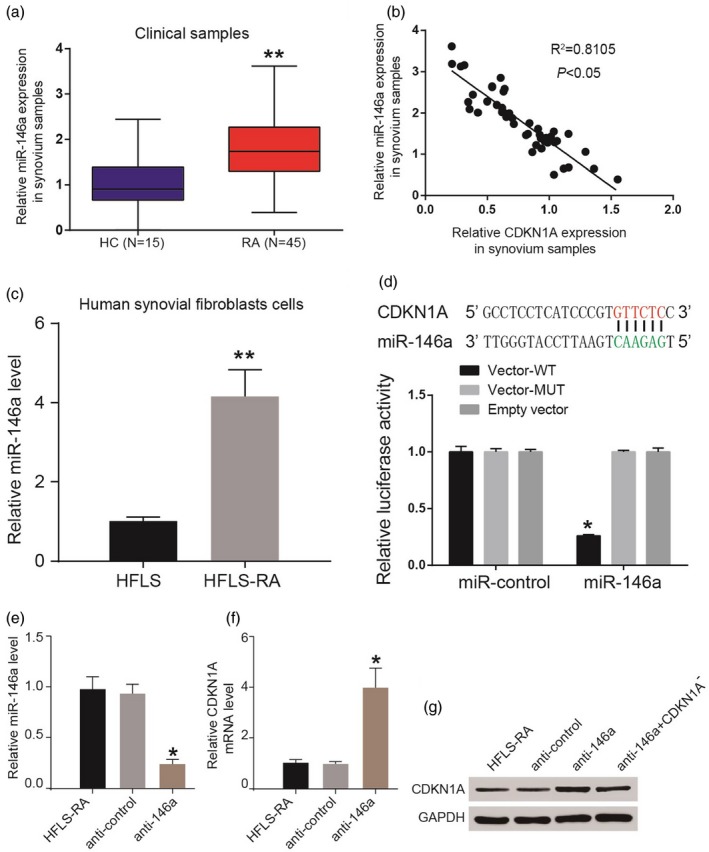
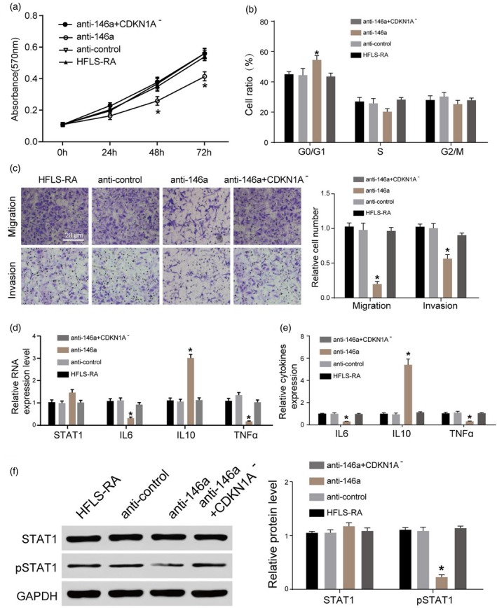
The objective of the present study was to evaluate the role of CDKN1A in rheumatoid arthritis (RA). Related gene expression data screened from Gene Expression Omnibus (GEO) were processed with network analysis. Protein-protein interaction was analysed through string database. Quantitative reverse transcription-polymerase chain reaction (qRT-PCR) was used to measure mRNA and microRNA expression. Cell proliferation and cell cycle were tested by MTT assay and flow cytometry, respectively. Transwell migration and invasion assay was used to test cell migration and invasion. CDKN1A screened by bioinformatics methods showed differential expression in RA cells compared with healthy controls (HC), and was at an important position in the protein-protein interaction network of RA. Compared with the HC group, CDKN1A was down-regulated in human RA synovium tissues and human fibroblast-like synoviocytes (HFLS). Contrary to CDKN1A silencing, CDKN1A over-expression significantly inhibited the proliferation and invasion of HFLS-RA, arrested HFLS-RA in G0/G1 phase and down-regulated the expressions of tumour necrosis factor (TNF)-α and interleukin (IL)-6, while it up-regulated the expression of IL-10. CDKN1A over-expression could also suppress phosphorylated signal transducers and activators of transcription 1 (pSTAT-1) expression. MiR-146a, highly expressed in RA tissues, could regulate CDKN1A negatively. Anti-146a suppressed cell proliferation and invasion, and at the same time enhanced IL-10 expression but inhibited IL-6, TNF-α and pSTAT-1 expression. The results indicated that CDKN1A over-expression, which could be enhanced by miR-146a suppression, inhibited the proliferation of invasion in HFLS-RA. This was probably a result of suppressed pSTAT-1, IL-6 and TNF-α expression and enhanced IL-10 expression.
Keywords: CDKN1A; MiR-146a; human fibroblast-like synoviocytes; rheumatoid arthritis.
© 2018 British Society for Immunology.
Figures
Similar articles
-
Down-regulation of microRNA-142-3p inhibits the aggressive phenotypes of rheumatoid arthritis fibroblast-like synoviocytes through inhibiting nuclear factor-κB signaling.Biosci Rep. 2019 Jul 8;39(7):BSR20190700. doi: 10.1042/BSR20190700. Print 2019 Jul 31. Biosci Rep. 2019. Retraction in: Biosci Rep. 2020 Sep 30;40(9):BSR-20190700_RET. doi: 10.1042/BSR-20190700_RET. PMID: 31239367 Free PMC article. Retracted.
-
Interleukin (IL)-23 p19 expression induced by IL-1beta in human fibroblast-like synoviocytes with rheumatoid arthritis via active nuclear factor-kappaB and AP-1 dependent pathway.Rheumatology (Oxford). 2007 Aug;46(8):1266-73. doi: 10.1093/rheumatology/kem055. Epub 2007 Jun 14. Rheumatology (Oxford). 2007. PMID: 17569750
-
The promoting effect of MMP13 on mediating the development of HFLS-RA by the target of miR-19a through IL-17 signaling pathway.J Cell Biochem. 2020 Oct;121(10):4282-4294. doi: 10.1002/jcb.29609. Epub 2020 Jan 21. J Cell Biochem. 2020. Retraction in: J Cell Biochem. 2021 Dec;122(12):1974. doi: 10.1002/jcb.30182. PMID: 31960999 Retracted.
-
miR-124a as a key regulator of proliferation and MCP-1 secretion in synoviocytes from patients with rheumatoid arthritis.Ann Rheum Dis. 2011 Mar;70 Suppl 1:i88-91. doi: 10.1136/ard.2010.138669. Ann Rheum Dis. 2011. PMID: 21339227 Review.
-
TLRs Play Crucial Roles in Regulating RA Synoviocyte.Endocr Metab Immune Disord Drug Targets. 2020;20(8):1156-1165. doi: 10.2174/1871530320666200427115225. Endocr Metab Immune Disord Drug Targets. 2020. PMID: 32338225 Review.
Cited by
-
Bioinformatic identification and validation of autophagy-related genes in rheumatoid arthritis.Clin Rheumatol. 2023 Mar;42(3):741-750. doi: 10.1007/s10067-022-06399-2. Epub 2022 Oct 11. Clin Rheumatol. 2023. PMID: 36220923
-
Myocardial Infarction-Associated Extracellular Vesicle-Delivered miR-208b Affects the Growth of Human Umbilical Vein Endothelial Cells via Regulating CDKN1A.Biomed Res Int. 2021 Jun 5;2021:9965639. doi: 10.1155/2021/9965639. eCollection 2021. Biomed Res Int. 2021. PMID: 34195287 Free PMC article.
-
Analysis of the Potential Link Between Dermatomyositis and Cancer.J Inflamm Res. 2024 Dec 3;17:10163-10182. doi: 10.2147/JIR.S480744. eCollection 2024. J Inflamm Res. 2024. PMID: 39649426 Free PMC article.
-
Identification of autophagy-related genes in osteoarthritis articular cartilage and their roles in immune infiltration.Front Immunol. 2023 Nov 27;14:1263988. doi: 10.3389/fimmu.2023.1263988. eCollection 2023. Front Immunol. 2023. PMID: 38090564 Free PMC article.
-
Differential Expression Profiles of the Transcriptome and miRNA Interactome in Synovial Fibroblasts of Rheumatoid Arthritis Revealed by Next Generation Sequencing.Diagnostics (Basel). 2019 Aug 18;9(3):98. doi: 10.3390/diagnostics9030098. Diagnostics (Basel). 2019. PMID: 31426562 Free PMC article.
References
-
- Scott DL, Wolfe F, Huizinga TW. Rheumatoid arthritis. Lancet 2010; 376:1094–108. - PubMed
-
- Svarts N. Etiology and pathogenesis of rheumatoid arthritis. Ter Arkh 1975; 47:19–24. - PubMed
-
- Firestein GS. Invasive fibroblast‐like synoviocytes in rheumatoid arthritis. Passive responders or transformed aggressors? Arthritis Rheum 1996; 39:1781–90. - PubMed
-
- Meinecke I, Rutkauskaite E, Gay S, Pap T. The role of synovial fibroblasts in mediating joint destruction in rheumatoid arthritis. Curr Pharm Des 2005; 11:563–8. - PubMed
MeSH terms
Substances
LinkOut - more resources
Full Text Sources
Other Literature Sources
Medical
