. 2018 Aug;40(8):e12566.
doi: 10.1111/pim.12566.

Increased susceptibility to oral Trichuris muris infection in the specific absence of CXCR5+ CD11c+ cells

Affiliations

Increased susceptibility to oral Trichuris muris infection in the specific absence of CXCR5+ CD11c+ cells

Barry M Bradford et al. Parasite Immunol. 2018 Aug.

Abstract

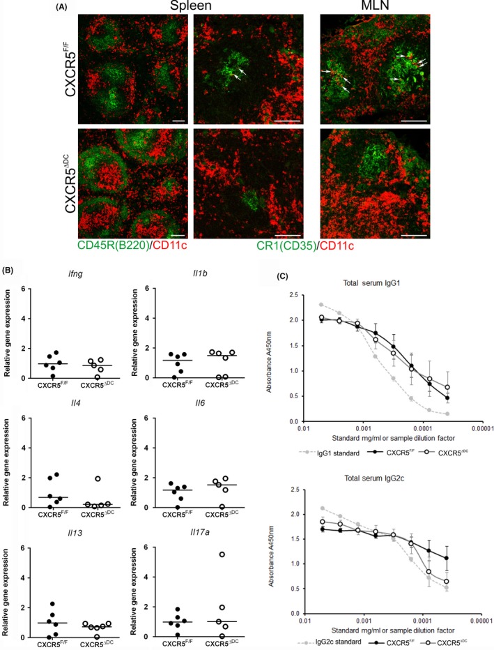
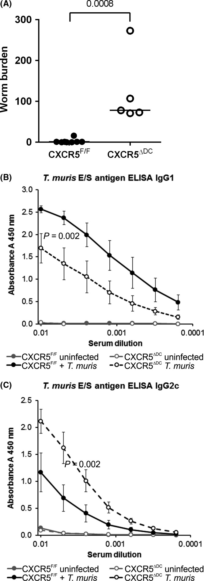
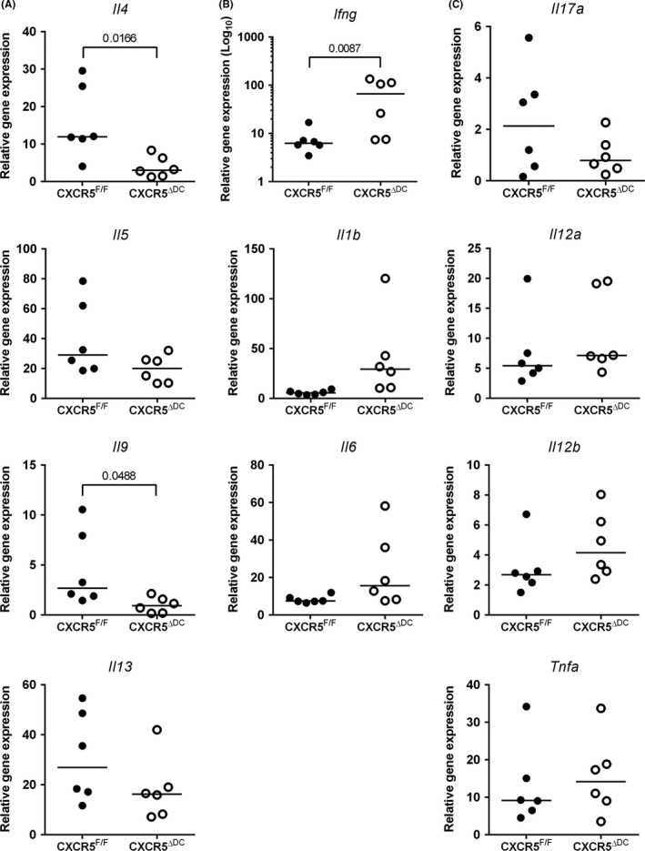
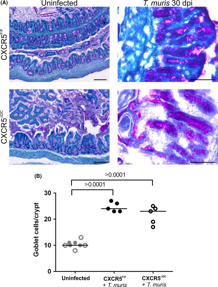
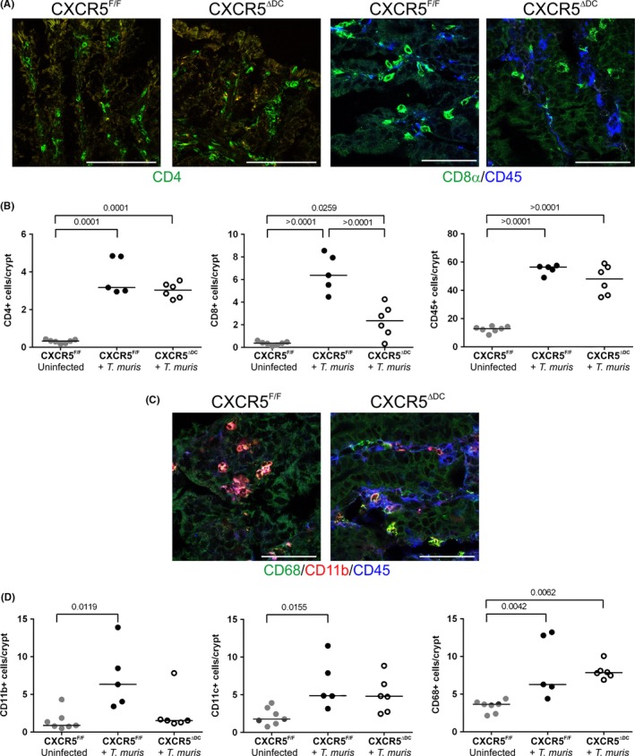
Trichuris muris is a natural mouse helminth pathogen which establishes infection specifically in the caecum and proximal colon. The rapid expulsion of T. muris in resistant mouse strains is associated with the induction of a protective T helper cell type 2 (Th2)-polarized immune response. Susceptible mouse strains, in contrast, mount an inappropriate Th1 response to T. muris infection. Expression of the chemokine CXCL13 by stromal follicular dendritic cells attracts CXCR5-expressing cells towards the B-cell follicles. Previous studies using a complex in vivo depletion model have suggested that CXCR5-expressing conventional dendritic cells (cDC) help regulate the induction of Th2-polarized responses. Here, transgenic mice with CXCR5 deficiency specifically restricted to CD11c+ cells were used to determine whether the specific absence CXCR5 on CD11c+ cells such as cDC would influence susceptibility to oral T. muris infection by affecting the Th1/Th2 balance. We show that in contrast to control mice, those which lacked CXCR5 expression on CD11c+ cells failed to clear T. muris infection and developed cytokine and antibody responses that suggested a disturbed Th1/Th2 balance with enhanced IFN-γ expression. These data suggest an important role of CXCR5-expressing CD11c+ cells such as cDC in immunity to oral T. muris infection.

Keywords: Trichuris muris; CXCR5; Th2; dendritic cell; helminth; mucosal immunity.


Figures

Figure 1
Figure 1
Altered positioning of CD11c+ cells in the secondary lymphoid organs of CXCR5Δ DC mice. A, Immunohistochemical (IHC) analysis of the distribution of B cells (CD45R/B220+  cells; green, left‐hand column), stromal FDC (CR1/CD35+ cells; green) and CD11c+ cells (red) in the spleen and MLN of uninfected CXCR5F/F control mice (upper panels) and CXCR5Δ DC mice (lower panels). In the MLN and spleens of CXCR5F/F mice, CD11c+ cells were occasionally detected within the FDC‐containing B‐cell follicles (arrows). Few, if any, CD11c+ cells were detected in the FDC‐containing B‐cell follicles of CXCR5Δ DC mice. Scale bar, 100 μm. B, qRTPCR analysis of cytokine‐encoding genes in mRNA from the MLN of uninfected CXCR5F/F control mice (closed circles) and CXCR5Δ DC mice (open circles). Horizontal bars, median. C, Similar levels of total IgG1 and IgG2c were detected in the serum of uninfected CXCR5F/F control mice (closed circles) and CXCR5Δ DC mice (open circles). Data were derived from 6 mice/group
Figure 2
Figure 2
Susceptibility to Trichuris muris infection is enhanced in the specific absence of CXCR5‐expressing cDC in CXCR5Δ DC mice. CXCR5F/F mice (n = 8, closed circles) and CXCR5Δ DC mice (n = 5, open circles) were orally infected with approximately 200 embryonated T. muris eggs and worm burdens in the large intestine compared at 30 dpi. Susceptibility to T. muris infection was significantly increased in CXCR5Δ DC mice when compared to CXCR5F/F control mice (< 0.008). (B&C) Serum T. muris E/S antigen‐specific IgG1 (B) and IgG2c (C) levels were determined by ELISA. When compared to T. muris‐infected CXCR5F/F mice, the sera of T. muris‐infected CXCR5Δ DC mice contained significantly lower levels of parasite‐specific IgG1 (B; < 0.002) and significantly higher levels of IgG2c (C; < 0.002)
Figure 3
Figure 3
The expression of Th1/Th2 cytokines in the MLN following Trichuris muris infection is altered in the specific absence of CXCR5‐expressing cDC. CXCR5F/F mice (n = 6) and CXCR5Δ DC mice (n = 6) were orally infected with approximately 200 embryonated T. muris eggs, and at 30 dpi, the expression of cytokine‐encoding genes in the MLN was compared by qRTPCR analysis. A, Comparison of the expression of mRNA encoding the Th2 cytokines IL‐4, IL‐5, IL‐9 and IL‐13. B, Comparison of the expression of mRNA encoding the Th1 cytokine IFN‐γ (Ifng) and proinflammatory cytokines IL‐1β (Il1b) and IL‐6. C, The expression levels of Il17a (encoding IL‐17) and D, Il21a, Il12b and Tnfa (encoding the IL‐12p35 and IL‐12p40 subunits, and TNF‐α, respectively) were similar in MLN from T. muris‐infected CXCR5F/F mice and CXCR5Δ DC mice. Gene expression data show the relative expression level in infected mice compared to uninfected CXCR5F/F control mice. Data were normalized so that the mean level in uninfected CXCR5F/F control mice was 1.0. Horizontal bars, median. Data were derived from MLN from 6 mice/group. CXCR5F/F mice (closed circles) and CXCR5Δ DC mice (open circles)
Figure 4
Figure 4
Goblet cell hyperplasia is unaltered in the proximal colons of Trichuris muris‐infected CXCR5Δ DC mice. A, Histological analysis of mucous‐secreting goblet cells (pink) in PAS‐stained sections from the colons of uninfected and T. muris‐infected CXCR5F/F mice and CXCR5Δ DC mice. Sections were counterstained with haematoxylin (blue). Scale bar, 100 μm. B, Following T. muris infection, a significant increase in goblet cell density was observed in the large intestines of CXCR5F/F mice (black closed circles) when compared to uninfected controls (grey closed circles). A similar significant increase in goblet cell density was also observed in T. muris‐infected CXCR5Δ DC mice (infected CXCR5Δ DC mice, black open circles; uninfected CXCR5Δ DC mice, grey open circles), which was not significantly different from that observed in T. muris‐infected CXCR5F/F mice. Data are derived from 14 to 37 crypts/mouse, from the intestines of 5‐8 mice/group. Horizontal bars, median. Data were analysed by ANOVA with Tukey's post hoc grouping
Figure 5
Figure 5
Influence of cDC‐specific CXCR5 deficiency on the abundance of leucocytes in the large intestinal lamina propria. A, IHC analysis of CD45+ cells (blue) and CD4+ lymphocytes (green) and CD+ lymphocytes in the lamina propria of the large intestines of Trichuris muris‐infected CXCR5F/F mice and CXCR5Δ DC mice. B, Dot plots show the numbers of CD45+ cells, CD4+ lymphocytes and CD+ lymphocytes/crypt. Horizontal bars, median. T. muris‐infected CXCR5F/F mice (closed circles) and T. muris‐infected CXCR5Δ DC mice (open circles). C, IHC analysis of the abundance of CD11b+ (red), CD11c+ (blue) and CD68+ mononuclear phagocytes in the lamina propria of the large intestines of T. muris‐infected CXCR5F/F mice and CXCR5Δ DC mice. D, Dot plots show the numbers of CD11b+, CD11c+ and CD68+ mononuclear phagocytes/crypt. Horizontal bars, median. T. muris‐infected CXCR5F/F mice (closed circles) and T. muris‐infected CXCR5Δ DC mice (open circles). Data are derived from 12 to 29 crypts/mouse, from the intestines of 5‐7 mice/group. Scale bars, 100 μm. All data were analysed by ANOVA with Tukey's post hoc grouping, with the exception of the abundance of CD11b+ cells which was analysed by Kruskal‐Wallis test

References

    1. Joeris T, Muller‐Luda K, Agace WW, Mowat AM. Diversity and functions of intestinal mononuclear phagocytes. Mucosal Immunol. 2017;10:845‐864. - PubMed
    1. Pulendran B, Smith JL, Caspary G, et al. Distinct dendritic cell subsets differentially regulate the class of immune response in vivo. Proc Natl Acad Sci USA. 1999;96:1036‐1041. - PMC - PubMed
    1. Persson EK, Uronen‐Hansson H, Semmrich M, et al. IRF4 transcription‐factor‐dependent CD103(+)CD11b(+) dendritic cells drive mucosal T helper 17 cell differentiation. Immunity. 2013;38:958‐969. - PubMed
    1. Schlitzer A, McGovern N, Teo P, et al. IRF4 transcription factor‐dependent CD11b+ dendritic cells in human and mouse control mucosal IL‐17 cytokine responses. Immunity. 2013;38:970‐983. - PMC - PubMed
    1. Satpathy AT, Briseno CG, Lee JS, et al. Notch2‐dependent classical dendritic cells orchestrate intestinal immunity to attaching‐and‐effacing bacterial pathogens. Nat Immunol. 2013;2013:937‐948. - PMC - PubMed

Publication types