

Sclerostin-Neutralizing Antibody Enhances Bone Regeneration Around Oral Implants
- PMID: 29921173
- PMCID: PMC6916116
- DOI: 10.1089/ten.TEA.2018.0013
Sclerostin-Neutralizing Antibody Enhances Bone Regeneration Around Oral Implants
Abstract
Background: Dental implants are an important option for replacement of missing teeth. A major clinical challenge is how best to accelerate bone regeneration and reduce the healing time for functional restoration after implant placement. A sclerostin-neutralizing antibody (Scl-Ab) has been shown to enhance alveolar bone formation and fracture repair. The aim of this study was to investigate the effects of systemic administration of Scl-Ab on dental implant osseointegration and bone regeneration in an experimental alveolar ridge tooth extraction model.
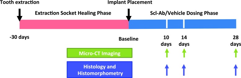
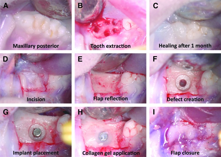
Materials and methods: To investigate the effects of Scl-Ab on bone regeneration and dental implant osseointegration, an experimental alveolar bone osteotomy rat model was adopted. One month after extraction of maxillary right first molars, osteotomy defects were created at the coronal aspect of each of the extraction sites, and 1 × 2-mm custom titanium implants were installed into the osteotomies. Coincident with implant placement, Scl-Ab was administered subcutaneously at a dose of 25 mg/kg twice weekly for 10-28 days and compared with a vehicle control. Animals were sacrificed 10, 14, and 28 days after surgery, and maxillae were harvested and analyzed by microcomputed tomography (microCT), histology, and histomorphometry.
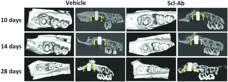
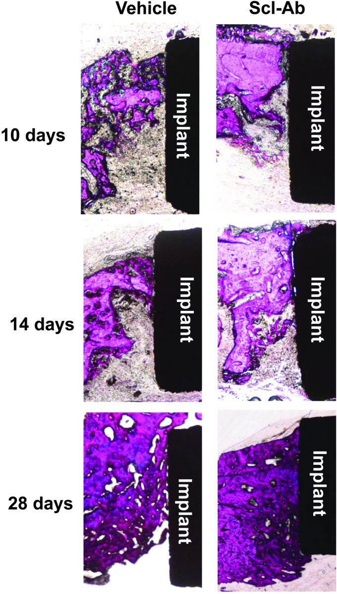
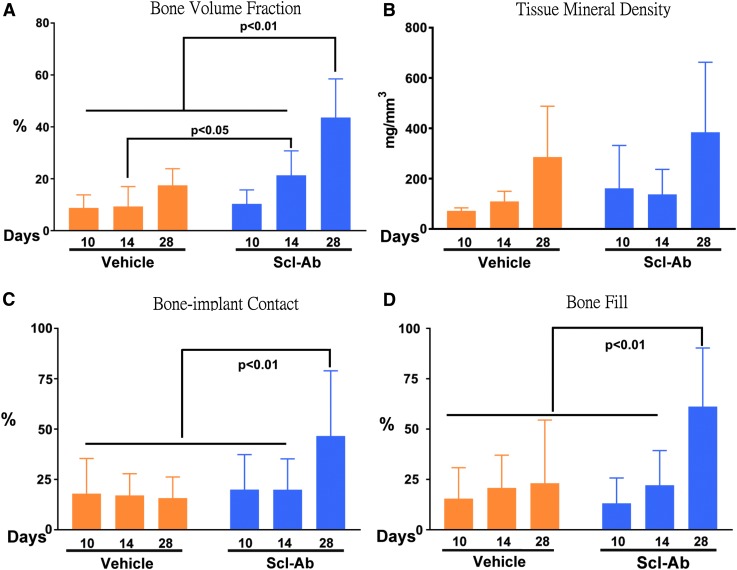
Results: microCT analysis demonstrated that the maxillary bone volume fraction was approximately 2- to 2.5-fold greater in Scl-Ab-treated animals compared with vehicle alone at days 14 and 28. Consistent with those findings, two-dimensional bone fill percentages within the coronal osteotomy sites were highest in Scl-Ab treatment groups at 28 days. In addition, bone-implant contact at 28 days was approximately twofold greater in the Scl-Ab group compared with the vehicle control.
Conclusions: These results indicate that systemic Scl-Ab administration enhances osseointegration and bone regeneration around dental implants. This approach offers potential as a treatment modality for patients with low bone mass or bone defects to achieve more predictable bone regeneration at alveolar bone defects and to enhance dental implant osseointegration.
Keywords: bone anabolics; bone repair; dental implants; osseointegration; osseous healing; sclerostin.
Conflict of interest statement
No competing financial interests exist.
Figures
References
-
- Buser D., Janner S.F., Wittneben J.G., Brägger U., Ramseier C.A., and Salvi G.E. 10-year survival and success rates of 511 titanium implants with a sandblasted and acid-etched surface: a retrospective study in 303 partially edentulous patients. Clin Implant Dent Relat Res 14, 839, 2012 - PubMed
-
- Wittneben J.G., Buser D., Salvi G.E., Bürgin W., Hicklin S., and Brägger U. Complication and failure rates with implant-supported fixed dental prostheses and single crowns: a 10-year retrospective study. Clin Implant Dent Relat Res 16, 356, 2014 - PubMed
-
- Mohan S., and Baylink D.J. Evidence that the inhibition of TE85 human bone cell proliferation by agents which stimulate cAMP production may in part be mediated by changes in the IGF-II regulatory system. Growth Regul 1, 110, 1991 - PubMed
-
- Le Guéhennec L., Soueidan A., Layrolle P., and Amouriq Y. Surface treatments of titanium dental implants for rapid osseointegration. Dent Mater 23, 844, 2007 - PubMed
-
- Gabet Y., Müller R., Levy J., et al. Parathyroid hormone 1–34 enhances titanium implant anchorage in low-density trabecular bone: a correlative micro-computed tomographic and biomechanical analysis. Bone 39, 276, 2006 - PubMed
Publication types
MeSH terms
Substances
Grants and funding
LinkOut - more resources
Full Text Sources
Other Literature Sources
Research Materials
Miscellaneous
