. 2018 Jun 19;19(1):7.
doi: 10.1186/s12867-018-0108-5.

Laser capture microdissection for transcriptomic profiles in human skin biopsies

Affiliations

Laser capture microdissection for transcriptomic profiles in human skin biopsies

Silvia Santoro et al. BMC Mol Biol. .

Abstract

Background: The acquisition of reliable tissue-specific RNA sequencing data from human skin biopsy represents a major advance in research. However, the complexity of the process of isolation of specific layers from fresh-frozen human specimen by laser capture microdissection, the abundant presence of skin nucleases and RNA instability remain relevant methodological challenges. We developed and optimized a protocol to extract RNA from layers of human skin biopsies and to provide satisfactory quality and amount of mRNA sequencing data.

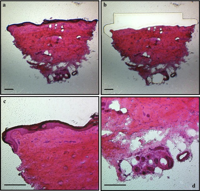
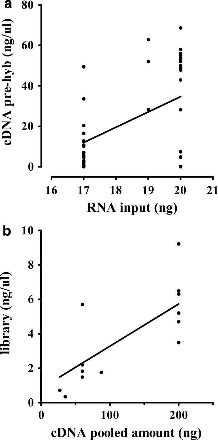
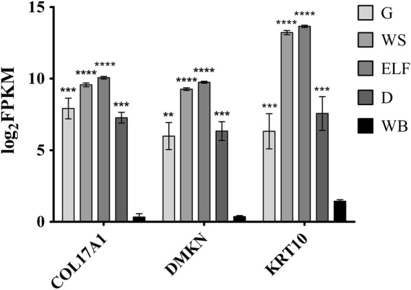
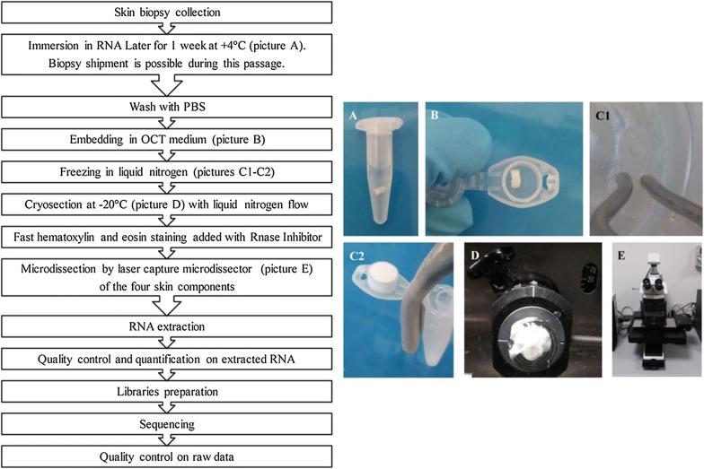
Results: The protocol includes steps of collection, embedding, freezing, histological coloration and relative optimization to preserve RNA extracted from specific components of fresh-frozen human skin biopsy of 14 subjects. Optimization of the protocol includes a preservation step in RNALater® Solution, the control of specimen temperature, the use of RNase Inhibitors and the time reduction of the staining procedure. The quality of extracted RNA was measured using the percentage of fragments longer than 200 nucleotides (DV200), a more suitable measurement for successful library preparation than the RNA Integrity Number (RIN). RNA was then enriched using the TruSeq® RNA Access Library Prep Kit (Illumina®) and sequenced on HiSeq® 2500 platform (Illumina®). Quality control on RNA sequencing data was adequate to get reliable data for downstream analysis.

Conclusions: The described implemented and optimized protocol can be used for generating transcriptomics data on skin tissues, and it is potentially applicable to other tissues. It can be extended to multicenter studies, due to the introduction of an initial step of preservation of the specimen that allowed the shipment of biological samples.

Keywords: Idiopathic neuropathy; Laser capture microdissection; RNA sequencing; Skin biopsy; Transcriptomics.


Figures

Fig. 1
Fig. 1
Representation of the four skin components stained with hematoxylin and eosin. a whole section: b dermis; c enriched layer of fibers extending for 200–300 μm from the surface layer of the skin and d glands. Magnification: ×4 (a, b) and ×10 (c, d). Scale bar = 200 μm
Fig. 2
Fig. 2
RNA/cDNA and cDNA/library correlation. Correlation between the RNA input and the yield of cDNA obtained before the hybridization step (a; p: 0.031, beta: 7.56 and r2: 0.22) and between the pooled amount used for cDNA hybridization and the yield of final libraries (b; p < 0.0001, beta: 0.02 and r2: 0.52)
Fig. 3
Fig. 3
RNA expression of genes enriched in skin. Graph of log2 FPKM (Fragments Per Kilobase Million) values of three genes known to be enriched in skin tissue (COL17A1: Collagen Type XVII Alpha 1 Chain; DMKN Dermokine; KRT10: Keratin 10) evaluated in each skin compartments and in whole blood (ELF enriched layer of fibers, G glands, D dermis, WS whole section, WB whole blood). Bar plot shows the mean ± standard error of the mean. Statistical significance is reported for each skin compartments compared to whole blood (**p: 0.0013; ***0.0001 ≤ p ≤ 0.0007; ****p < 0.0001)
Fig. 4
Fig. 4
Workflow of the steps of the protocol. The flowchart summarizes the steps of the protocol from the skin biopsy collection to the quality control check on raw data generated from sequencing

References

    1. Dong X, You Y, Wu JQ. Building an RNA sequencing transcriptome of the central nervous system. Neuroscientist. 2016;22(6):579–592. doi: 10.1177/1073858415610541. - DOI - PMC - PubMed
    1. Sheng Q, Vickers K, Zhao S, Wang J, Samuels DC, Koues O, Shyr Y, Guo Y. Multi-perspective quality control of Illumina RNA sequencing data analysis. Brief Funct Genomics. 2017;16(4):194–204. - PMC - PubMed
    1. Mo A, Jackson S, Devers TJ, Rosenberg DW. Targeted transcriptional profiling of microdissected biopsy specimens representing early colonic neoplasia. J Cell Biochem. 2016;117(12):2677–2681. doi: 10.1002/jcb.25644. - DOI - PubMed
    1. Emmert-Buck MR, Bonner RF, Smith PD, Chuaqui RF, Zhuang Z, Goldstein SR, Weiss RA, Liotta LA. Laser capture microdissection. Science. 1996;274(5289):998–1001. doi: 10.1126/science.274.5289.998. - DOI - PubMed
    1. Hu P, Zhang W, Xin H, Deng G. Single cell isolation and analysis. Front Cell Dev Biol. 2016;4:116. doi: 10.3389/fcell.2016.00116. - DOI - PMC - PubMed

Publication types