. 2018 Jun 19;14(1):195.
doi: 10.1186/s12917-018-1494-0.

Causes of mortality and morbidity in free-ranging mustelids in Switzerland: necropsy data from over 50 years of general health surveillance

Affiliations

Causes of mortality and morbidity in free-ranging mustelids in Switzerland: necropsy data from over 50 years of general health surveillance

E Akdesir et al. BMC Vet Res. .

Abstract

Background: Although mustelids occur worldwide and include a wide range of species, little is known about the diseases affecting them. Mustelids have regularly been submitted for post mortem investigation in the framework of the program for general wildlife health surveillance in Switzerland, which has been in place for nearly 60 years. We performed a retrospective analysis of the necropsy reports on mustelids submitted to the diagnostic service of the University of Bern. The aims of this study were to present an overview of the causes of mortality and morbidity observed in these carnivores, to assess differences among species, to assess changes in disease detection over the study period, and to describe the pathology of selected diseases.

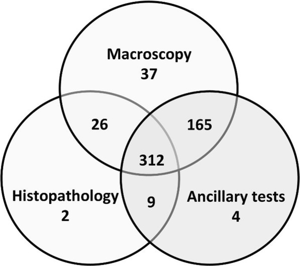
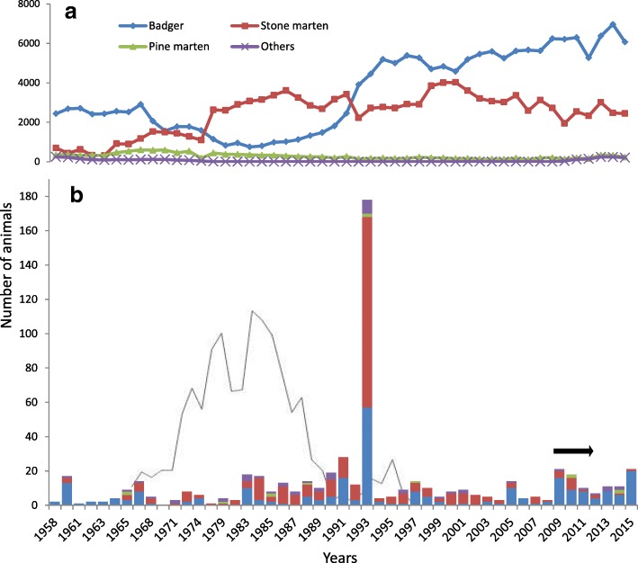
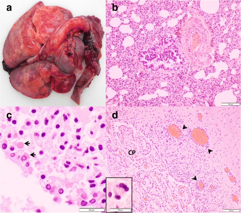
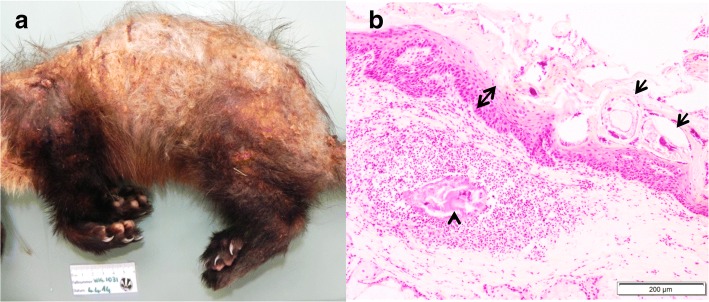
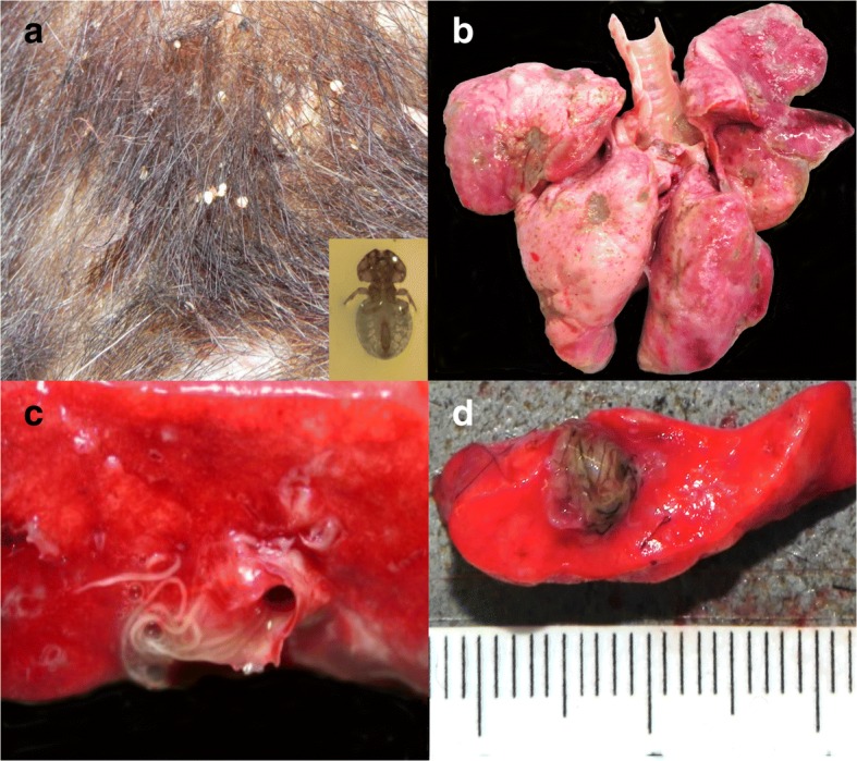
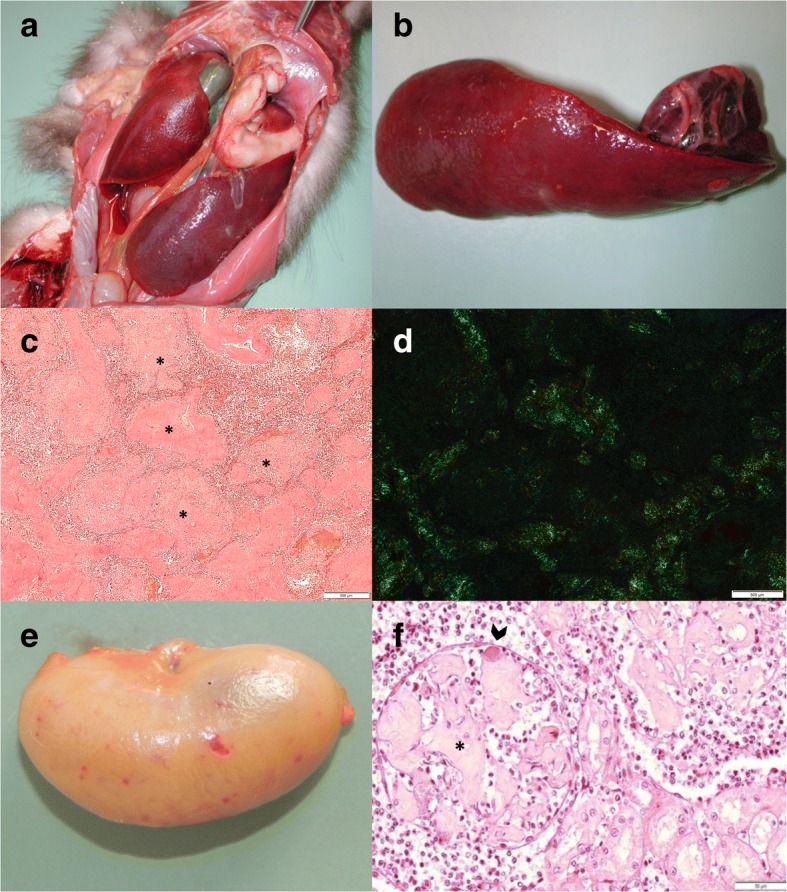
Results: Five hundred and sixty-six reports from 1958 to 2015 were analyzed. Most animals were stone martens (Martes foina, 46%) and badgers (Meles meles, 44%); the remaining species were polecats (Mustela putorius, 4.7%), pine martens (Martes martes, 2%), stoats (Mustela erminea, 1.4%), weasels (Mustela nivalis, 0.8%) and otters (Lutra lutra, 0.3%). Infectious diseases (n = 262) were frequent and were mostly bacterial or viral; non-infectious conditions (n = 169) were less common and were mostly traumatic or due to metabolic disorders. The most frequent diagnoses included distemper (75% were badgers), amyloidosis (96% were martens), bacterial respiratory infections (all mustelids), biting lice (badgers only) and pulmonary and gastro-intestinal helminths (all species). Less frequent diseases included histoplasmosis (badgers only), aspergillosis, toxoplasmosis, hepatozoonosis, and sarcoptic mange. Lesions due to infection with distemper virus were primarily appreciated in the respiratory tract and central nervous system; they presented species-specific characteristics such as necrosis in the ependyma in badgers and absence of syncytia in stone martens. Amyloidosis in martens was multisystemic in most cases and included both AA and AL amyloidosis; the main macroscopic change was severe splenomegaly.

Conclusion: Infectious diseases were the most frequent causes of morbidity and mortality of mustelids, with marked species-specific differences. Lung and skin were the most commonly affected organs. Contagious diseases such as canine distemper, sarcoptic mange and rabies in mustelids showed a similar temporal pattern as in red foxes (Vulpes vulpes), suggesting pathogen spillovers from foxes to mustelids.

Keywords: Amyloidosis; Bacteria; Badger; Canine distemper; Histoplasmosis; Marten; Parasites; Pathology; Sarcoptic mange; Virus.


Conflict of interest statement

Ethics approval and consent to participate

This study did not involve purposeful killing of animals. All samples originated from dead wildlife (found dead in the field, legally shot because of severe debilitation). According to the legislation of Switzerland (922.0 hunting law and 455 animal protection law, including legislation on animal experimentation; www.admin.ch) and the Principality of Liechtenstein (www.gesetze.li), no ethical approval or permit for animal experimentation was required.

Competing interests

The authors declare that they have no competing interests.

Publisher’s Note

Springer Nature remains neutral with regard to jurisdictional claims in published maps and institutional affiliations.

Figures

Fig. 1
Fig. 1
Investigations performed on the free-ranging mustelids submitted to the FIWI, Switzerland, 1958 to 2015. Ancillary tests: bacteriological, virological, parasitological and/or toxicological analyses
Fig. 2
Fig. 2
Free-ranging mustelids found dead, culled or hunted in Switzerland from 1958 to 2015. Diagram a shows the total number of dead mustelids, including those shot and found dead, which were recorded in Switzerland from 1958 to the winter 2014/15 (national hunting statistics). Diagram b shows the number of cases submitted to the FIWI from 1958 to 2015 (animals included in the present study), using the same color code for different mustelid species as in Diagram a. The black line in Diagram b represents the number of mustelids diagnosed positive for rabies at the Swiss Rabies Center. Rabies investigations began in 1967 and cases were recorded until 1997, when the disease was eradicated from Switzerland. The horizontal black arrow indicates the years of the national epidemic of canine distemper
Fig. 3
Fig. 3
Map of Switzerland depicting the geographical origin of the mustelids submitted to the FIWI, 1958-2015. Different mustelid species are indicated by dots of different colors: Green: Badgers, Red: Martens (including stone marten Martes foina, and pine marten Martes martes), Yellow: Others (polecat Mustela putorius, stoat Mustela ermine, weasel Mustela nivalis and Eurasian otter Lutra lutra). Areas with different shades of grey illustrate the case density or submission intensity per Swiss canton (number of submitted cases per ha). BE: Canton Bern; BS: Canton Basel-Stadt; BL: Canton Basel-Landschaft
Fig. 4
Fig. 4
Lesions associated with canine distemper virus infection in badgers. a Lungs with “wet-like” appearance, which are not collapsed and display multifocal red to dark red mottling. b Lung section, H&E stain: Severe interstitial pneumonia. c Brain section, H&E stain: Intracytoplasmic, eosinophilic inclusions (arrows) in the ependymal epithelium. d Brain section of the same animal, H&E stain: Multifocal, lymphoplasmacytic perivascular cuffs (arrow heads) and neuronal syncytia (inset), choroid plexus (CP)
Fig. 5
Fig. 5
Skin lesions associated with Histoplasma capsulatum infection in badgers. a Multiple, well-demarcated, ulcerated nodules disseminated on the ventral aspect of the body. b Close-up of a nodule. c Histological section of a nodule, PAS stain: Numerous round-oval, 1-3 μm diameter yeasts consistent with Histoplasma capsulatum (arches). d Histological section of a nodule, Grocott stain: Detailed morphology of Histoplasma capsulatum with evident narrow-based budding
Fig. 6
Fig. 6
Skin lesions associated with Sarcoptes scabiei in martens. a Pine marten: Hair loss and skin crust formation affecting mainly the dorsal aspect of the body. b Stone marten, histological section (H&E stain): Mites (arrows) embedded in sero-cellular crusts on the hyperplastic epidermis (double headed arrow). Remnants of a mite (arrow head) in the dermis with focal, suppurative dermatitis and dermal edema
Fig. 7
Fig. 7
Common ecto- and endoparasitism in free-ranging mustelids from Switzerland. a Close-up of the skin of a badger with severe Trichodectes melis infestation with hair loss and crust formation. A close up of T. melis is shown in the inset. b Lungs of a badger with multifocal, light tan to gray indented areas associated with Crenosoma sp. c Cut surface of the lungs of a badger with numerous intrabronchial lungworms, Crenosoma vulpis. d Cut surface of a polecat’s lung with a cluster of lungworms, Filaroides sp.
Fig. 8
Fig. 8
Lesions associated with amyloidosis in martens. a Pine marten, abdominal organs in situ: Severely enlarged spleen. b Same pine marten as in a, extracted spleen: Severe splenomegaly, increased consistency and rubber-like texture. c) Stone marten, Congo red stain, spleen: Severe deposition of amyloid (asterisk) around the blood vessels. d Same stone marten as c, Congo red stain, spleen: Apple-green birefringence under polarized light. e Pine marten, kidney: Diffuse tan discoloration with petechial hemorrhages. f Stone marten, histological section of a kidney (H&E stain): The glomeruli are partially obstructed by abundant deposit of eosinophilic, fibrillary material consistent with amyloid (asterisk) and a bacterial embolus within the lumen of the glomerular capillary (arrow head)

References

    1. Musteliden HJ. Säugetiere Schweiz Verbreit. Biol. Ökol. Basel: DSAN und Birkhäuser; 1995. pp. 367–402.
    1. The IUCN red list of threatened species. http://www.iucnredlist.org/ (2016-3). Accessed 13 Feb 2017.
    1. Young J. Movement patterns of two New Zealand mustelids : implications for predator pest management. Lincoln University; 1998. https://researcharchive.lincoln.ac.nz/handle/10182/2464. Accessed 13 Feb 2017.
    1. Gallagher J, Clifton-Hadley RS. Tuberculosis in badgers; a review of the disease and its significance for other animals. Res Vet Sci. 2000;69:203–217. doi: 10.1053/rvsc.2000.0422. - DOI - PubMed
    1. Holmala K, Kauhala K. Ecology of wildlife rabies in Europe. Mammal Rev. 2006;36:17–36. doi: 10.1111/j.1365-2907.2006.00078.x. - DOI