

Splenogonadal fusion - a rare cause of scrotal swelling: a case report
- PMID: 29921313
- PMCID: PMC6011191
- DOI: 10.1186/s13256-018-1712-1
Splenogonadal fusion - a rare cause of scrotal swelling: a case report
Abstract
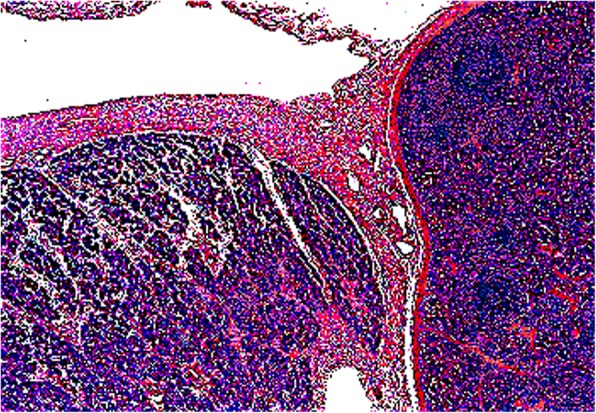
Background: Splenogonadal fusion is a rare and benign condition. Diagnosis is challenging for clinicians. Despite its indolence, diagnosis is often confirmed after orchidectomy. Surgery is mandatory, particularly to rule out the extremely rare association with malignancy.
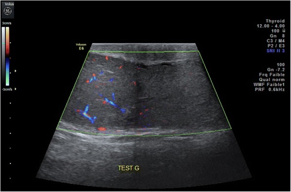
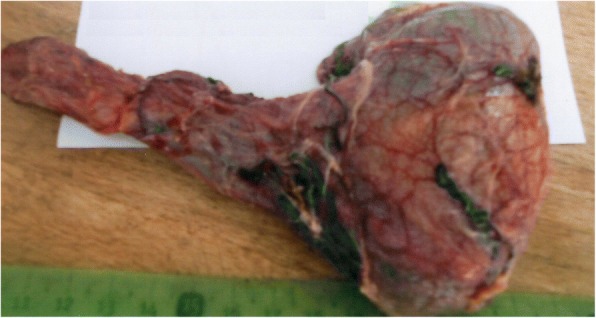
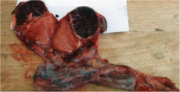
Case presentation: We report a case of splenogonadal fusion in a 38-year-old North African man presenting a palpable scrotal mass. We describe clinical aspects, pathogenic hypothesis, radiological features, as well as surgical management principles.
Conclusions: Splenogonadal fusion is rarely suspected and diagnosed preoperatively. A diagnosis is made once an ectopic testicular mass is associated with cryptorchidism and suggestive radiological signs. A better knowledge of the clinical and radiological features of splenogonadal fusion provides an opportunity for conservative surgery.
Keywords: Congenital abnormalities; Orchiectomy; Spleen; Testis.
Conflict of interest statement
Ethics approval and consent to participate
No ethics committee approval is required at our institution for a case report involving a limited number of patients.
Consent for publication
Written informed consent was obtained from the patient for publication of this case report and any accompanying image. A copy of the written consent is available for review by the Editor-in-Chief of this journal.
Competing interests
The authors declare that they have no competing interests.
Publisher’s Note
Springer Nature remains neutral with regard to jurisdictional claims in published maps and institutional affiliations.
Figures
References
-
- Boestrom E. Demonstration eines Praparates von Verwachsung der MilZ mit dem linken Hoden, vol. 149. Freiburg: Gellschaft deutscher Naturforscher und Artze Verhandlungen der 56 Versammlung; 1883.
Publication types
MeSH terms
LinkOut - more resources
Full Text Sources
Other Literature Sources
Medical
