. 2018 Jun 19;9(1):167.
doi: 10.1186/s13287-018-0910-5.

Inhibition of the RhoGTPase Cdc42 by ML141 enhances hepatocyte differentiation from human adipose-derived mesenchymal stem cells via the Wnt5a/PI3K/miR-122 pathway: impact of the age of the donor

Affiliations

Inhibition of the RhoGTPase Cdc42 by ML141 enhances hepatocyte differentiation from human adipose-derived mesenchymal stem cells via the Wnt5a/PI3K/miR-122 pathway: impact of the age of the donor

Diana Chaker et al. Stem Cell Res Ther. .

Abstract

Background: Human adipose-derived mesenchymal stem cells (hADSCs) are promising cells that may promote hepatocyte differentiation (Hep-Dif) and improve liver function, but the involvement of Cdc42, a key small RhoGTPase which plays a crucial role in aging, is still not well established. We hypothesized that the inhibition of Cdc42 may rescue the hepatogenic potential of hADSCs derived from aged donors.

Methods: hADSCs isolated from 61 women of different ages were cultured for evaluation of the proliferation of cells, adherence, apoptosis, immunomodulation, immunophenotyping, multipotency, gene expression, and cell function during Hep-Dif. Inhibition of Cdc42 by ML141 was realized during two phases: initiation (days -2 to 14 (D-2/14)) from undifferentiated to hepatoblast-like cells, or maturation (days 14 to 28 (D14/28)) from undifferentiated to hepatocyte-like cells. Mechanistic insights of the Wnt(s)/MAPK/PI3K/miR-122 pathways were studied.

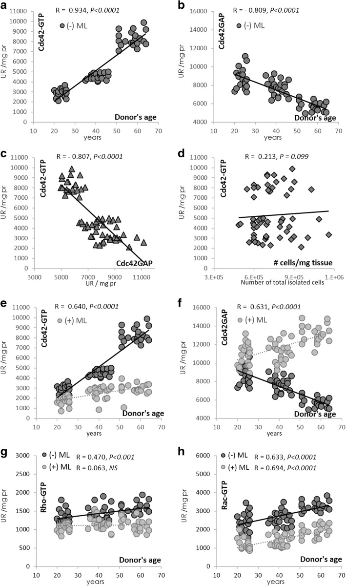
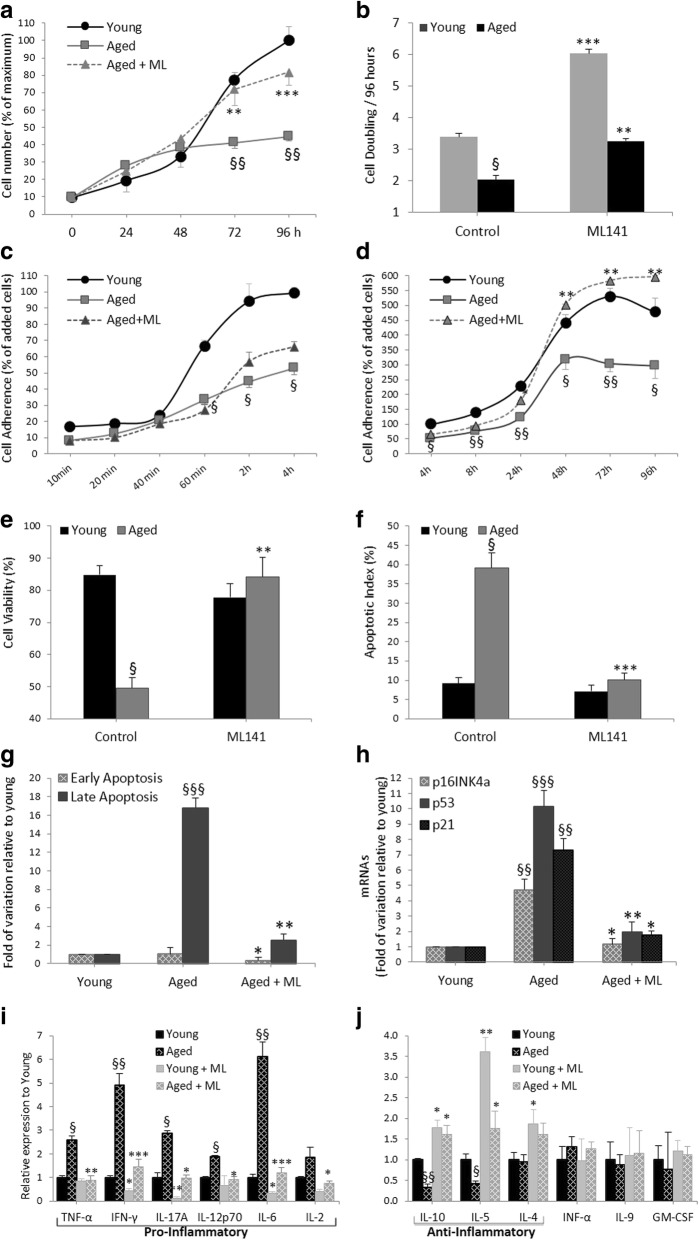
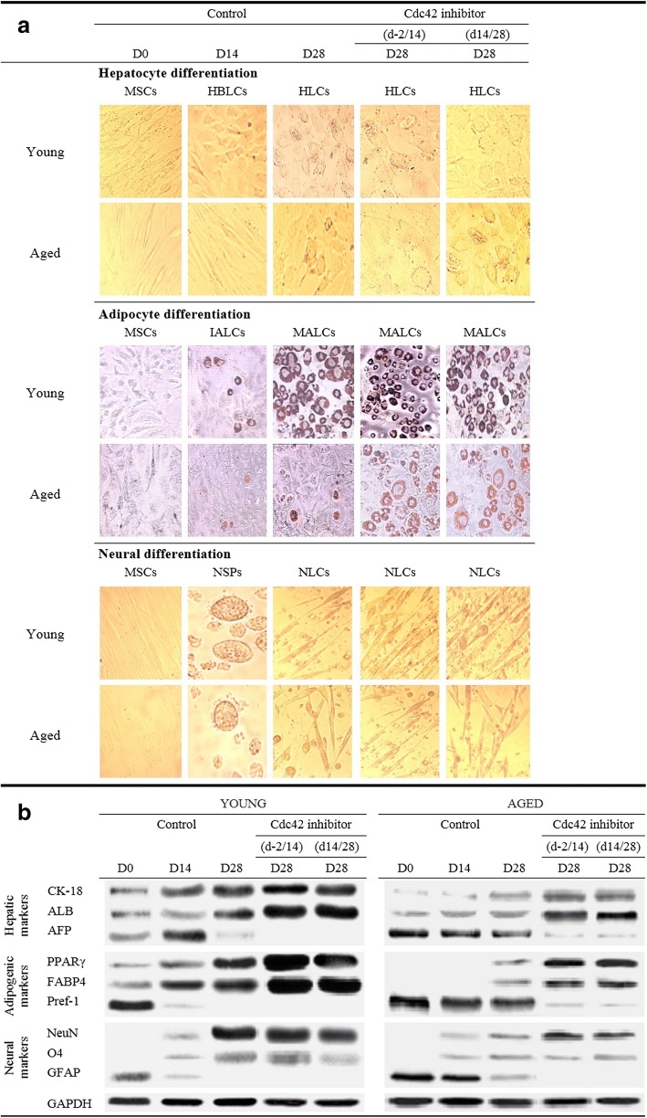
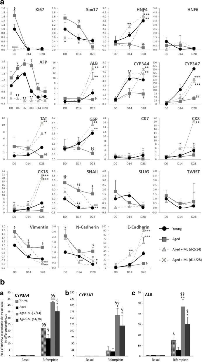
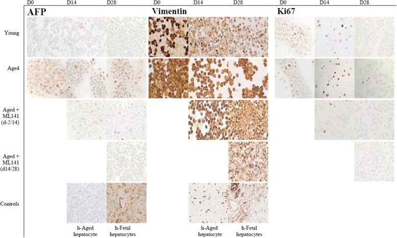
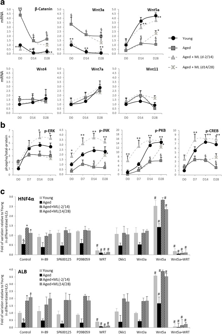
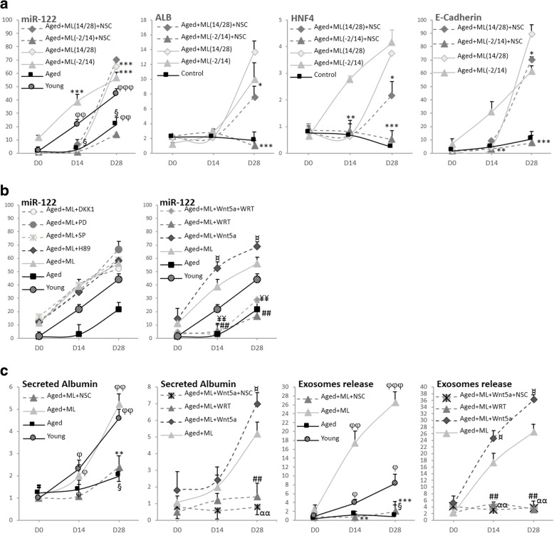
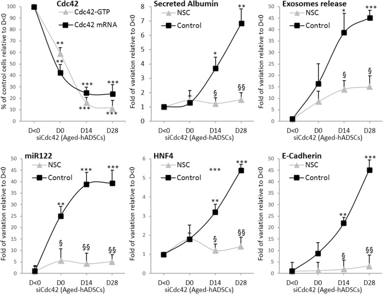
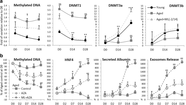
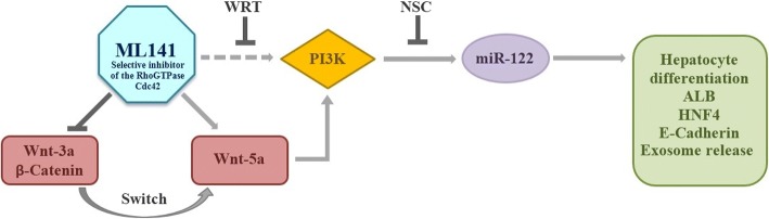
Results: Cdc42 activity in undifferentiated hADSCs showed an age-dependent significant increase in Cdc42-GTP correlated to a decrease in Cdc42GAP; the low potentials of cell proliferation, doubling, adherence, and immunomodulatory ability (proinflammatory over anti-inflammatory) contrary to the apoptotic index of the aged group were significantly reversed by ML141. Aged donor cells showed a decreased potential for Hep-Dif which was rescued by ML141 treatment, giving rise to mature and functional hepatocyte-like cells as assessed by hepatic gene expression, cytochrome activity, urea and albumin production, low-density lipoprotein (LDL) uptake, and glycogen storage. ML141-induced Hep-Dif showed an improvement in mesenchymal-epithelial transition, a switch from Wtn-3a/β-catenin to Wnt5a signaling, involvement of PI3K/PKB but not the MAPK (ERK/JNK/p38) pathway, induction of miR-122 expression, reinforcing the exosomes release and the production of albumin, and epigenetic changes. Inhibition of PI3K and miR-122 abolished completely the effects of ML141 indicating that inhibition of Cdc42 promotes the Hep-Dif through a Wnt5a/PI3K/miR-122/HNF4α/albumin/E-cadherin-positive action. The ML141(D-2/14) protocol had more pronounced effects when compared with ML141(D14/28); inhibition of DNA methylation in combination with ML141(D-2/14) showed more efficacy in rescuing the Hep-Dif of aged hADSCs. In addition to Hep-Dif, the multipotency of aged hADSC-treated ML141 was observed by rescuing the adipocyte and neural differentiation by inducing PPARγ/FABP4 and NeuN/O4 but inhibiting Pref-1 and GFAP, respectively.

Conclusion: ML141 has the potential to reverse the age-related aberrations in aged stem cells and promotes their hepatogenic differentiation. Selective inhibition of Cdc42 could be a potential target of drug therapy for aging and may give new insights on the improvement of Hep-Dif.

Keywords: Adipose derived mesenchymal stem cells; Aging; Cdc42; Exosomes release; Hepatocyte differentiation; MAPK; ML141; PI3K; Wnt; miR122.


Conflict of interest statement

Ethics approval and consent to participate

All clinical investigations on human samples have been conducted according to the principles expressed in the Declaration of Helsinki (https://www.wma.net/policies-post/wma-declaration-of-helsinki-ethical-principles-for-medical-research-involving-human-subjects/) as revised in 2008 (https://www.wma.net/wp-content/uploads/2016/11/DoH-Oct2008.pdf). All donors gave written informed consent. Samples were procured by Dr. Ibrahim El Achkar and by the Nancy University Hospital Clinical Investigation Center (ethical approval number, CPP3.07.01, France).

Competing interests

The authors declare that they have no competing interests.

Publisher’s Note

Springer Nature remains neutral with regard to jurisdictional claims in published maps and institutional affiliations.

Figures

Fig. 1
Fig. 1
Cdc42 activity increases with age. hADSCs were isolated from lipoaspirates and cultured for 96 h and then treated for 24 h with or without the Cdc42/Rac1 GTPase inhibitor ML141 (ML; 10 μM). Cell lysates from undifferentiated hADSCs were used for the Pull-down assays of Cdc42-GTP (500 μg) and Cdc42GAP (100 μg). Correlations between donor age and Cdc42-GTP (a,e), donor age and Cdc42GAP (b,f), Cdc42-GTP and Cdc42GAP (c), Cdc42-GTP and cell yield/mg of collected adipose tissue (d), donor age and Rho-GTP (g), and donor age and Rac-GTP (h). Results represent the mean ± SEM of two independent experiments realized on the 61 subjects after normalization to GAPDH. Pearson correlation coefficient (R) and the P value are shown
Fig. 2
Fig. 2
Pharmacological targeting of Cdc42 activity by ML141 (ML) on the yield, growth, adherence, apoptosis, and immunomodulatory characteristics of hADSCs. Cells derived from young and aged subjects were cultured for 96 h. Cells were treated with or without ML141 (10 μM, 24 h). Proliferative potential as indicated by cell number (a) and time for population doubling (b). Kinetics of hADSC adherence as evaluated by the number of adhered cells and expressed as the percentage of applied cells (c,d). Cell viability (e) and apoptotic index (f) as assessed by labeled cells with Annexin V/propidium iodide (PI)/7AAD. The cell viability and apoptotic index were expressed respectively as the percentage of Annexin V(−)/PI(−)/7AAD(−) or Annexin V(+) cells divided by total cells. g Early from late-apoptotic cells were identified as Annexin V(+)/PI(−)/7AAD(−) vs Annexin V(+)/PI(+)/7AAD(+), respectively. h Real-time quantitative RT-PCR determination of p16INK4a, p53, and p21 mRNA levels. The results are expressed as fold variations ± SEM over the young group (R) after normalization to β-actin. i,j Cell culture supernatants from undifferentiated hADSCs were collected and undiluted samples were analyzed for the detection of cytokines as indicated in the Methods. Three to five measures were realized by group and results are the mean ± SEM presented in fold of variation relative to young. The cytokine limits of detection (pg/ml) were: tumor necrosis factor (TNF)-α (2.17), interferon (IFN)-γ 6.26, IFN-α (10.4), granulocyte/macrophage colony-stimulating factor (GM-CSF) (0.20), interleukin (IL)-2 (1.52), IL-4 (34.2), IL-5 (0.27), IL-6 (0.077), IL-9 (32.7), IL-10 (2.76), IL-12 (3.44), and IL-17A (0.7). The results represent individual expression per subject (young (n = 19), aged (n = 20) and aged+ML141 (n = 20)) and are the means of two independent experiments performed in triplicate. §*P < 0.05, §§**P < 0.01, §§§***P < 0.001; §aged versus young and *aged treated with ML141 versus untreated
Fig. 3
Fig. 3
Impact of ML141 on hepatic, adipogenic, and neurogenic differentiation of young and aged-derived hADSCs. hADSCs were isolated from young or aged subjects and subjected to hepatocyte/adipocyte/neural differentiation as indicated in the Methods for 28 days. Cells were collected at day 0 (MSCs) at the moment of induction of the differentiation, and days 14 and 28 of the differentiation. Cells were incubated with or without 10 μM ML141 following two protocols of treatment: from day −2 to day 14 of the differentiation (d-2/14) and from day 14 to day 28 of the differentiation (d14/28). The effect of ML141 was evaluated at day 28 of the differentiation. a Representative images of morphological cell changes from: undifferentiated MSCs at D0, hepatoblast-like cells (HBLCs) or immature adipocyte-like cells (IALCs) or neurospheres (NSPs) at D14, hepatocyte-like cells (HLCs) or mature adipocyte-like cells (MALCs) or neural-like cells (NLCs) at D28. Cells from adipocyte differentiation were stained with oil-red O. b Hepatic/adipogenic/neurogenic marker expression: cell lysates (80–150 μg of protein) were separated by SDS-PAGE and immunoblotted with antibodies raised against cytokeratin (CK)-18, albumin (ALB), alpha fetoprotein (AFP), peroxisome proliferator activated receptor (PPAR)γ, FABP4, Pref-1, NeuN, O4, GFAP, and GAPDH. Protein expression profiling was determined during differentiation at D0/14/28; representative blots are shown
Fig. 4
Fig. 4
Impact of pharmacological targeting of Cdc42 activity by ML141 (ML) on the gene expression profile during Hep-Dif. hADSCs were isolated from young and aged subjects and were induced to Hep-Dif for 28 days with or without ML141 (10 μM) for the indicated time of incubation. RNAs were collected at D0/14/28 and mRNA levels of the studied genes were quantified by RT-qPCR. a Gene expression profile of hepatic and mesenchymal-to-epithelial transition markers as well as epigenetic markers (the DNA methyltransferases, DNMTs). b Rifampicin responsiveness of hepatocyte-like cells: RNAs were collected at day 28 of the differentiation after cell treatment with rifampicin (20 μM, 24 h). Expression levels of CYP3A4, CYP3A7, and albumin (ALB) genes are shown. The results are expressed as fold variation relative to controls (without rifampicin) after normalization to GAPDH, and are the mean ± SEM of two independent experiments performed twice, each in duplicate. D0 = day of induction of the differentiation. d-2/14, d14/28: cells were treated with ML141 from day −2 to day 14, or day 14 to day 28, respectively. §*P < 0.05, §§**P < 0.01, ***P < 0.001; §aged versus young or rifampicin versus basal and *aged treated with ML141 versus untreated
Fig. 5
Fig. 5
Extinction of alpha fetoprotein (AFP), Vimentin, and Ki67 from hepatocyte-like cells derived from aged ADSCs treated with ML141. Cells derived from young and aged groups were differentiated and treated with or without ML141 as indicated in Fig. 4 and evaluated for AFP (fetal hepatoblast marker), Vimentin (EMT marker), and Ki67 (proliferation marker) by immunohistochemistry (IHC) as described in the Methods. Cells were examined microscopically and phase-contrast images were captured. Cultures of primary human (h) fetal and adult hepatocytes were assessed as positive controls and negative controls, respectively
Fig. 6
Fig. 6
Pharmacological targeting of Cdc42 activity by ML141 (ML) induces rifampicin responsiveness and restores cell function. hADSCs derived from young and aged subjects were differentiated and treated with or without ML141 as indicated in Fig. 4. a–c Secreted albumin, urea production, and low-density lipoprotein (LDL) uptake: supernatants of cultured cells and the lysates were collected at D0/D14/D28 of the differentiation for the quantification of albumin, urea, and LDL uptake as indicated in the Methods. Results are expressed per ng/ml (for the production of albumin and urea) and RFU (fluorescent LDL uptake) and are presented as fold variation relative to young at day D0, and are the mean ± SEM of several measures (5, 6 and 10, respectively). d Cytochrome P450 (CYP3A4) activity at day 28 of the Hep-Dif after induction with rifampicin (20 μM, 24 h). The results are expressed as fold variation relative to basal (without rifampicin), and are the mean ± SEM of three independent experiments performed in duplicate. e hepatocyte-like cells derived from aged ADSCs treated with ML141 exhibit hepatic-specific function of glycogen storage. Cells were evaluated for glycogen storage capacity (pink color) using periodic acid-Schiff (PAS) staining as described in the Methods. Cells were examined microscopically and phase-contrast images were captured. Cultures of HepG2 cells were assessed as positive controls. §*P < 0.05, §§**P < 0.01; §rifampicin versus controls or aged versus young and *aged treated with ML141 versus untreated
Fig. 7
Fig. 7
ML141 (ML) impact on hepatocyte differentiation is dependent of PI3K and Wnt5a signaling. hADSCs were induced to Hep-Dif for 28 days with or without ML141 (10 μM) for the indicated time of incubation (d-2/14 or d14/28). Cells were treated 24 h before adding ML141, and maintained with ML141, with: 1) inhibitors of PKA (H-89, 5μM), JNK (SP600125, 10μM), ERK (PD98059, 50μM), and PI3K (Wortmannin, 10μM); or 2) Wnt-antagonist Dkk1 (200 ng/ml, 24 h), Wnt3a (50 ng/ml, 24 h), and Wnt5a (100 ng/ml, 24 h). a mRNA expression of Wnt(s) and β-catenin expressed as fold variation relative to young at D0 after normalization to GAPDH. b Cell lysates (80–150 μg of protein) were separated by SDS-PAGE and immunoblotted with antibodies raised against phospho and total ERK/JNK/PKB/CREB. Protein expression profiling was determined during differentiation at D0/14/28 and results are expressed as fold variation of phospho/total levels relative to young at D0 after normalization to GAPDH. c Impact of H-89/SP/PD/WRT/Dkk1/Wnt3a/Wnt5a on the mRNA expression of the hepatic markers hepatocyte nuclear factor (HNF)4 and albumin (ALB) at D28 (hepatocyte-like cells; HLCs): results are expressed as fold variation relative to young untreated cells. Results are the mean ± SEM of three independent experiments performed in duplicate realized on 19 (young) and 20 (aged, aged+ML141) subjects. §*#P < 0.05, §§**## P < 0.01; §aged versus young, *aged treated with ML141 versus control, and #WRT or Wnt5a-treated cells versus control. WRT Wortmannin
Fig. 8
Fig. 8
miR-122 selective inhibitor (NSC5476 (NSC)) abolished the effects of ML141 (ML). hADSCs were induced to Hep-Dif for 28 days with or without ML141 (10 μM) for the indicated time of incubation (D-2/14 or D14/28). Cells were incubated as indicated in Fig. 7 (with H-89, SP, PD, WRT, Wnt5a) and with or without NSC5476 (NSC, 5 μM) 24 h before adding ML141 and maintained in parallel to ML141. a RNAs were collected at D0/14/28 and mRNA levels of hepatocyte nuclear factor (HNF)4α/albumin (ALB)/E-cadherin/miR-122 genes were determined by RT-qPCR: the results are expressed as fold variation relative to D0 (or young at D0) after normalization to GAPDH. b Effects of H-89, SP, PD, WRT, Wnt5a, and NSC on the expression of miR-122 mRNA on the ML(−2/14) treated group. c Effects of Wnt5a, WRT, and NSC on the levels of secreted albumin and released exosomes on the ML141(−2/14) treated group. ¥P < 0.05, aged+ML(14/28) + NSC versus same group without NSC. The results are expressed as fold variation relative to D0 (or young at D0). Results are the mean ± SEM of three independent experiments performed in duplicate realized on 19 (young) and 20 (aged, aged+ML) subjects. φ§*¤P < 0.05, φφ§§**## ¥¥ααP < 0.01, φφφ§§§***P < 0.001. φD14/D28 versus D0, §aged versus young, *aged+ML + NSC versus aged+ML, ¤aged+ML + Wnt5a versus aged+ML, #aged+ML + WRT versus aged+ML, ¥aged+ML + Wnt5a + WRT versus aged+ML, and αaged + ML + Wnt5a + NSC versus aged+ML. PD PD98059, SP SP600125, WRT Wortmannin
Fig. 9
Fig. 9
Induction of Hep-Dif in Cdc42 knockdown aged human adipose-derived mesenchymal stem cells (hADSCs). hADSCs derived from aged subjects were transfected with an Cdc42-targeting siRNA (siCdc42) at day –2 (D–2) and then differentiated at day 0 (D0) with 1% FBS. Cells were incubated with or without NSC5476 (NSC, 5 μM) 24 h before launching the differentiation and maintained along the Hep-Dif. Cdc42 mRNA and Cdc42-GTP protein levels as well as key factors were assessed during hepatocyte differentiation of Cdc42-knockdown cells from D<0 to D28: secreted albumin protein, released exosomes, miR122 mRNA, hepatocyte nuclear factor (HNF)4 mRNA, and E-cadherin mRNA. Results are normalized as previously to the expression of the housekeeping gene and are the mean ± SEM of three independent experiments and are expressed as the percentage or fold variation of control relative to day D–2 (considered as D<0). *P < 0.05, **P < 0.01, ***P < 0.005, D0/D14/D28 vs D < 0; §P < 0.05, §§P < 0.01, NSC vs control
Fig. 10
Fig. 10
Global methylation status and involvement of DNA methyltransferases (DNMTs) in Hep-Dif of hADSCs. hADSCs derived from young and aged subjects were differentiated as indicated in Fig. 4. Cells were incubated with or without ML141 (ML, 10 μM) from day (D)−2 to D14 of the differentiation. a Methylation status of hADSCs derived from the young, aged, and ML141-treated aged groups were analyzed for the indicated time of Hep-Dif; expressions of DNMTs mRNA are shown. b hADSCs derived from aged subjects were subjected to Hep-Dif in the presence or absence of 5-azacytidine (AZA, 20 μM) for 48 h (from day 0 to day 2 of the differentiation)); ML141 was added from day −2 to day 14: methylation status, hepatocyte nuclear factor (HNF)4α mRNA, secreted albumin protein, and released exosomes are shown. Results are normalized as previously to the expression of the housekeeping gene and are the mean ± SEM of three independent experiments and are expressed as the percentage or fold variation of control relative to day D0. §*¤P < 0.05, §§**¤¤P < 0.01, ***P < 0.005; §aged vs young, *aged+ML vs aged, and ¤AZA or AZA + ML vs control
Fig. 11
Fig. 11
Model for the regulation of ML141-induced hepatocyte differentiation. In hADSCs, ML141 induced the hepatocyte differentiation by a mechanism involving the Wnt5a/PI3K/miR-122 signaling pathway, and regulated positively the hepatic specific genes and function, importantly the exosome release. ALB albumin, HNF hepatocyte nuclear factor, NSC NSC5476, PI3K phosphatidylinositol-3 kinase, WRT Wortmannin

References

    1. Goodell MA, Rando TA. Stem cells and healthy aging. Science. 2015;350(6265):1199–1204. doi: 10.1126/science.aab3388. - DOI - PubMed
    1. Kim H, Kisseleva T, Brenner DA. Aging and liver disease. Curr Opin Gastroenterol. 2015;31(3):184–191. doi: 10.1097/MOG.0000000000000176. - DOI - PMC - PubMed
    1. Brunet A, Rando TA. Interaction between epigenetic and metabolism in aging stem cells. Curr Opin Cell Biol. 2017;45:1–7. doi: 10.1016/j.ceb.2016.12.009. - DOI - PMC - PubMed
    1. Liu L, Rando TA. Manifestations and mechanisms of stem cell aging. J Cell Biol. 2011;193(2):257–266. doi: 10.1083/jcb.201010131. - DOI - PMC - PubMed
    1. Beane OS, Fonseca VC, Cooper LL, Koren G, Darling EM. Impact of aging on the regenerative properties of bone marrow-, muscle-, and adipose-derived mesenchymal stem/stromal cells. PLoS One. 2014;9(12):e115963. doi: 10.1371/journal.pone.0115963. - DOI - PMC - PubMed

Publication types