. 2018 Aug 10;293(32):12491-12501.
doi: 10.1074/jbc.RA118.002443. Epub 2018 Jun 19.

Loading of malonyl-CoA onto tandem acyl carrier protein domains of polyunsaturated fatty acid synthases

Affiliations

Loading of malonyl-CoA onto tandem acyl carrier protein domains of polyunsaturated fatty acid synthases

Omar Santín et al. J Biol Chem. .

Abstract

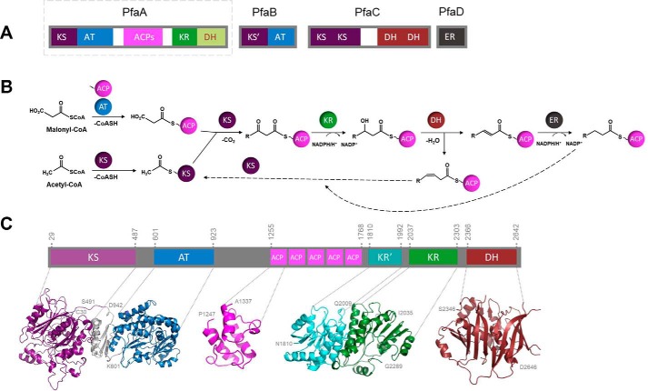
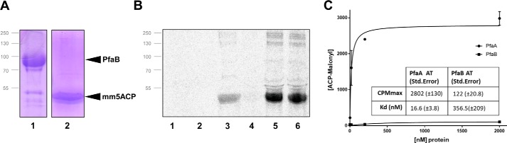
Omega-3 polyunsaturated fatty acids (PUFA) are produced in some unicellular organisms, such as marine gammaproteobacteria, myxobacteria, and thraustochytrids, by large enzyme complexes called PUFA synthases. These enzymatic complexes resemble bacterial antibiotic-producing proteins known as polyketide synthases (PKS). One of the PUFA synthase subunits is a conserved large protein (PfaA in marine proteobacteria) that contains three to nine tandem acyl carrier protein (ACP) domains as well as condensation and modification domains. In this work, a study of the PfaA architecture and its ability to initiate the synthesis by selecting malonyl units has been carried out. As a result, we have observed a self-acylation ability in tandem ACPs whose biochemical mechanism differ from the previously described for type II PKS. The acyltransferase domain of PfaA showed a high selectivity for malonyl-CoA that efficiently loads onto the ACPs domains. These results, together with the structural organization predicted for PfaA, suggest that this protein plays a key role at early stages of the anaerobic pathway of PUFA synthesis.

Keywords: acyl carrier protein (ACP); acyltransferase; fatty acid synthase (FAS); polyketide; polyunsaturated fatty acid (PUFA).


Conflict of interest statement

The authors declare that they have no conflicts of interest with the contents of this article

Figures

Figure 1.
Figure 1.
PfaA analysis and general biochemical route. A, general scheme of the pfa genetic cluster of M. marina. Each domain is represented with a specific color. Domain regions correspond to KS, AT, acyl carrier proteins (5ACP), KR, DH, and ER. PfaA that will be further analyzed is squared with a dashed line. B, overview of the biochemical route for omega-3 PUFA synthesis. The dashed arrows represent the beginning of a new synthesis cycle. C, PfaA domain organization. Prediction of folding domains within the PfaA protein sequence made with InterPro. Each domain is represented with a specific color. Structural predictions made with Phyre2 of each domain are shown in the lower panel using the same color code. The first and last amino acid of each domain prediction is indicated in the figure. Only the structure of one of the five ACPs has been represented as they are similar.
Figure 2.
Figure 2.
Ligand-binding assays of mm5ACP, mmKS-AT, and mmAT domains. A, 12% SDS-PAGE gel containing 20 μg of purified mm5ACP in its holo- (lane 1) and apo- (lane 2) forms. B, 20 μm mm5ACP incubated with [14C]malonyl-CoA and [14C]acetyl-CoA and analyzed by 12% radio-SDS-PAGE. Lanes 1, apo-5ACP + malonyl-CoA; 2, apo-5ACP + acetyl-CoA; 3, holo-5ACP + malonyl-CoA; 4, holo-5ACP + acetyl-CoA. C, 12% SDS-PAGE gel containing purified mmKS-AT (lane 1), holo-mm5ACP (lane 2), mmKS*(C229A)-AT (lane 3), and mmAT (lane 4). D, same domains incubated with [14C]malonyl-CoA and [14C]acetyl-CoA, and analyzed by 12% radio-SDS-PAGE. Lanes 1, mmKS-AT + acetyl-CoA; 2, mmKS-AT + malonyl-CoA; 3, holo-5ACP + acetyl-CoA; 4, holo-5ACP + malonyl-CoA; 5, holo-mm5ACP + mmKS-AT + acetyl-CoA; 6, holo-mm5ACP + mmKS-AT + malonyl-CoA; 7, holo-mm5ACP + mmKS-AT + acetyl-CoA + malonyl-CoA; E, quantification of the protein band intensities of holo-mm5ACP + malonyl-CoA (D, lane 4) and holo-mm5ACP + mmKS-AT + malonyl-CoA (D, lane 6) by densitometric scanning, normalized with respect to the molarity of ACP in each sample. F, mmKS(C229A)-AT mutant and mmAT isolated domain were incubated with [14C]malonyl-CoA and [14C]acetyl-CoA, and analyzed by 12% radio-SDS-PAGE. Lanes 1, mmKS(C229A)-AT + acetyl-CoA; 2, mmKS(C229A)-AT + malonyl-CoA; 3, holo-mm5ACP + mmKS(C229A)-AT + malonyl-CoA; 4, holo-mm5ACP + mmKS(C229A)-AT + malonyl-CoA + acetyl-CoA; 5, mmAT + acetyl-CoA; 6, mmAT + malonyl-CoA; 7, holo-mm5ACP + mmAT + acetyl-CoA; 8, holo-mm5ACP + mmAT + malonyl-CoA. Protein mapping positions are indicated in A and C by black arrows.
Figure 3.
Figure 3.
Comparative analysis of representative ACP domains from different organisms. A, multiple sequence alignment of representative ACPs from PKSII, omega-3 PUFA synthases, and FAS. Alignment names correspond to actinorhodin PKSII ACP (Act) from S. coelicolor (UniProt accession number Q02054), oxytetracycline PKSII ACP (Oxyt) from Streptomyces rimosus (accession number P43677), frenolicin PKSII ACP (Fren) from Streptomyces roseofulvus (accession number Q54996), PfaA ACP1 (Morit) from M. marina (accession number Q9RA21), Pfa ACP1 (Enhy) from Enhygromyxa salina (accession number A0A0C2CTM9), Pfa1 ACP1 (Schizo) from Schizochytrium ATCC 20888 (accession number AAK72879), ACP (E. coli) from E. coli FAS (accession number P0A6A8), and ACP (Strept) from Streptomyces coelicolor (accession number P72393), ACP (Plasm) from P. falciparum (accession number O77077), and ACP (Brass) from B. napus (accession no. P17650). Identical residues are shown in white on a red background, whereas similar residues are shown in red. The position of the “D” motif, characteristic of self-acylating ACPs, has been marked with a black arrow. Left panel indicates with a color code the type of organism in each sequence. Position of α-helices is represented with gray boxes at the bottom of the alignment. B, predicted structure of mmACP1 from M. marina. The four residues selected for the alanine screening are highlighted in green and the active serine in red. C, phylogram representation of all the previous sequences, showing evolutive divergence between them.
Figure 4.
Figure 4.
Ligand-binding assays of mmACP1 and mm5ACP with mmKS-AT. A, 12% radio-SDS-PAGE gel containing 2.2 μg of mmKS-AT and 0.32, 0.64, 1.6, or 3.2 μg (lanes 1–4) of mmACP1 or 0.8, 0.8, 2.0, or 4.0 μg (lanes 5–8) of mm5ACP incubated 1 h with [14C]malonyl-CoA. Protein mapping positions are indicated by black arrows. B, quantification of the protein band intensities was by densitometric scanning, normalized with respect to the molarity of ACP in each sample.
Figure 5.
Figure 5.
Binding assays of mmPfaB and mm5ACP. A, 12% SDS-PAGE containing the purified mmPfaB (lane 1) and holo-mm5ACPs proteins (lane 2). Protein mapping positions are indicated with black arrows. B, mmPfaB and holo-mm5ACPs were incubated with radiolabeled substrates [14C]malonyl-CoA and [14C]acetyl-CoA and analyzed by 12% radio-SDS-PAGE. Lanes 1, mmPfaB + acetyl-CoA; 2, mmPfaB + malonyl-CoA; 3, holo-mm5ACP + malonyl-CoA; 4, holo-mm5ACP + mmPfaB + acetyl-CoA; 5, holo-mm5ACP + mmPfaB + malonyl-CoA; 6, holo-mm5ACP + mmPfaB + acetyl-CoA + malonyl-CoA. C, determination of ATs dissociation constants. The AT concentration dependence of [14C]malonyl-ACP formation was measured by titration experiments. Concentration of protein (PfaA or PfaB AT domains) was progressively increased and the reaction product (malonyl-mm5ACP) was measured with a scintillation counter.
Figure 6.
Figure 6.
Multiple sequence alignment of AT protein regions implicated in substrate recognition. Alignment of representative AT protein regions from PKS and omega-3 PUFA synthases is shown. Sequences correspond to epothilone PKS AT2 domain (Epot) from Sorangium cellulosum (accession number Q9KIZ8), dynemicin PKS DynE8 AT domain (Ene) from Micromonospora chersina (accession number Q84HI8), DEBS ATs 1 and 6 (DEBS1 and DEBS6) from Saccharopolyspora erythraea (accession number Q03131 and Q03132), PfaA AT domains (Mor_A, Col_A, and Phot_A) from M. marina, Colwellia psychrerythraea, and Photobacterium profundum (accession numbers Q9RA21, Q47ZG8, and Q93CG8, respectively), PfaB AT domains (Mor_B, Col_B, and Phot_B) from M. marina, Colwellia psychrerythraea, and Photobacterium profundum (accession numbers Q9RA20, Q47ZG9, and Q93CG7, respectively). Identical residues are shown in white on a red background, whereas similar residues are shown in red. The position of the “S” catalytic motif has been marked with a black arrow, the XAXH motif, the determinant of specificity was marked with orange arrows, and the histidine of the GHSXGE motif was marked with a gray arrow. The color legend indicates the selectivity against malonyl (MAT) or methylmalonyl (MMAT) of the previously known domains.

References

    1. Lee J. M., Lee H., Kang S., and Park W. J. (2016) Fatty acid desaturases, polyunsaturated fatty acid regulation, and biotechnological advances. Nutrients 8, E23 10.3390/nu8010023 - DOI - PMC - PubMed
    1. Tapiero H., Ba G. N., Couvreur P., and Tew K. D. (2002) Polyunsaturated fatty acids (PUFA) and eicosanoids in human health and pathologies. Biomed. Pharmacother. 56, 215–222 10.1016/S0753-3322(02)00193-2 - DOI - PubMed
    1. Yoshida K., Hashimoto M., Hori R., Adachi T., Okuyama H., Orikasa Y., Nagamine T., Shimizu S., Ueno A., and Morita N. (2016) Bacterial long-chain polyunsaturated fatty acids: their biosynthetic genes, functions, and practical use. Mar. Drugs 14, E94 10.3390/md14050094 - DOI - PMC - PubMed
    1. Gemperlein K., Rachid S., Garcia R. O., Wenzel S. C., and Müller R. (2014) Polyunsaturated fatty acid biosynthesis in myxobacteria: different PUFA synthases and their product diversity. Chem. Sci. 5, 1733–1741 10.1039/c3sc53163e - DOI
    1. Okuyama H., Orikasa Y., Nishida T., Watanabe K., and Morita N. (2007) Bacterial genes responsible for the biosynthesis of eicosapentaenoic and docosahexaenoic acids and their heterologous expression. Appl. Environ. Microbiol. 73, 665–670 10.1128/AEM.02270-06 - DOI - PMC - PubMed

Publication types

MeSH terms