. 2018 Jun 19;9(3):e00989-18.
doi: 10.1128/mBio.00989-18.

Cytokinin Signaling in Mycobacterium tuberculosis

Affiliations

Cytokinin Signaling in Mycobacterium tuberculosis

Marie I Samanovic et al. mBio. .

Abstract

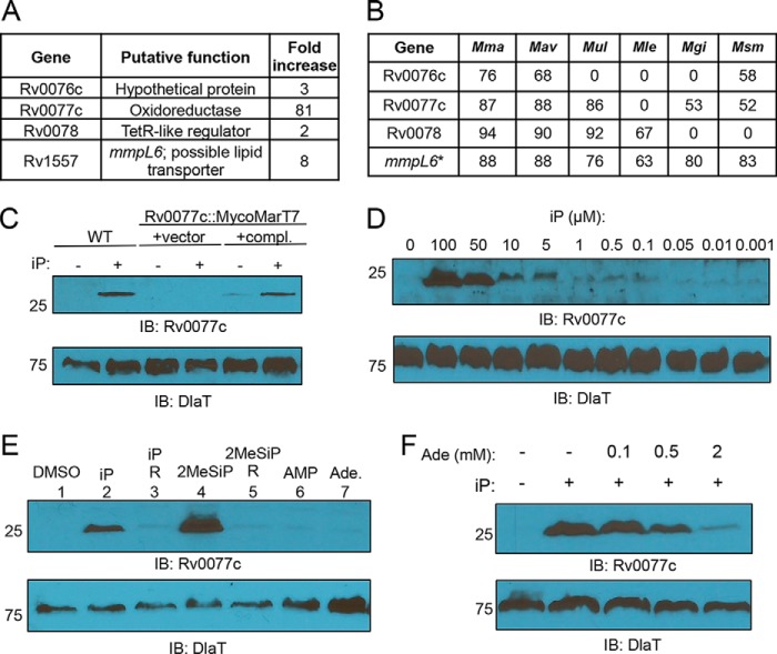
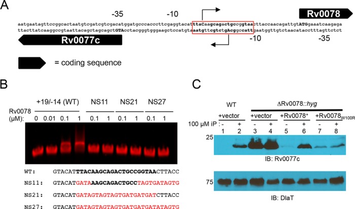
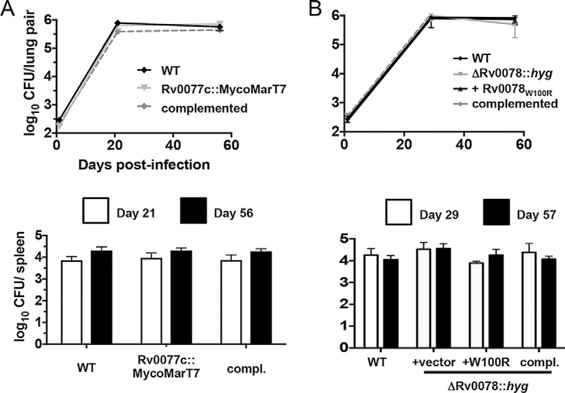
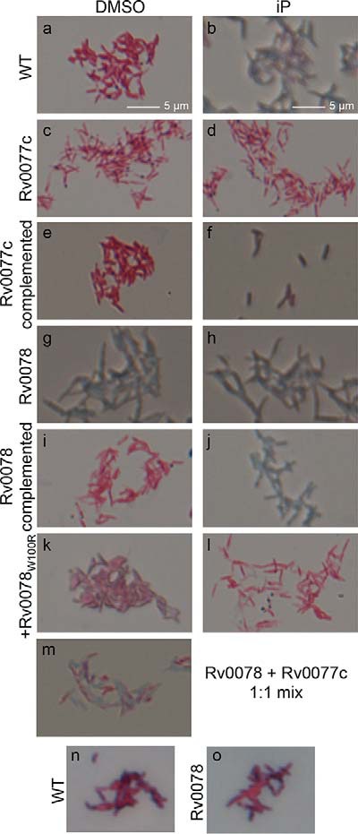
It was recently reported that the human-exclusive pathogen Mycobacterium tuberculosis secretes cytokinins, which had only been known as plant hormones. While cytokinins are well-established, adenine-based signaling molecules in plants, they have never been shown to participate in signal transduction in other kingdoms of life. M. tuberculosis is not known to interact with plants. Therefore, we tested the hypothesis that cytokinins trigger transcriptional changes within this bacterial species. Here, we show cytokinins induced the strong expression of the M. tuberculosis gene Rv0077c. We found that Rv0077c expression is repressed by a TetR-like transcriptional repressor, Rv0078. Strikingly, cytokinin-induced expression of Rv0077c resulted in a loss of acid-fast staining of M. tuberculosis While acid-fast staining is thought to be associated with changes in the bacterial cell envelope and virulence, Rv0077c-induced loss of acid-fastness did not affect antibiotic susceptibility or attenuate bacterial growth in mice, consistent with an unaltered mycolic acid profile of Rv0077c-expressing cells. Collectively, these findings show cytokinins signal transcriptional changes that can affect M. tuberculosis acid-fastness and that cytokinin signaling is no longer limited to the kingdom Plantae.IMPORTANCE Cytokinins have only previously been known as plant hormones. The discovery that they can be used as signaling molecules outside of plants broadens the repertoire of small molecules that can potentially affect gene expression in all domains of life.

Keywords: Mycobacterium tuberculosis; acid-fast staining; cytokinin; regulation; signaling.


Figures

FIG 1
FIG 1
Cytokinins induce the expression of Rv0077c in M. tuberculosis. (A) Genes significantly regulated by the presence of 100 µM iP for 5 h as analyzed by RNA-Seq. (B) Percentage of identity between M. tuberculosis H37Rv proteins and proteins of targeted mycobacterial genomes, including M. marinum (Mma), M. avium (Mav), M. ulcerans (Mul), M. leprae (Mle), M. gilvum (Mgi), and M. smegmatis (Msm). An asterisk indicates mmpL6 encodes a truncated protein in H37Rv, unlike in the other mycobacterial species in the table. (C) Immunoblot for Rv0077c in total cell lysates of WT M. tuberculosis. “compl.” indicates complemented. iP was used at a final concentration of 100 µM when added. (D) Dose-dependent production of Rv0077c protein. Bacteria were incubated with cytokinin at the indicated concentrations for 24 h. (E) Only cytokinins, and not closely related molecules, induce the production of Rv0077c. Each compound was added to a final concentration of 100 µM. “R” indicates the riboside form of the preceding indicated cytokinin. (F) Adenine inhibits the induction of Rv0077c by 100 µM iP. For all panels, we added an equal volume of DMSO to samples where iP was not added. For all immunoblots (IB), we stripped the membranes and incubated them with antibodies to dihydrolipoamide acyltransferase (DlaT) to confirm equal loading of total lysates. Molecular weight standards are indicated to the left of the blots and are in kilodaltons (kDa). Ade, adenine.
FIG 2
FIG 2
Rv0078 represses the expression of Rv0077c. (A) The putative transcriptional start sites (+1) of Rv0077c and Rv0078 as determined by 5′ RACE and represented as bent arrows. The predicted start codons are in capital boldface letters. (B) EMSA analysis identifies a putative repressor binding site. Probe sequence +19/−14 refers to positions relative to Rv0077c position +1. In boldface is the presumed binding site. Mutated residues are in red. Not shown at the end of each probe is a sequence for annealing to a fluorescent tag (Table S1). Rv0078 was purified under native conditions from E. coli. (C) Deletion and disruption of Rv0078 result in the constitutive expression of Rv0077c. Total cell lysates were prepared and separated on a 10% SDS-PAGE gel. IB, immunoblot. The membrane was stripped and then incubated with antibodies to DlaT to confirm equal loading of samples. Molecular weight standards are indicated to the left of the blots and are in kilodaltons (kDa).
FIG 3
FIG 3
Crystal structure of Rv0078 in complex with DNA. (A) ITC of Rv0078 binding to the +13/−8 DNA probe. The binding stoichiometry, ΔH, and KD are marked. (B) Overall structure of Rv0078-DNA complex in cartoon view. Two Rv0078 dimers (“central” and “peripheral”) bind to one DNA molecule. (C) The distance between two DNA-binding domains decreases by ~6 Å when bound to DNA. The DNA-free Rv0078 is in green, and the DNA-bound Rv0078 is in magenta. (D) Interactions between the LBD (magenta) and DBD (green) in a monomer. (E) The ligand binding pocket (magenta mesh) of Rv0078 is enclosed by a four-helix bundle (helices α5 to α8).
FIG 4
FIG 4
Rv0078-DNA interactions determined by X-ray crystallography. The hydroxyl groups of Thr37 (A), Thr47 (B), and Tyr52 (C) interact with the backbone phosphate with a distance of 2.6 Å. (D) Arg48 interacts with guanidine with a distance of 2.6 Å. The 2FoFc maps are contoured at the 1σ level. (E) A schematic representation of Rv0078-DNA contacts. Residues of the central dimer are labeled in red, and residues of the peripheral dimer are in purple. (F) EMSA using WT DNA probe and Rv0078 with mutations in Thr37, Thr47, Arg48, or Tyr52. (G, left panel) EMSA of Rv0078 with DNA probes with G-to-A substitutions in the central dimer binding region (S1), peripheral dimer binding region (S2), or in both DNA regions (S3). The white arrowhead marks the partial shift with the S2 probe at high protein concentration. (G, right panel) Sequences of the four DNA probes used in EMSAs. The nucleotides contacting the central dimer are boxed in red, and those contacting the peripheral dimer are boxed in purple.
FIG 5
FIG 5
Loss of Rv0077c or Rv0078 does not attenuate bacterial survival in mice. (A) Bacterial CFU after infection of C57BL/6J mice with the WT, Rv0077c mutant, and complemented strains. (B) Bacterial CFU after infection of C57BL/6J mice with the WT, Rv0078 mutant, and complemented strains. For both panels, data in each are from a single experiment that is representative of two independent experiments. Error bars indicate the standard error of the mean.
FIG 6
FIG 6
Induction of Rv0077c expression results in loss of acid-fast staining. M. tuberculosis strains were examined by Ziehl-Neelsen staining. The magnification is 63-fold. Scale bars are applicable to images in panels a to m. See text for details. “Rv0077c” indicates the Rv0077c::MycoMarT7 strain and “Rv0078” indicates the ΔRv0078::hyg strain.

References

    1. WHO 2017. Tuberculosis. World Health Organization, Geneva, Switzerland: http://www.who.int/mediacentre/factsheets/fs104/en/.
    1. Cerda-Maira FA, Pearce MJ, Fuortes M, Bishai WR, Hubbard SR, Darwin KH. 2010. Molecular analysis of the prokaryotic ubiquitin-like protein (Pup) conjugation pathway in Mycobacterium tuberculosis. Mol Microbiol 77:1123–1135. doi:10.1111/j.1365-2958.2010.07276.x. - DOI - PMC - PubMed
    1. Darwin KH, Ehrt S, Gutierrez-Ramos JC, Weich N, Nathan CF. 2003. The proteasome of Mycobacterium tuberculosis is required for resistance to nitric oxide. Science 302:1963–1966. doi:10.1126/science.1091176. - DOI - PubMed
    1. Gandotra S, Lebron MB, Ehrt S. 2010. The Mycobacterium tuberculosis proteasome active site threonine is essential for persistence yet dispensable for replication and resistance to nitric oxide. PLoS Pathog 6:e1001040. doi:10.1371/journal.ppat.1001040. - DOI - PMC - PubMed
    1. Gandotra S, Schnappinger D, Monteleone M, Hillen W, Ehrt S. 2007. In vivo gene silencing identifies the Mycobacterium tuberculosis proteasome as essential for the bacteria to persist in mice. Nat Med 13:1515–1520. doi:10.1038/nm1683. - DOI - PMC - PubMed

Publication types

MeSH terms

LinkOut - more resources