. 2018 Jul 18;38(29):6427-6438.
doi: 10.1523/JNEUROSCI.3174-17.2018. Epub 2018 Jun 19.

The State of the NIH BRAIN Initiative

Affiliations

The State of the NIH BRAIN Initiative

Walter Koroshetz et al. J Neurosci. .

Abstract

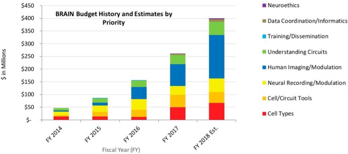
The BRAIN Initiative arose from a grand challenge to "accelerate the development and application of new technologies that will enable researchers to produce dynamic pictures of the brain that show how individual brain cells and complex neural circuits interact at the speed of thought." The BRAIN Initiative is a public-private effort focused on the development and use of powerful tools for acquiring fundamental insights about how information processing occurs in the central nervous system (CNS). As the Initiative enters its fifth year, NIH has supported >500 principal investigators, who have answered the Initiative's challenge via hundreds of publications describing novel tools, methods, and discoveries that address the Initiative's seven scientific priorities. We describe scientific advances produced by individual laboratories, multi-investigator teams, and entire consortia that, over the coming decades, will produce more comprehensive and dynamic maps of the brain, deepen our understanding of how circuit activity can produce a rich tapestry of behaviors, and lay the foundation for understanding how its circuitry is disrupted in brain disorders. Much more work remains to bring this vision to fruition, and the National Institutes of Health continues to look to the diverse scientific community, from mathematics, to physics, chemistry, engineering, neuroethics, and neuroscience, to ensure that the greatest scientific benefit arises from this unique research Initiative.


Figures

Figure 1.
Figure 1.
Thanks to strong bipartisan support in Congress and the Innovation Funds of the 21st Century Cures Act, the budget for the BRAIN Initiative at NIH has increased each fiscal year (FY) since 2014, bringing the total investment to: $46M in FY2014, $85M in FY2015, $155M in FY2016, and $262M in FY2017. With the guidance of the BRAIN 2025 strategic plan and the expertise of BRAIN Institutes and Centers' advisory councils, scientific program staff at NIH have distributed funds across the 7 scientific goals and overarching principles of the Initiative. NIH grants are usually funded incrementally (i.e., one award for each year of the project period). Some BRAIN awards in FY2017 and FY2018 are multi-year funded awards, where funding for the entire project period (more than one year) is provided in the first fiscal year and comparing investment with previous years can become complex. The FY2018 bar represents a very preliminary estimate.
Figure 2.
Figure 2.
A, BRAIN Initiative scientists are using whole-brain activity imaging and connectomics to chart a detailed diagram of zebrafish brain circuits. Here is a reconstruction of myelinated axons found in one brain (see also Movie 1). Courtesy of the F. Engert laboratory (Harvard University). Reproduced with permission of Springer. B, New high-throughput imaging methods developed by BRAIN researchers offer unprecedented capabilities for imaging tissue molecular architectures. This is an example of array tomography of mouse cortex. Courtesy of the S. Smith laboratory (Stanford University).
Figure 3.
Figure 3.
The BRAIN Initiative supports innovations in the next generation of human imaging technologies, as well as projects for novel noninvasive neuromodulation technologies. This magnetic resonance angiography highlights the vasculature throughout the human brain in high resolution, without the use of any contrast agent, on a 7 tesla MRI scanner. Courtesy of J.R. Plimeni and L.L. Wald (Massachusetts General Hospital).

References

    1. Abdelfattah AS, Farhi SL, Zhao Y, Brinks D, Zou P, Ruangkittisakul A, Platisa J, Pieribone VA, Ballanyi K, Cohen AE, Campbell RE (2016) A bright and fast red fluorescent protein voltage indicator that reports neuronal activity in organotypic brain slices. J Neurosci 36:2458–2472. 10.1523/JNEUROSCI.3484-15.2016 - DOI - PMC - PubMed
    1. Ai-Awami AK, Beyer J, Haehn D, Kasthuri N, Lichtman JW, Pfister H, Hadwiger M (2016) NeuroBlocks: visual tracking of segmentation and proofreading for large connectomics projects. IEEE Trans Vis Comput Graph 22:738–746. 10.1109/TVCG.2015.2467441 - DOI - PubMed
    1. Alivisatos AP, Chun M, Church GM, Greenspan RJ, Roukes ML, Yuste R (2012) The brain activity map project and the challenge of functional connectomics. Neuron 74:970–974. 10.1016/j.neuron.2012.06.006 - DOI - PMC - PubMed
    1. Aljadeff J, Lansdell BJ, Fairhall AL, Kleinfeld D (2016) Analysis of neuronal spike trains, deconstructed. Neuron 91:221–259. 10.1016/j.neuron.2016.05.039 - DOI - PMC - PubMed
    1. Antic SD, Hines M, Lytton WW (2018) Embedded ensemble encoding hypothesis: The role of the “Prepared” cell. J Neurosci Res. Advance online publication. Retrieved Apr. 6, 2018. doi: 10.1002/jnr.24240. 10.1002/jnr.24240 - DOI - DOI - PMC - PubMed

LinkOut - more resources