Comparative Study
. 2018 Aug;29(8):2081-2088.
doi: 10.1681/ASN.2017111210. Epub 2018 Jun 19.

Region-Based Convolutional Neural Nets for Localization of Glomeruli in Trichrome-Stained Whole Kidney Sections

Affiliations
Comparative Study

Region-Based Convolutional Neural Nets for Localization of Glomeruli in Trichrome-Stained Whole Kidney Sections

John D Bukowy et al. J Am Soc Nephrol. 2018 Aug.

Abstract

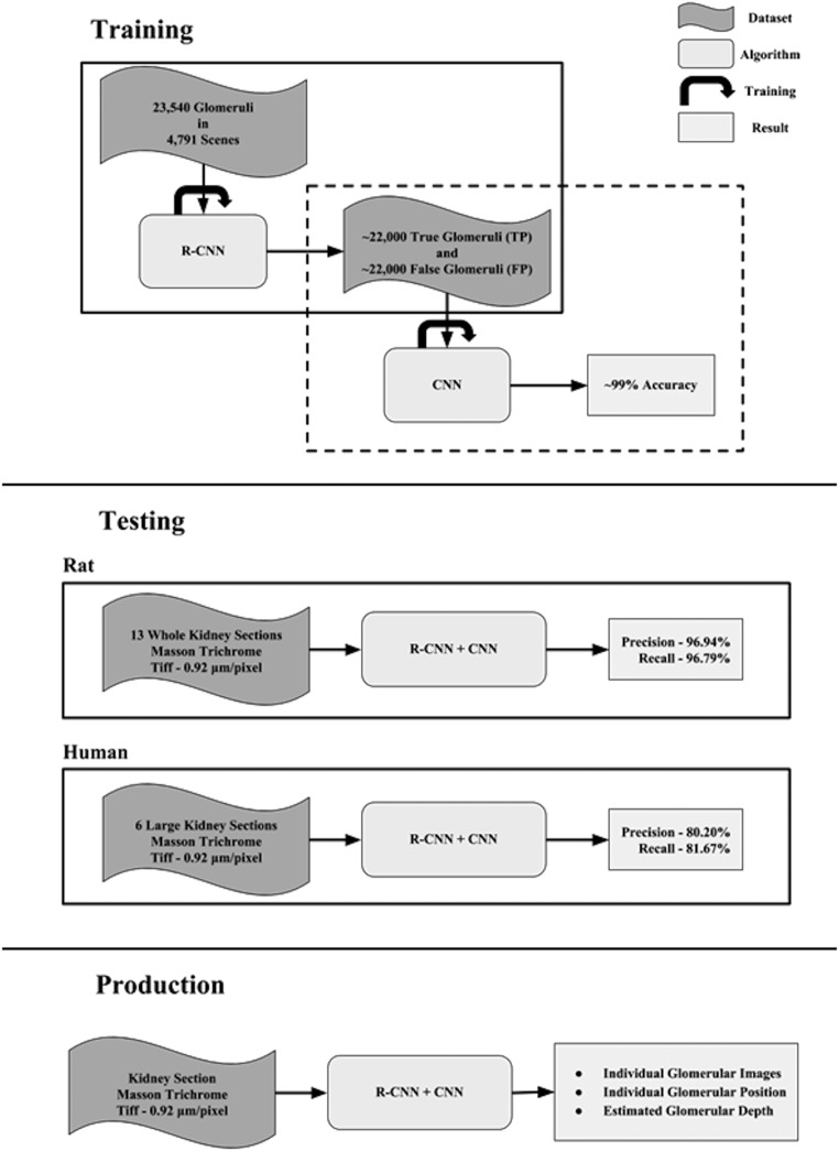
Background Histologic examination of fixed renal tissue is widely used to assess morphology and the progression of disease. Commonly reported metrics include glomerular number and injury. However, characterization of renal histology is a time-consuming and user-dependent process. To accelerate and improve the process, we have developed a glomerular localization pipeline for trichrome-stained kidney sections using a machine learning image classification algorithm.Methods We prepared 4-μm slices of kidneys from rats of various genetic backgrounds that were subjected to different experimental protocols and mounted the slices on glass slides. All sections used in this analysis were trichrome stained and imaged in bright field at a minimum resolution of 0.92 μm per pixel. The training and test datasets for the algorithm comprised 74 and 13 whole renal sections, respectively, totaling over 28,000 glomeruli manually localized. Additionally, because this localizer will be ultimately used for automated assessment of glomerular injury, we assessed bias of the localizer for preferentially identifying healthy or damaged glomeruli.Results Localizer performance achieved an average precision and recall of 96.94% and 96.79%, respectively, on whole kidney sections without evidence of bias for or against glomerular injury or the need for manual preprocessing.Conclusions This study presents a novel and robust application of convolutional neural nets for the localization of glomeruli in healthy and damaged trichrome-stained whole-renal section mounts and lays the groundwork for automated glomerular injury scoring.

Keywords: Renal pathology; glomerular disease; glomerulus; kidney disease; renal injury; renal morphology.


Figures

Figure 1.
Figure 1.
Color normalization process virtually separates stains to normalize tissue samples and decrease signal associated with fibrosis. (A) Color deconvolution vectors were extracted from this subset of pixels using a method adapted from Macenko et al. To decrease computation time, a small region with “pure” examples of red and blue staining was selected from the larger image. Color deconvolution vectors were solved for each renal section. (B and C) After deconvolving, the original image with color vectors derived from (A) virtual stain separation approximated (B) red stain contribution from (C) blue stain contribution. (D) Using only the grayscale image associated with the red staining, histogram equalization was performed over the tissue section, increasing contrast for glomerular detection (dark puncta near surface).
Figure 2.
Figure 2.
Testing of localizer and proposed workflow shows high precision and recall in rat and human test data. (A) Example scene of a rat kidney within the test set. Cyan boxes denote true positives or where there is >50% overlap of the predicted region of interest with a ground truth box of 150×150 pixels placed over the center of a glomerulus. Magenta boxes show false negatives and show relative size of the ground truth bounding box. Red boxes show false positives or where the algorithm incorrectly identified background tissue as a glomerulus. The representative field shows the variability of glomerular injury and tubular damage. (B) Precision-recall curve of all observations within the test dataset (5447 glomeruli). Values were achieved by sweeping the probability threshold associated with glomerular candidate output of the trained region–based convolutional neural net. (Inset) Magnified precision-recall curve with probability threshold between 0.86 and 0.92. (C) Example scene of a human kidney within the expanded test set, with examples of glomerular and tubular injury. Color code is identical to that in A. (D) Precision-recall curve for all human observations within the expanded test set (1173 glomeruli). (Inset) Magnified precision-recall curve with probability threshold between 0.88 and 0.94.
Figure 3.
Figure 3.
Characterization of distance from renal surface to glomeruli within whole kidney sections shows separation between individuals in heterogeneous rat stock. (A) Empirically determined probability distribution functions describing the probability of a glomerulus given a distance from the renal surface. Sixteen individual rats are plotted in order of skewness from least to most (top to bottom). Skewness describes the “lean” of a probability density function. (B) Cumulative distribution functions of log-normal distributions fit to the two extreme animals of plot A (top row [gray line]; bottom row [black line]). The dashed lines show the 95% confidence intervals. Inset shows cumulative distribution functions from 0% to 50% probability.
Figure 4.
Figure 4.
Training, testing, and production workflow of the described glomerular localizer. Training describes the datasets used for the total training of the classifiers. Solid and dashed outlines show separate training efforts. The output from the region-based convolutional neural network (R-CNN) was used for training of the secondary convolutional neural network (CNN). When the training dataset for the CNN was enriched with examples that the first-stage R-CNN failed to identify correctly, the secondary classifier becomes more robust against errors. Training of the localizer was performed solely on rat tissue specimens but provides impressive localization on human kidney samples.

Comment in

  • AI: What Have You Done for Us Lately?
    Torres R, Olson E. Torres R, et al. J Am Soc Nephrol. 2018 Aug;29(8):2031-2032. doi: 10.1681/ASN.2018050566. Epub 2018 Jun 19. J Am Soc Nephrol. 2018. PMID: 29921720 Free PMC article. No abstract available.

References

    1. Kakimoto T, Okada K, Hirohashi Y, Relator R, Kawai M, Iguchi T, et al. .: Automated image analysis of a glomerular injury marker desmin in spontaneously diabetic Torii rats treated with losartan. J Endocrinol 222: 43–51, 2014 - PubMed
    1. Evans LC, Petrova G, Kurth T, Yang C, Bukowy JD, Mattson DL, et al. .: Increased perfusion pressure drives renal T-cell infiltration in the dahl salt-sensitive rat. Hypertension 70: 543–551, 2017 - PMC - PubMed
    1. Kumar V, Wollner C, Kurth T, Bukowy JD, Cowley AW Jr: Inhibition of mammalian target of rapamycin complex 1 attenuates salt-induced hypertension and kidney injury in dahl salt-sensitive rats. Hypertension 70: 813–821, 2017 - PMC - PubMed
    1. Venkatachalam MA, Griffin KA, Lan R, Geng H, Saikumar P, Bidani AK: Acute kidney injury: A springboard for progression in chronic kidney disease. Am J Physiol Renal Physiol 298: F1078–F1094, 2010 - PMC - PubMed
    1. Bertram JF, Soosaipillai MC, Ricardo SD, Ryan GB: Total numbers of glomeruli and individual glomerular cell types in the normal rat kidney. Cell Tissue Res 270: 37–45, 1992 - PubMed

Publication types