. 2018 Jun 6;8(6):52.
doi: 10.1038/s41408-018-0087-2.

Leukemic stem cell signatures identify novel therapeutics targeting acute myeloid leukemia

Affiliations

Leukemic stem cell signatures identify novel therapeutics targeting acute myeloid leukemia

Isabelle Laverdière et al. Blood Cancer J. .

Abstract

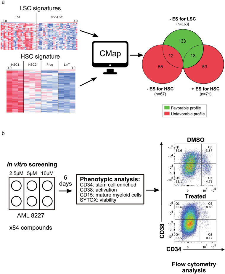
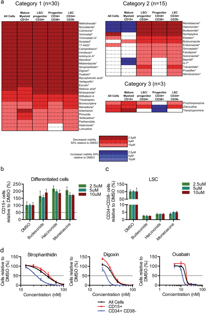
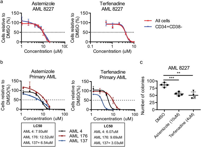
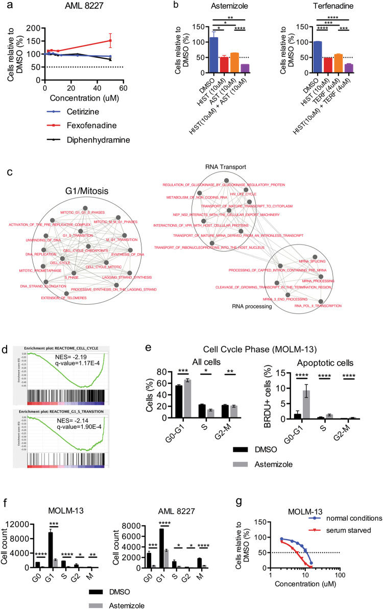
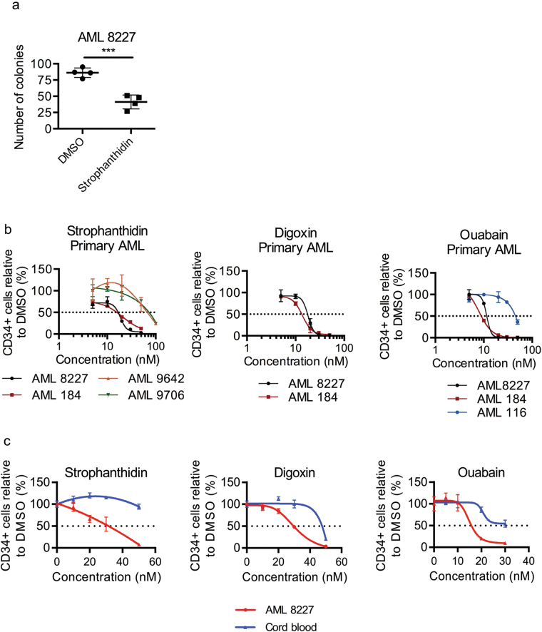
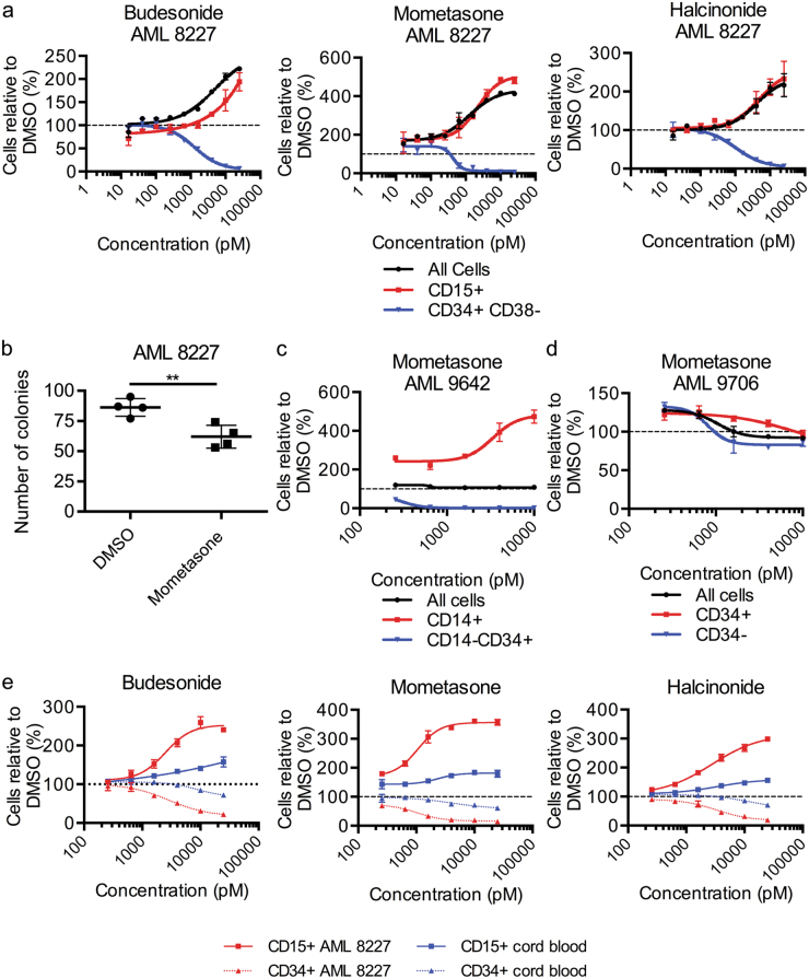
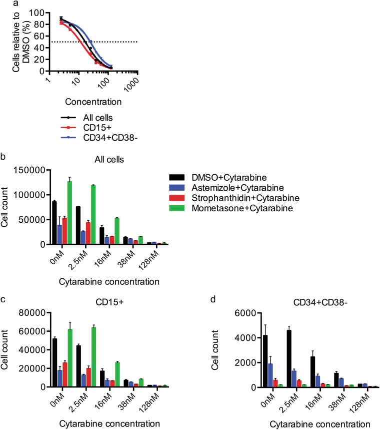
Therapy for acute myeloid leukemia (AML) involves intense cytotoxic treatment and yet approximately 70% of AML are refractory to initial therapy or eventually relapse. This is at least partially driven by the chemo-resistant nature of the leukemic stem cells (LSCs) that sustain the disease, and therefore novel anti-LSC therapies could decrease relapses and improve survival. We performed in silico analysis of highly prognostic human AML LSC gene expression signatures using existing datasets of drug-gene interactions to identify compounds predicted to target LSC gene programs. Filtering against compounds that would inhibit a hematopoietic stem cell (HSC) gene signature resulted in a list of 151 anti-LSC candidates. Using a novel in vitro LSC assay, we screened 84 candidate compounds at multiple doses and confirmed 14 drugs that effectively eliminate human AML LSCs. Three drug families presenting with multiple hits, namely antihistamines (astemizole and terfenadine), cardiac glycosides (strophanthidin, digoxin and ouabain) and glucocorticoids (budesonide, halcinonide and mometasone), were validated for their activity against human primary AML samples. Our study demonstrates the efficacy of combining computational analysis of stem cell gene expression signatures with in vitro screening to identify novel compounds that target the therapy-resistant LSC at the root of relapse in AML.


Conflict of interest statement

The authors declare that they have no conflict of interest.

Figures

Fig. 1
Fig. 1. In silico and in vitro screening for drugs preferentially targeting LSCs over HSCs.
a Schematic illustrating the in silico screen process for identifying anti-LSC compounds. The Venn diagram indicates the number of candidate compounds that target LSCs without harming HSCs (n = 133) or enhance the HSC-signature (n = 18). The + or – ES indicates positive or negative enrichment score. b Experimental design of the in vitro screening and analysis process of 84 candidate compounds
Fig. 2
Fig. 2. In vitro validation of anti-leukemic compounds from an in silico screen against AML 8227.
a Summary of all compounds that affected at least one population in 8227 AML cells. Red denotes decreased viability of at least 50% at the indicated concentration and blue denotes increased viability of at least 50% at the indicated concentration; experiment performed in duplicate. An ‘*’ indicates that the compound was retested. b, c Confirmation of the effect of 9 compounds shown to be preferential for CD34+ cells on b the CD15+ terminally differentiated blast population and c the CD34+ CD38− LSC-containing population. d Viability of bulk and CD34+ CD38− AML 8227 cells treated with the three cardiac glycosides, strophanthidin, digoxin and ouabain at indicated concentrations for 6 days. Data are representative of three independent experiments performed in triplicate and displays the mean ± s.d.
Fig. 3
Fig. 3. The H1-antihistamines astemizole and terfenadine have anti-leukemic properties against all cells within the AML hierarchy.
a Viability of bulk and CD34+ CD38− AML 8227 cells treated with astemizole and terfenadine at indicated concentrations for 6 days. Data are representative of three independent experiments performed in triplicate and displays the mean ± s.d. b Viability of 3 additional primary AML samples (AML 4, AML 176 and AML 137) treated with astemizole and terfenadine at indicated concentrations for 4 days. Data represent the mean ± s.d.; experiment performed in triplicate. c Colony formation assay of AML 8227 after 4 days of treatment with 10 μM astemizole and 4 μM terfenadine. Data are representative of three independent experiments performed in quadruplicate and display mean ± s.d.; *p ≤ 0.05, **p ≤ 0.01, ***p ≤ 0.001, ****p ≤ 0.0001
Fig. 4
Fig. 4. Astemizole initiates apoptosis in the G0/G1 cell cycle phase independently of the H1 receptor.
a Viability of AML 8227 cells treated with the H1-antihistamines cetirizine, fexofenadine and diphenhydramine up to 50 μM for 6 days. b Viability of AML 8227 after the addition of 10 μM histamine to 10 μM astemizole or 4 μM terfenadine treatment for 4 days. c Enrichment map of CMap expression data revealing astemizole modulated pathways. Nodes (circles) represent gene sets and edges (lines) represent shared genes. d GSEA plot of negative enrichment of cell cycle and G1/S transition using the gene expression signature of cells treated with astemizole from CMap. e Apo-BrdU TUNEL assay on MOLM-13 cells treated with 10 μM astemizole for 24 h. f Cell cycle analysis using Ki-67 on MOLM-13 and 8227 cells with 10 μM astemizole for 24 h. g Viability of MOLM-13 cell growth in serum-starved (0% FBS) or normal conditions (20% FBS) after 24 h of treatment with indicated doses of astemizole. Data are representative of at least two independent experiments performed in triplicate and represent the mean ± s.d.; **p ≤ 0.01, ***p ≤ 0.001
Fig. 5
Fig. 5. The cardiac glycosides strophanthidin, digoxin and ouabain are cytotoxic against primitive AML cells.
a Colony formation of AML 8227 treated with 30 nM of strophanthidin for 4 days. Data are representative of two independent experiments performed in quadruplicate and represents mean ± s.d. b Viability of additional primary AML treated with strophanthidin, digoxin and ouabain at indicated concentrations for 4 days. Data represent mean ± s.d; experiment performed in triplicate. c Viability of CD34+ cord blood cells and CD34+ 8227 cells treated with strophanthidin, digoxin and ouabain at indicated concentrations for 4 days. Data are representative of at least two independent experiments performed in triplicate and represent the mean ± s.d. ***p ≤ 0.001
Fig. 6
Fig. 6. The glucocorticoids budesonide, mometasone and halcinonide differentiate AML samples with limited toxicity to HSPCs.
a Viability of bulk, CD15+ (mature blast) and CD34+ CD38− AML 8227 cells treated with budesonide, mometasone and halcinonide at indicated concentrations for 6 days. Data are representative of at least three independent experiments performed in triplicate and represents the mean ± s.d. b Colony formation of AML 8227 treated with 1 nM of mometasone for 4 days. Data are representative of two independent experiments performed in duplicate and represents mean ± s.d. c, d Viability of c bulk, CD14+ (mature blast) and CD14– CD34+ AML 9642 cells and d bulk, CD34– and CD34+ AML 9706 cells treated with indicated concentrations of mometasone for 4 days. Data represent mean ± s.d; experiment performed in triplicate. e Viability of CD34+ and CD15+ mature cord blood cells, as well as CD34+ and CD15+ AML 8227 cells treated with budesonide, mometasone and halcinonide at indicated concentrations for 4 days. Data are representative of two independent experiments performed in triplicate and represents the mean ± s.d.; **p ≤ 0.01, ***p ≤ 0.001
Fig. 7
Fig. 7. Candidate compounds potentiate the effect of cytarabine in AML 8227.
a Viability of bulk, CD15+ (mature blast) and CD34+ CD38− AML 8227 cells treated with cytarabine at indicated concentrations for 6 days. bd Viability of b bulk, c CD15+ and d CD34+ CD38− AML 8227 treated with cytarabine in combination with either DMSO, 10 µM astemizole, 30 nM strophanthidin or 10 nM mometasone for 6 days. Data are representative of three independent experiments performed in triplicate and represent the mean ± s.d.

References

    1. Lowenberg B, et al. Cytarabine dose for acute myeloid leukemia. N. Engl. J. Med. 2011;364:1027–1036. doi: 10.1056/NEJMoa1010222. - DOI - PubMed
    1. Dohner H, et al. Diagnosis and management of acute myeloid leukemia in adults: recommendations from an international expert panel, on behalf of the European LeukemiaNet. Blood. 2010;115:453–474. doi: 10.1182/blood-2009-07-235358. - DOI - PubMed
    1. Dohner H, et al. Diagnosis and management of AML in adults: 2017 ELN recommendations from an international expert panel. Blood. 2017;129:424–447. doi: 10.1182/blood-2016-08-733196. - DOI - PMC - PubMed
    1. de Rooij JD, Zwaan CM, van den Heuvel-Eibrink M. Pediatric AML: from biology to clinical management. J. Clin. Med. 2015;4:127–149. doi: 10.3390/jcm4010127. - DOI - PMC - PubMed
    1. Tallman MS, Gilliland DG, Rowe JM. Drug therapy for acute myeloid leukemia. Blood. 2005;106:1154–1163. doi: 10.1182/blood-2005-01-0178. - DOI - PubMed

Publication types

MeSH terms

Grants and funding