

Extramedullary hematopoiesis secondary to malignant solid tumors: a case report and literature review
- PMID: 29922090
- PMCID: PMC5997179
- DOI: 10.2147/CMAR.S161746
Extramedullary hematopoiesis secondary to malignant solid tumors: a case report and literature review
Abstract
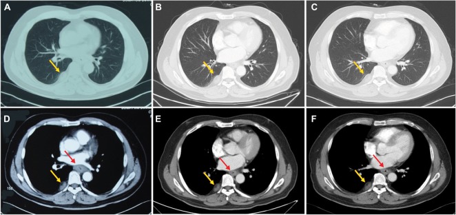
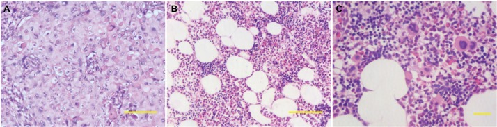
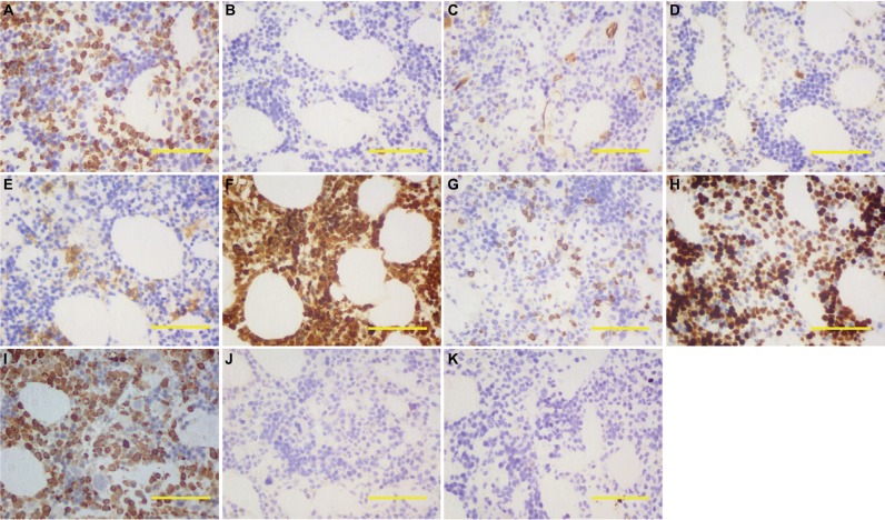
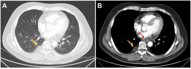
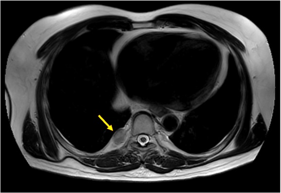
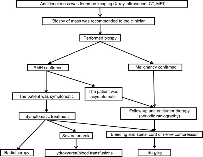
Extramedullary hematopoiesis (EMH) usually occurs in hematological disease, but more rarely develops in cases of malignant solid tumors. Due to its features on computed tomography (CT) and magnetic resonance imaging (MRI) that are atypical, EMH in tumor patients might easily be misdiagnosed as metastasis leading to the improper TNM staging and inappropriate therapy. Here, we reported the first case of pleural EMH occurring in a patient with esophageal carcinoma whose pleural lesion was first diagnosed as metastasis and confirmed EMH after the needle biopsy. In addition, a retrospective review was conducted by analyzing patients presented with EMH with malignant solid tumors from PubMed and Medline databases. A total of 42 solid tumor patients with EMH were enrolled, and breast cancer was the most common (n=13, 31.0%), followed by renal carcinoma (n=7, 16.7%) and lung cancer (n=6, 14.3%). A wide variety of body sites may be affected by EMH in malignant solid tumor patients, of which the lymph nodes (n=8, 19.0%) and liver (n=7, 16.7%) were the most common, followed by the kidney (n=6, 14.3%). All patients were diagnosed with EMH by excision, biopsy, or autopsy. Treatment strategies for EMH included surgery (n=25, 59.5%), hydroxyurea (n=1, 2.4%), and blood transfusions (n=2, 4.8%); a further 14 patients (33.3%) were subjected to clinical observation without intervention. Of the patients for whom outcome was reported, 10 patients maintained a good performance status (23.8%) and a further six patients died from the malignant tumor. This was the first study to summarize the presentations of EMH in malignant solid tumors, and our findings might provide some useful guidance for clinical practice, especially for treating patients harboring nonresponse lesions during the antitumor treatment.
Keywords: biopsy; cancer; extramedullary hematopoiesis; imaging features; sarcoma.
Conflict of interest statement
Disclosure The authors report no conflicts of interest in this work.
Figures
References
-
- Meykler S, Obstfeld A, Jhala N, Vergara N, Gupta PK. Pleural mass forming extramedullary hematopoiesis masquerading as a malignant neoplasm. Diagn Cytopathol. 2015;43(12):996–999. - PubMed
-
- Ardakani NM, Kumarasinghe MP, Spagnolo DV, Stewart CJ. Extramed-ullary hematopoiesis associated with organizing peritoneal hemorrhage: a report of 5 cases in patients presenting with primary gynecologic disorders. Int J Gynecol Pathol. 2014;33(3):317–322. - PubMed
Publication types
LinkOut - more resources
Full Text Sources
Other Literature Sources
