. 2018 Jun 5:12:47.
doi: 10.3389/fnana.2018.00047. eCollection 2018.

A Quantitative Tractography Study Into the Connectivity, Segmentation and Laterality of the Human Inferior Longitudinal Fasciculus

Affiliations

A Quantitative Tractography Study Into the Connectivity, Segmentation and Laterality of the Human Inferior Longitudinal Fasciculus

Sandip S Panesar et al. Front Neuroanat. .

Abstract

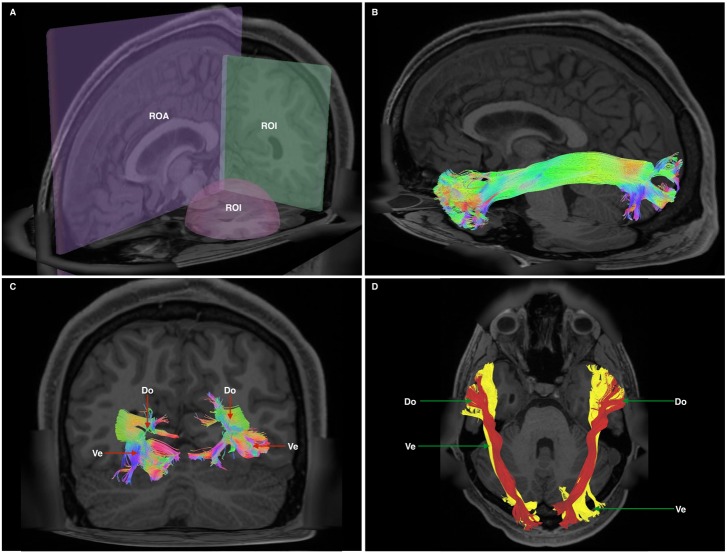
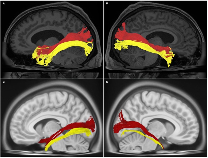
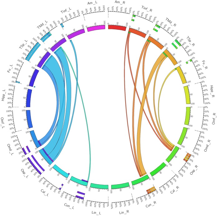
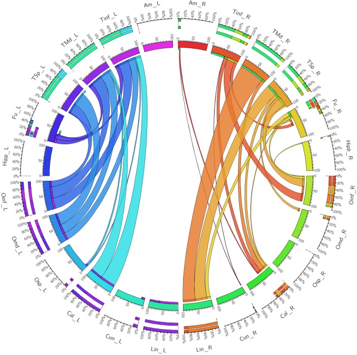
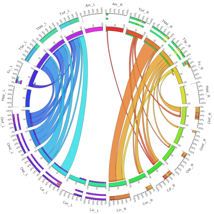
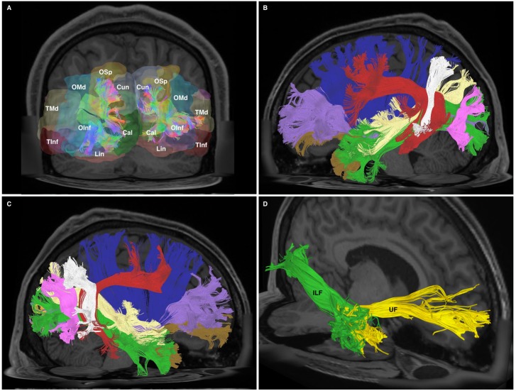
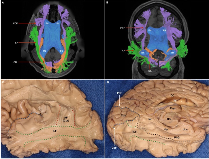
The human inferior longitudinal fasciculus (ILF) is a ventral, temporo-occipital association tract. Though described in early neuroanatomical works, its existence was later questioned. Application of in vivo tractography to the neuroanatomical study of the ILF has generally confirmed its existence, however, consensus is lacking regarding its subdivision, laterality and connectivity. Further, there is a paucity of detailed neuroanatomic data pertaining to the exact anatomy of the ILF. Generalized Q-Sampling imaging (GQI) is a non-tensor tractographic modality permitting high resolution imaging of white-matter structures. As it is a non-tensor modality, it permits visualization of crossing fibers and accurate delineation of close-proximity fiber-systems. We applied deterministic GQI tractography to data from 30 healthy subjects and a large-volume, averaged diffusion atlas, to delineate ILF anatomy. Post-mortem white matter dissection was also carried out in three cadaveric specimens for further validation. The ILF was found in all 60 hemispheres. At its occipital extremity, ILF fascicles demonstrated a bifurcated, ventral-dorsal morphological termination pattern, which we used to further subdivide the bundle for detailed analysis. These divisions were consistent across the subject set and within the atlas. We applied quantitative techniques to study connectivity strength of the ILF at its anterior and posterior extremities. Overall, both morphological divisions, and the un-separated ILF, demonstrated strong leftward-lateralized connectivity patterns. Leftward-lateralization was also found for ILF volumes across the subject set. Due to connective and volumetric leftward-dominance and ventral location, we postulate the ILFs role in the semantic system. Further, our results are in agreement with functional and lesion-based postulations pertaining to the ILFs role in facial recognition.

Keywords: inferior longitudinal fasciculus; non-tensor tractography; semantic anatomy; tractography; white matter anatomy.


Figures

FIGURE 1
FIGURE 1
(A) Cutaway view showing all three radiological planes (axial, sagittal, and coronal) with ROIs and ROAs used. Spherical ROI is placed within the anterior temporal lobe white matter, whilst the rectangular ROI is placed in a coronal plane at the approximate position of the temporo-occipital junction. The ROA was placed in the mid-sagittal plane to exclude any fibers crossing the midline. (B) Generated whole, undivided ILF bundle after manual removal of spurious fibers belonging to other white matter systems and prior to separation. Color assignment is directional. (C) A posterior-coronal view demonstrating the posterior terminations of both left and right ILF bundles. Visible clearly is the bifurcated arrangement of each whole-ILF bundle which were subsequently used as geographical landmarks to separate the ILF further. Do, dorsal; Ve, ventral. (D) A superior-axial view demonstrating left and right ILF bundles following separation of the ILF into its respective subdivisions. The sub-fascicles have been individually colored. Do, dorsal (red); Ve, ventral (yellow).
FIGURE 2
FIGURE 2
(A) Left-hemispheric, sagittal-view representation of a subdivided ILF in a single subject from the subject-specific analysis. DL, dorsal (red); VL, ventral (yellow). (B) Right-hemispheric, sagittal-view representation of a subdivided ILF in a single subject from the subject-specific analysis. DL, dorsal (red); VL, ventral (yellow). (C) Left-hemispheric, sagittal-view representation of a subdivided ILF in the HCP 1021 atlas. DL, dorsal (red); VL, ventral (yellow). (D) Right-hemispheric, sagittal-view representation of a subdivided ILF in the HCP 1021 atlas. DL, dorsolateral (red); VL, ventral (yellow).
FIGURE 3
FIGURE 3
A connectogram representing bilateral connectivity patterns of the dorsal ILF subfascicle. Atlas regions are listed around the circumference of the connectogram. TInf, inferior temporal gyrus; TMd, middle temporal gyrus; TSp, superior temporal gyrus; Fu, fusiform gyrus; OInf, inferior occipital gyrus; OMd, middle occipital gyrus; OSp, superior occipital gyrus; Cal, calcarine gyrus; Cun, cuneus; Lin, lingual gyrus. Left and Right hemispheric connections are demonstrated with a suffix _L or _R.
FIGURE 4
FIGURE 4
A connectogram representing bilateral connectivity patterns of the ventral ILF subfascicle. Atlas regions are listed around the circumference of the connectogram. TInf, inferior temporal gyrus; TMd, middle temporal gyrus; TSp, superior temporal gyrus; Fu, fusiform gyrus; OInf, inferior occipital gyrus; OMd, middle occipital gyrus; OSp, superior occipital gyrus; Cal, calcarine gyrus; Cun, cuneus; Lin, lingual gyrus. Left and Right hemispheric connections are demonstrated with a suffix _L or _R.
FIGURE 5
FIGURE 5
A connectogram representing bilateral connectivity patterns of the whole, unseparated ILF. Atlas regions are listed around the circumference of the connectogram. TInf, inferior temporal gyrus; TMd, middle temporal gyrus; TSp, superior temporal gyrus; Fu, fusiform gyrus; OInf, inferior occipital gyrus; OMd, middle occipital gyrus; OSp, superior occipital gyrus; Cal, calcarine gyrus; Cun, cuneus; Lin, lingual gyrus. Left and Right hemispheric connections are demonstrated with a suffix _L or _R.
FIGURE 6
FIGURE 6
(A) Posterior-coronal view demonstrating occipito-temporal terminations of all divisions of the ILF. OSp, superior occipital gyrus; OMd, middle occipital gyrus; OInf, inferior occipital gyrus; Lin, lingual gyrus; Cun, cuneus; Cal, calcarine gyrus; TMd, middle temporal gyrus; TInf, inferior temporal gyrus. (B) Left-hemispheric sagittal-view representation of relational white matter tracts. Tracts are colored separately. IFOF, purple; Claustrum, blue; AF, red; PAT, white; MdLF, yellow; ILF, green; UF, brown; VOF, pink. (C) Right-hemispheric sagittal-view representation of relational white matter tracts. Tracts are colored separately. IFOF, purple; Claustrum, blue; AF, red; PAT, white; MdLF, yellow; ILF, green; UF, brown; VOF, pink. (D) An oblique-frontal view of the ILF (green) and UF (yellow) with focus of anterior temporal terminations. Occupying the medial white matter of the anterior temporal lobe are UF ventral terminations. The lateral white matter of the anterior temporal lobe consists of fibers which are part of the ILF.
FIGURE 7
FIGURE 7
(A) Superior axial-view representation of tracts contributing to the sagittal stratum and their relation to the ventricular system. The ventricles are represented in blue as a 3-dimensional structure superimposed upon the axial T1 image. AH, anterior horn of the lateral ventricle; Bo, body of the lateral ventricle. IFOF, purple; ILF, green; OR, orange. (B) Inferior para-axial representation of tracts contributing to the sagittal stratum and their relation to the ventricular system. The ventricles are represented in blue as a 3-dimensional structure superimposed upon the axial T1 image. AH, anterior horn of the lateral ventricle; TH, temporal horn. IFOF, purple; ILF, green; ML, Meyer’s loop, passing over the superior surface of the temporal horn. (C) White matter dissection within a post-mortem specimen. Lateral-view. A portion of the Sylvian fissure is visible (blue broken line). At the temporal and parietal lobe, overlying cortex, U-fibers have been removed to expose the white matter of the ILF (demarcated by green broken lines). Some fibers of the dorsal portion of the AF, which overlies the ILF, are still visible (labeled AF Cut). (D) White matter dissection within a post-mortem specimen. View of infero-medial hemispheric surface. The corpus callosum (CC) and 3rd ventricle (3V) have been transected to achieve this view. The medial surface of the left thalamus is visible, which is situated superior to the superior brainstem (BS), which has also been transected. The cuneus (Cun) is a triangular-shaped lobe when viewed from the medial hemispheric surface. It is bounded superiorly by the parieto occipital fissure (PoF) and inferiorly by the calcarine fissure (CaF). Superficial structures have been removed to expose the ILF (between broken green (inferior) and black (superior) lines. Superior to the broken black line is the hippocampus (Hip), the calcar avis (CA). Dorsal to these structures are the isthmus (Ist), parahippocampal gyrus (PhG), and the uncus (Unc).

References

    1. Allison T., McCarthy G., Nobre A., Puce A., Belger A. (1994). Human extrastriate visual cortex and the perception of faces, words, numbers, and colors. Cereb. Cortex 4 544–554. 10.1093/cercor/4.5.544 - DOI - PubMed
    1. Bauer R. M. (1984). Autonomic recognition of names and faces in prosopagnosia: a neuropsychological application of the guilty knowledge test. Neuropsychologia 22 457–469. 10.1016/0028-3932(84)90040-X - DOI - PubMed
    1. Behrens T. E., Sporns O. (2012). Human connectomics. Curr. Opin. Neurobiol. 22 144–153. 10.1016/j.conb.2011.08.005 - DOI - PMC - PubMed
    1. Benson D. F. (1974). Visual agnosia-prosopagnosia: a clinicopathologic correlation. Arch. Neurol. 30 307–310. 10.1001/archneur.1974.00490340035007 - DOI - PubMed
    1. Bonilha L., Hillis A. E., Hickok G., den Ouden D. B., Rorden C., Fridriksson J. (2017). Temporal lobe networks supporting the comprehension of spoken words. Brain J. Neurol. 140 2370–2380. 10.1093/brain/awx169 - DOI - PMC - PubMed