

Propolis Extract for Onychomycosis Topical Treatment: From Bench to Clinic
- PMID: 29922236
- PMCID: PMC5996904
- DOI: 10.3389/fmicb.2018.00779
Propolis Extract for Onychomycosis Topical Treatment: From Bench to Clinic
Abstract
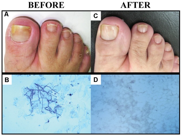
Onychomycosis is a chronic fungal infection of nails, commonly caused by dermatophyte fungi, primarily species of Trichophyton. Because of the limited drug arsenal available to treat general fungal infections and the frequent failure of onychomycosis treatment, the search for new therapeutic sources is essential, and topical treatment with natural products for onychomycosis has been encouraged. Propolis, an adhesive resinous compound produced by honeybees (Apis mellifera), has shown multiple biological properties including significant antifungal and anti-biofilm activities in vitro. In spite of promising in vitro results, in vivo results have not been reported so far. This study assessed an ethanol propolis extract (PE) as a topical therapeutic option for onychomycosis, including its characterization in vitro and its applicability as a treatment for onychomycosis (from bench to clinic). The in vitro evaluation included analysis of the cytotoxicity and the antifungal activity against the planktonic cells and biofilm formed by Trichophyton spp. We also evaluated the capacity of PE to penetrate human nails. Patients with onychomycosis received topical PE treatments, with a 6-month follow-up period. The results of the in vitro assays showed that PE was non-toxic to the cell lines tested, and efficient against both the planktonic cells and the biofilm formed by Trichophyton spp. The results also showed that PE is able to penetrate the human nail. The results for PE applied topically to treat onychomycosis were promising, with complete mycological and clinical cure of onychomycosis in 56.25% of the patients. PE is an inexpensive commercially available option, easy to obtain and monitor. Our results indicated that PE is a promising natural compound for onychomycosis treatment, due to its ability to penetrate the nail without cytotoxicity, and its good antifungal performance against species such as Trichophyton spp. that are resistant to conventional antifungals, both in vitro and in patients.
Keywords: antifungal activity; dermatophytosis; ex vivo nail model; natural products; permeation property; photoacoustic spectroscopy; propolis.
Figures


References
-
- Agüero M. B., Svetaz L., Baroni V., Lima B., Luna L., Zacchino S., et al. (2014). Urban propolis from San Juan province (Argentina): Ethnopharmacological uses and antifungal activity against Candida and dermatophytes. Ind. Crops Prod. 57 166–173. 10.1016/j.indcrop.2014.03.009 - DOI
-
- Aly R., Gupta A. K., Winter T., Zane L. T., Vlahovic T. (2017). Tavaborole in difficult-to-treat onychomycosis cases: a post-hoc assessment of phase III subjects. J. Drugs Dermatol. 16 1016–1021. - PubMed
-
- Ames F. Q., Sato F., de Castro L. V., de Arruda L. L. M., da Rocha B. A., Cuman R. K. N., et al. (2017). Evidence of anti-inflammatory effect and percutaneous penetration of a topically applied fish oil preparation: a photoacoustic spectroscopy study. J. Biomed. Opt. 22:55003. 10.1117/1.JBO.22.5.055003 - DOI - PubMed
LinkOut - more resources
Full Text Sources
Other Literature Sources
Miscellaneous