. 2018 Jun 5:9:1180.
doi: 10.3389/fmicb.2018.01180. eCollection 2018.

Comparative Genomic Analysis of Re-emergent Human Adenovirus Type 55 Pathogens Associated With Adult Severe Community-Acquired Pneumonia Reveals Conserved Genomes and Capsid Proteins

Affiliations

Comparative Genomic Analysis of Re-emergent Human Adenovirus Type 55 Pathogens Associated With Adult Severe Community-Acquired Pneumonia Reveals Conserved Genomes and Capsid Proteins

Zetao Cheng et al. Front Microbiol. .

Abstract

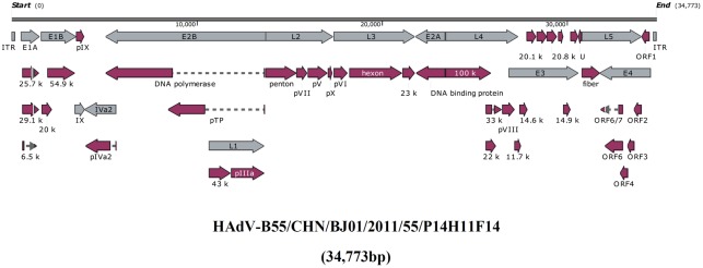
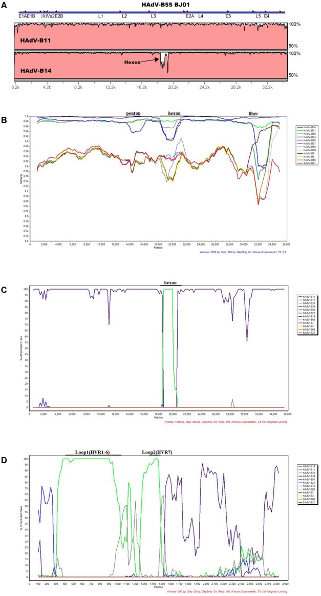
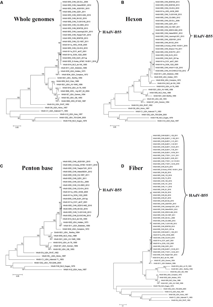
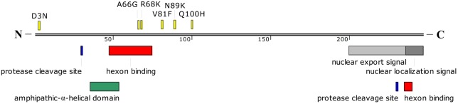
Human adenovirus type 55 (HAdV-B55) is a recently identified acute respiratory disease (ARD) pathogen in HAdV species B with a recombinant genome between renal HAdV-B11 and respiratory HAdV-B14. Since HAdV-B55 first appeared in China school in 2006, no more ARD cases associated with it had been reported until 2011, when there was an outbreak of adult severe community-acquired pneumonia (CAP) in Beijing, China. Reported here is the bioinformatics analysis of the re-emergent HAdV-B55 responsible for this outbreak. Recombination and protein sequence analysis re-confirmed that this isolate (BJ01) was a recombinant virus with the capsid hexon gene from HAdV-B11. The selection pressures for the three capsid proteins, i.e., hexon, penton base, and fiber genes, were all negative, along with very low non-synonymous (dN) and synonymous (dS) substitutions/site (<0.0007). Phylogenetic analyses of the whole genome and the three major capsid genes of HAdV-B55 revealed the close phylogenetic relationship among all HAdV-B55 strains. Comparative genomic analysis of this re-emergent HAdV-B55 strain (BJ01; 2011) with the first HAdV-B55 strain (QS-DLL; 2006) showed the high genome identity (99.87%), including 10 single-nucleotide non-synonymous substitutions, 11 synonymous substitutions, 3 insertions, and one deletion in non-coding regions. The major non-synonymous substitutions (6 of 10) occurred in the protein pVI in its L3 region, which protein has different functions at various stages of an adenovirus infection, and may be associated with the population distribution of HAdV-B55 infection. No non-synonymous substitutions were found in the three major capsid proteins, which proteins are responsible for type-specific neutralizing antibodies. Comparative genomic analysis of the re-emergent HAdV-B55 strains associated with adult severe CAP revealed conserved genome and capsid proteins, providing the foundation for the development of effective vaccines against this pathogen. This study also facilitates the further investigation of HAdV-B55 epidemiology, molecular evolution, patterns of pathogen emergence and re-emergence, and the predication of genome recombination between adenoviruses.

Keywords: China; comparative genomics; human adenovirus type 55; recombination; severe community-acquired pneumonia.


Figures

FIGURE 1
FIGURE 1
Transcriptional map and genome organization of HAdV-B55 strain BJ01. The genome is indicated by two black horizontal lines marked at 10,000 bp intervals. Early, intermediate, and late transcription units are designated by gray arrows, while the red designates coding regions. Arrows reflect the transcriptional orientation of the coding transcripts.
FIGURE 2
FIGURE 2
Genome and recombination analysis of HAdV-B55 Strain BJ01. (A) Global pairwise alignment of HAdV-B55 (BJ01) with HAdV-B11 and HAdV-B14 using mVISTA LAGAN. Transcription units are shown above the graph by black arrows relative to their position and orientation in the HAdV genome. The y-axis represents the sequence similarity and the genome positions are indicated on the x-axis. (B) Simplot and (C) Bootscan analysis of the complete genomes of strain BJ01 compared to other species B adenoviruses. (D) Bootscan analysis of the hexon genes compared with other HAdV-B types. Parameters are as follows: 100 repetitions; Kimura distance model; neighbor-joining tree model; (B) 2,000-bp window, 200-bp step; (C) 1,000-bp window, 200-bp step; (D) 200-bp window, 20-bp step. Genome nucleotide positions are noted along the x-axis, and the sequence similarities are indicated along the y-axis. The landmarks above each graph demonstrate the approximate positions of the major capsid protein genes. (B)/(C)/(D): Main genomes used were marked in the graph. Colors: violet, HAdV-B14; bright green, HAdV-B11; dark gray, HAdV-B35; blue, HAdV-B34; pink, HAdV-B50; blue green, HAdV-B21; gray-violet, HAdV-B16; light gray, HAdV-B68; dull-red, HAdV-B7; yellow, HAdV-B3, brown, HAdV-B66; bright red, SAdV-B21.
FIGURE 3
FIGURE 3
Phylogenetic analysis of HAdV-B55 strain BJ01. The HAdV nucleotide sequences of the whole genome (A), hexon (B), penton base (C), and fiber (D) genes, were analyzed for their phylogenetic relationships. For reference, taxon names include the corresponding GenBank accession number, country of isolation, strain name, year of isolation, and genome type. Bootstrapped, maximum likelihood trees with 1,000 replicates were constructed using the MEGA 5.05 software (see Footnote 5). and by applying default parameters, with a maximum-composite-likelihood method. Bootstrap numbers shown at the nodes indicate the percentages of 1,000 replications producing the clade. The scale bar indicates units of nucleotide substitutions per site. The sequences used for phylogenic analyses are retrieved from GenBank and summarized in Table 1. Sequences from strain BJ01 are noted for reference (formula image).
FIGURE 4
FIGURE 4
Schematic representation of HAdV-B55 protein VI. The amino acid mutations between HAdV-B55 strains BJ01 and QS-DLL are indicated in yellow. Sites for cleavage by the protease, nuclear export signal, and nuclear localization signal are also indicated in different colors. The hexon binding domains are located between amino acid residues 48 to 74 and 235 to 239. A predicted amphipathic-α-helical domain is also shown. The location of these domains was estimated from the previous report by Wiethoff et al. (2005).

References

    1. Alissa Alkhalaf M., Al Qurashi Y. M., Guiver M., Cooper R. J. (2014). Genome sequences of three species d adenoviruses isolated from AIDS patients. Genome Announc. 2:e01267-13. 10.1128/genomeA.01267-13 - DOI - PMC - PubMed
    1. Alkhalaf M. A., Guiver M., Cooper R. J. (2015). Genome stability of adenovirus types 3 and 7 during a simultaneous outbreak in Greater Manchester. UK. J. Med. Virol. 87 117–124. 10.1002/jmv.23969 - DOI - PubMed
    1. Brudno M., Do C. B., Cooper G. M., Kim M. F., Davydov E., Green E. D., et al. (2003). LAGAN and Multi-LAGAN: efficient tools for large-scale multiple alignment of genomic DNA. Genome Res. 13 721–731. 10.1101/gr.926603 - DOI - PMC - PubMed
    1. Cao B., Huang G. H., Pu Z. H., Qu J. X., Yu X. M., Zhu Z., et al. (2014). Emergence of community-acquired adenovirus type 55 as a cause of community-onset pneumonia. Chest 145 79–86. 10.1378/chest.13-1186 - DOI - PMC - PubMed
    1. Chen Y., Liu F., Wang C., Zhao M., Deng L., Zhong J., et al. (2016). Molecular identification and epidemiological features of human adenoviruses associated with acute respiratory infections in hospitalized children in Southern China, 2012-2013. PLoS One 11:e0155412. 10.1371/journal.pone.0155412 - DOI - PMC - PubMed

LinkOut - more resources