. 2018 Jun;10(1):39-44.

Use of 4D Computer Tomographic Angiography to Accurately Identify Distal Internal Carotid Artery Occlusions and Pseudo-Occlusions: Technical Note

Affiliations

Use of 4D Computer Tomographic Angiography to Accurately Identify Distal Internal Carotid Artery Occlusions and Pseudo-Occlusions: Technical Note

Stephan A Munich et al. J Vasc Interv Neurol. 2018 Jun.

Abstract

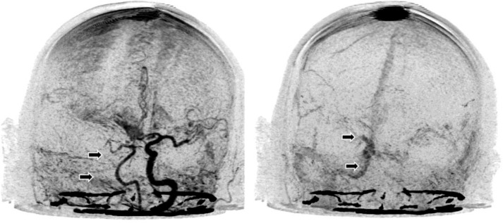
Background and purpose: Traditional methods of computed tomographic angiography (CTA) can be unreliable in detecting carotid artery pseudo-occlusions or in accurately locating the site of carotid artery occlusion. With these methods, lack of adequate distal runoff due to pseudo-occlusion or intracranial occlusion can result in the inaccurate diagnoses of complete occlusion or cervical carotid occlusion, respectively. The site of carotid occlusion has important therapeutic and interventional considerations. We present several cases in which 4D CTA was utilized to accurately and noninvasively diagnose carotid pseudo-occlusion and intracranial internal carotid artery (ICA) occlusion.

Methods: We identified five patients who presented to our institute with ischemic stroke symptoms and evaluated images from traditional CTA protocols and 4D CTA protocols in each of these patients, comparing diagnoses rendered by each imaging technique.

Results: In two patients, traditional CTA suggested the presence of complete ICA occlusion. However, 4D CTA demonstrated pseudo-occlusion. Similarly, in three patients, traditional CTA demonstrated cervical ICA occlusion, whereas the 4D CTA demonstrated intracranial ICA occlusion.

Conclusion: 4D CTA may be a more effective noninvasive imaging technique than traditional CTA to detect intracranial carotid artery occlusions and carotid artery pseudo-occlusions. Accurate, rapid, and noninvasive diagnosis of carotid artery lesions may help tailor and expedite endovascular intervention.

Keywords: carotid terminus occlusion; computed tomographic angiography; imaging; internal carotid artery; pseudo-occlusion.


Figures

Figure 1
Figure 1. (Patient 4 in Table 1). [color (top)] 3D reconstruction of traditional CTA image acquisition demonstrating no filling of the visible segments of the left ICA (from the distal cervical segment to the ICA terminus). [middle (left)] Sagittal reformat of traditional CTA demonstrating no evidence of left ICA filling from the midcervical segment through its entrance into the skull base (arrows). This suggests proximal ICA occlusion. [middle (center)] Lateral view of early arterial phase DSA of the cervical left ICA. Filling of the distal ICA (arrows) is not seen in this early arterial phase due to blood flow stagnation caused by an ICA terminus occlusion. [middle (right)] Lateral view of late venous phase DSA of the cervical left ICA. Weak filling is seen in the distal cervical ICA (arrows) and as the ICA enters the skull base. This demonstrates that the site of occlusion is not in the proximal ICA as suggested by the CTA, but rather in the intracranial ICA. [bottom (left)] Anteroposterior (AP) view of early arterial phase 4D CTA. This image roughly corresponds to the time of image acquisition of traditional CTA. Consequently, this image resembles that of the 3D CTA reconstruction (top, color). No filling of the left ICA is noted in the distal cervical and intracranial segments (arrows). [Bottom (center)] AP views of early venous phase 4D CTA. Again, the distal cervical and intracranial ICA is not seen (arrows). [Bottom (right)] AP views of late venous phase 4D CTA. the left Ica is seen filling anterogradely in delayed fashion (arrow). However, there is no filling at the ICA terminus, suggesting that this is the site of occlusion. the severely delayed filling is likely due to the lack of distal runoff, which creates a stagnant column of blood. This was confirmed using DSA (D, image on the right in the previous composite).
Figure 2
Figure 2. (Patient 5 in Table 1). [top (color)] 3D reconstruction of traditional CTA image demonstrating no filling of the right ICA or its branch vessels. [middle (left)] Sagittal reformat of traditional CTA image demonstrating no filling of the distal right ICA (arrows). Faint contrast filling is noted proximally, which appears to stop at the midcervical ICA. [middle (center)] Lateral view of midarterial phase Dsa of the right cervical Ica. the "pseudo-dissection phenomenon" described by Siddiq et al. [2] can be seen as decreased caliber of both the proximal and distal Ica. [D (right)] Lateral view of late arterial phase DSA demonstrating a right carotid terminus occlusion (arrow). There is no blood flow distal to the occlusion, representing complete occlusion (TICI 0). [bottom (left)] Anteroposterior (Ap) view of peak arterial phase 4D CTA. This roughly corresponds to the time of image acquisition of traditional CTA (a, color). There is no filling of the right ICA (arrows). [Bottom (right)] AP views of very late venous phase 4D CTA. Contrast material can be seen within the petrous, cavernous, and supraclinoid right ICA (arrows). No contrast is seen distal to the carotid terminus, which was confirmed with DSA to be the side of occlusion (middle, image on the right in the previous composite).

Similar articles

References

    1. Kniemeyer HW, et al. Pseudo- and segmental occlusion of the internal carotid artery: a new classification, surgical treatment and results. Eur J Vasc Endovasc Surg. 1996;12:310–320. Available from: https://secureacademic.oup.com/licensing/index.html?doi=10.1093%252fons%.... - PubMed
    1. Siddiq F, et al. Occurrence and prognostic significance of cervical pseudodissection phenomenon associated with acute intracranial internal carotid artery occlusion. J Neuroimaging. 2013;23:384–390. Available from: http://www.ncbi.nlm.nih.gov/pubmed/23228049. - PubMed
    1. Orrison WW, Jr, et al. Whole-brain dynamic CT angiography and perfusion imaging. Clin Radiol. 2011;66:566–574. Available from: http://www.ncbi.nlm.nih.gov/pubmed/21371698. - PubMed
    1. Berkhemer OA, et al. A randomized trial of intraarterial treatment for acute ischemic stroke. N Engl J Med. 2015;372:11–20. Available from: http://www.ncbi.nlm.nih.gov/pubmed/25517348. - PubMed
    1. Furst G, et al. Reliability and validity of noninvasive imaging of internal carotid artery pseudo-occlusion. Stroke. 1999;30:1444–1449. Available from: http://www.ncbi.nlm.nih.gov/pubmed/10390321. - PubMed

LinkOut - more resources