

The effects of high glucose condition on rat tenocytes in vitro and rat Achilles tendon in vivo
- PMID: 29922457
- PMCID: PMC5987694
- DOI: 10.1302/2046-3758.75.BJR-2017-0126.R2
The effects of high glucose condition on rat tenocytes in vitro and rat Achilles tendon in vivo
Abstract
Objectives: The aim of this study was to investigate the effect of hyperglycaemia on oxidative stress markers and inflammatory and matrix gene expression within tendons of normal and diabetic rats and to give insights into the processes involved in tendinopathy.
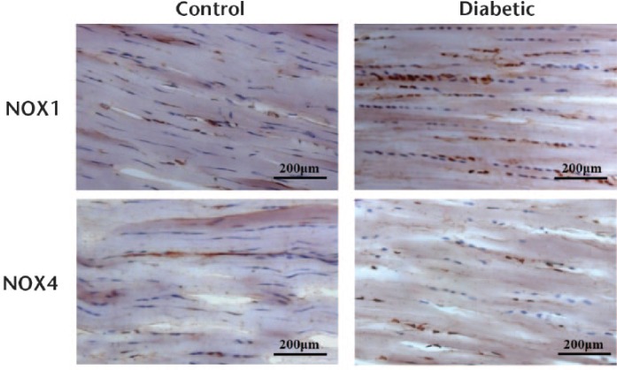
Methods: Using tenocytes from normal Sprague-Dawley rats, cultured both in control and high glucose conditions, reactive oxygen species (ROS) production, cell proliferation, messenger RNA (mRNA) expression of NADPH oxidase (NOX) 1 and 4, interleukin-6 (IL-6), matrix metalloproteinase (MMP)-2, tissue inhibitors of matrix metalloproteinase (TIMP)-1 and -2 and type I and III collagens were determined after 48 and 72 hours in vitro. In an in vivo study, using diabetic rats and controls, NOX1 and 4 expressions in Achilles tendon were also determined.
Results: In tenocyte cultures grown under high glucose conditions, gene expressions of NOX1, MMP-2, TIMP-1 and -2 after 48 and 72 hours, NOX4 after 48 hours and IL-6, type III collagen and TIMP-2 after 72 hours were significantly higher than those in control cultures grown under control glucose conditions. Type I collagen expression was significantly lower after 72 hours. ROS accumulation was significantly higher after 48 hours, and cell proliferation after 48 and 72 hours was significantly lower in high glucose than in control glucose conditions. In the diabetic rat model, NOX1 expression within the Achilles tendon was also significantly increased.
Conclusion: This study suggests that high glucose conditions upregulate the expression of mRNA for NOX1 and IL-6 and the production of ROS. Moreover, high glucose conditions induce an abnormal tendon matrix expression pattern of type I collagen and a decrease in the proliferation of rat tenocytes.Cite this article: Y. Ueda, A. Inui, Y. Mifune, R. Sakata, T. Muto, Y. Harada, F. Takase, T. Kataoka, T. Kokubu, R. Kuroda. The effects of high glucose condition on rat tenocytes in vitro and rat Achilles tendon in vivo. Bone Joint Res 2018;7:362-372. DOI: 10.1302/2046-3758.75.BJR-2017-0126.R2.
Keywords: High glucose; Oxidative stress; Tendinitis.
Figures


Similar articles
-
In vitro and in vivo tenocyte-protective effectiveness of dehydroepiandrosterone against high glucose-induced oxidative stress.BMC Musculoskelet Disord. 2021 Jun 5;22(1):519. doi: 10.1186/s12891-021-04398-z. BMC Musculoskelet Disord. 2021. PMID: 34090401 Free PMC article.
-
Evaluation of apocynin in vitro on high glucose-induced oxidative stress on tenocytes.Bone Joint Res. 2020 May 16;9(1):23-28. doi: 10.1302/2046-3758.991.BJR-2019-0074.R1. eCollection 2020 Jan. Bone Joint Res. 2020. PMID: 32435452 Free PMC article.
-
Quercetin treatment protects the Achilles tendons of rats from oxidative stress induced by hyperglycemia.BMC Musculoskelet Disord. 2022 Jun 10;23(1):563. doi: 10.1186/s12891-022-05513-4. BMC Musculoskelet Disord. 2022. PMID: 35689230 Free PMC article.
-
Exploring the In Vivo Anti-Inflammatory Actions of Simvastatin-Loaded Porous Microspheres on Inflamed Tenocytes in a Collagenase-Induced Animal Model of Achilles Tendinitis.Int J Mol Sci. 2018 Mar 12;19(3):820. doi: 10.3390/ijms19030820. Int J Mol Sci. 2018. PMID: 29534523 Free PMC article.
-
Light stimulation on tenocytes: A systematic review of in vitro studies.Porto Biomed J. 2022 Sep 9;7(4):e176. doi: 10.1097/j.pbj.0000000000000176. eCollection 2022 Jul-Aug. Porto Biomed J. 2022. PMID: 36186115 Free PMC article. Review.
Cited by
-
Evaluating the role of type 2 diabetes mellitus in rotator cuff tendinopathy: Development and analysis of a novel rat model.Front Endocrinol (Lausanne). 2022 Oct 10;13:1042878. doi: 10.3389/fendo.2022.1042878. eCollection 2022. Front Endocrinol (Lausanne). 2022. PMID: 36299460 Free PMC article.
-
Long-duration type 1 diabetes is associated with deficient cortical bone mechanical behavior and altered matrix composition in human femoral bone.J Bone Miner Res. 2024 Dec 31;40(1):87-99. doi: 10.1093/jbmr/zjae184. J Bone Miner Res. 2024. PMID: 39561104
-
Growth factor and macromolecular crowding supplementation in human tenocyte culture.Biomater Biosyst. 2021 Jan 30;1:100009. doi: 10.1016/j.bbiosy.2021.100009. eCollection 2021 Mar. Biomater Biosyst. 2021. PMID: 36825160 Free PMC article.
-
Roles of Oxidative Stress in Acute Tendon Injury and Degenerative Tendinopathy-A Target for Intervention.Int J Mol Sci. 2022 Mar 25;23(7):3571. doi: 10.3390/ijms23073571. Int J Mol Sci. 2022. PMID: 35408931 Free PMC article. Review.
-
Effects of adipose-derived mesenchymal stem cell conditioned medium on human tenocytes exposed to high glucose.Ther Adv Musculoskelet Dis. 2024 Jan 8;16:1759720X231214903. doi: 10.1177/1759720X231214903. eCollection 2024. Ther Adv Musculoskelet Dis. 2024. PMID: 38204801 Free PMC article.
References
-
- Yosipovitch G, Yosipovitch Z, Karp M, Mukamel M. Trigger finger in young patients with insulin dependent diabetes. J Rheumatol 1990;17:951-952. - PubMed
-
- Noble J, Heathcote JG, Cohen H. Diabetes mellitus in the aetiology of Dupuytren’s disease. J Bone Joint Surg [Br] 1984;66-B:322-325. - PubMed
-
- Pourmemari MH, Shiri R. Diabetes as a risk factor for carpal tunnel syndrome: a systematic review and meta-analysis. Diabet Med 2016;33:10-16. - PubMed
-
- Balci N, Balci MK, Tüzüner S. Shoulder adhesive capsulitis and shoulder range of motion in type II diabetes mellitus: association with diabetic complications. J Diabetes Complications 1999;13:135-140. - PubMed
LinkOut - more resources
Full Text Sources
Other Literature Sources
Research Materials
Miscellaneous
