Review
. 2018 Jun 5:8:166.
doi: 10.3389/fcimb.2018.00166. eCollection 2018.

Recent Advances in Replication and Infection of Human Parvovirus B19

Affiliations
Review

Recent Advances in Replication and Infection of Human Parvovirus B19

Safder S Ganaie et al. Front Cell Infect Microbiol. .

Abstract

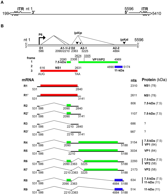
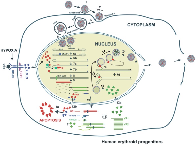
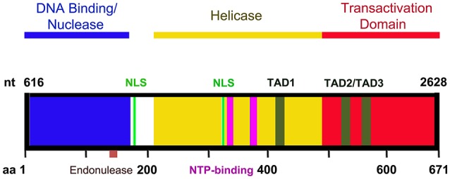
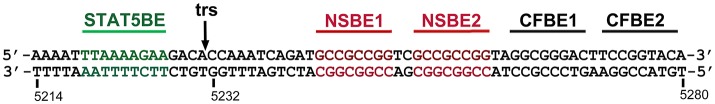
Parvovirus B19 (B19V) is pathogenic to humans and causes bone marrow failure diseases and various other inflammatory disorders. B19V infection exhibits high tropism for human erythroid progenitor cells (EPCs) in the bone marrow and fetal liver. The exclusive restriction of B19V replication to erythroid lineage cells is partly due to the expression of receptor and co-receptor(s) on the cell surface of human EPCs and partly depends on the intracellular factors essential for virus replication. We first summarize the latest developments in the viral entry process and the host cellular factors or pathways critical for B19V replication. We discuss the role of hypoxia, erythropoietin signaling and STAT5 activation in the virus replication. The B19V infection-induced DNA damage response (DDR) and cell cycle arrest at late S-phase are two key events that promote B19V replication. Lately, the virus infection causes G2 arrest, followed by the extensive cell death of EPCs that leads to anemia. We provide the current understanding of how B19V exploits the cellular resources and manipulate pathways for efficient virus replication. B19V encodes a single precursor mRNA (pre-mRNA), which undergoes alternate splicing and alternative polyadenylation to generate at least 12 different species of mRNA transcripts. The post-transcriptional processing of B19V pre-mRNA is tightly regulated through cis-acting elements and trans-acting factors flanking the splice donor or acceptor sites. Overall, in this review, we focus on the recent advances in the molecular virology and pathogenesis of B19V infection.

Keywords: DNA replication; RNA processing; erythroid precursor cells; human; infection; parvovirus B19.


Figures

Figure 1
Figure 1
Transcription map of Parvovirus B19. (A) Linear ssDNA genome of B19V. The genome is flanked by two inverted terminal repeats (ITRs), containing unpaired and mismatched bases, shown as bulges and bubbles, respectively. (B) Double stranded replicative form of B19V genome. The viral promoter denoted as P6 transcribes a single precursor mRNA (pre-mRNA). Pre-mRNA has two donor sites (D1 and D2) and four acceptor sites (A1-1, A1-2, A2-1, and A2-2). Using alternative splicing and polyadenylation, pre-mRNA is processed into at least 12 different mRNAs (only R1–R9 shown here). Mature mRNAs polyadenylate at (pA)p or (pA)d sites. At least five different proteins are known to be encoded by different species of mRNA transcripts. Different colors indicate the use of different open reading frames for the translation of proteins. Question marks indicate mRNAs encoding unknown proteins.
Figure 2
Figure 2
Proposed model of B19V life cycle. B19V infects human erythroid progenitor cells. The virus first interacts with globoside (Step 1) and undergoes a conformational change that exposes VP1u which subsequently binds an unknown co-receptor (Step 1). Thereupon, the virus is endocytosed and somehow escapes the lysosomal route and enters the nucleus (Step 3). Inside the nucleus, the virion uncoats and releases the ssDNA genome (Step 4). Using the 3′OH of the left ITR, the second strand is synthesized to form a functional origin of replication (Step 5). Next, EPO and hypoxia activates and increases pSTAT5, which interacts with MCM and then binds Ori region. NS1 binding to NS1BE is critical for nicking ssDNA at trs and for helicase activity (step 6). The nicking creates a new 3′ OH end to continue DNA replication that results into duplex replicative intermediate (Step 7). The dsDNA form also transcribes a single pre-mRNA that is processed into various mRNAs which are exported to cytoplasm for translation (Step 8). VP1/2 assemble into trimers to form capsids, which are transported back to the nucleus (Step 9). Through strand displacement, ssDNA is packaged into capsids, which probably requires NS1 (step 10). NS1 and 11-kDa in the cytoplasm induce apoptosis (Step 11). After multiplication, the virions are released though cell lysis.
Figure 3
Figure 3
A diagram of NS1 functional domains. The N-terminus (amino acid 2-176) of NS1 possesses DNA binding and endonuclease activity. The endonuclease motif resides between amino acids 137 and 145. The central region of NS1 exhibits putative helicase activity. Transactivation activity is restricted to the C-terminus of NS1. NS1 carries two nuclear localizing signals, between amino acids 177–179 and 316–321 (NLS, in green). Three putative transactivation domains have been identified in the C-terminus of the NS1 protein: TAD1 (aa 416–424), TAD2 (aa 523–531), and TAD3 (aa 566–574). The central region also contains two NTP binding motifs between amino acids 323–378 and 367–378 (NTP binding motifs, in purple).
Figure 4
Figure 4
A diagram of the B19V minimal origin of viral DNA replication (Ori). B19V has a 67-bp long minimum origin of DNA replication (Ori) at each end of the genome. Ori harbors two NS1 binding elements (NSBE1&2, in red), one STAT5 binding element (STAT5BE, in green), a terminal resolution site (trs, black), and two potential cellular factor binding elements (CFBE1&2). Question marks denote two unidentified host factors binding Ori.
Figure 5
Figure 5
NS1 is a multi-functional protein. The B19V NS1 multimer binds the dsDNA form of the genome at NSBE1-2 via N-terminus region (5–7), but nicks ssDNA at trs and covalently attaches to the 5′ end. NS1 induces a DNA damage response that is essential for virus replication. The virus replication process leads to the activation of ATR, ATM and DNA-PKcs. However, the activation of ATR and DNA-PKcs, but not ATM, is essential for virus replication. NS1 transactivates its P6 promoter with the assistance of Sp1/Sp3. NS1 is a global transactivator and regulates ~1,700 genes. NS1 induces apoptosis through the activation of caspases 2/6/8 and TNF-α. The central region of NS1 protein exhibits putative helicase activity.

References

    1. Adamson-Small L. A., Ignatovich I. V., Laemmerhirt M. G., Hobbs J. A. (2014). Persistent parvovirus B19 infection in non-erythroid tissues: possible role in the inflammatory and disease process. Virus Res. 190, 8–16. 10.1016/j.virusres.2014.06.017 - DOI - PubMed
    1. Anderson L. J., Tsou C., Parker R. A., Chorba T. L., Wulff H., Tattersall P., et al. (1986). Detection of antibodies and antigens of human parvovirus B19 by enzyme-linked immunosorbent assay. J. Clin. Microbiol. 24, 522–526. - PMC - PubMed
    1. Anderson S., Momoeda M., Kawase M., Kajigaya S., Young N. S. (1995). Peptides derived from the unique region of B19 parvovirus minor capsid protein elicit neutralizing antibodies in rabbits. Virology 206, 626–632. 10.1016/S0042-6822(95)80079-4 - DOI - PubMed
    1. Beard C., St Amand J., Astell C. R. (1989). Transient expression of B19 parvovirus gene products in COS-7 cells transfected with B19-SV40 hybrid vectors. Virology 172, 659–664. 10.1016/0042-6822(89)90211-0 - DOI - PubMed
    1. Blundell M. C., Astell C. R. (1989). A GC-box motif upstream of the B19 parvovirus unique promoter is important for in vitro transcription. J. Virol. 63, 4814–4823. - PMC - PubMed

Publication types

MeSH terms