

Molecular Evidence of Human Fasciolosis Due to Fasciola gigantica in Iran: A Case Report
- PMID: 29922619
- PMCID: PMC6005974
Molecular Evidence of Human Fasciolosis Due to Fasciola gigantica in Iran: A Case Report
Abstract
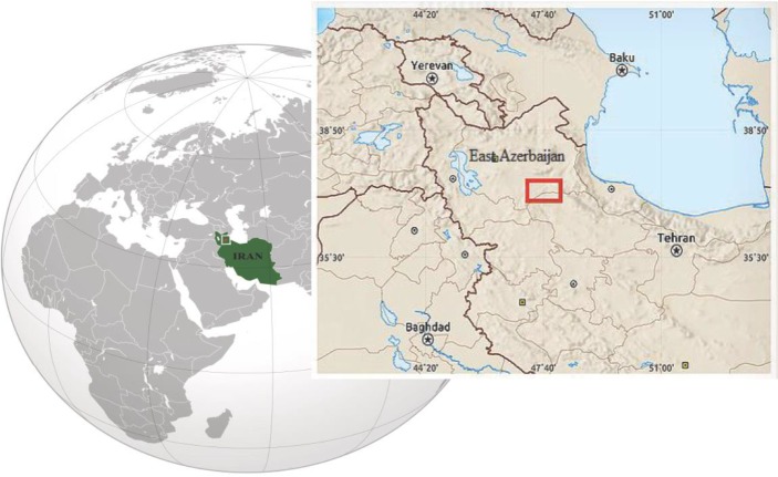
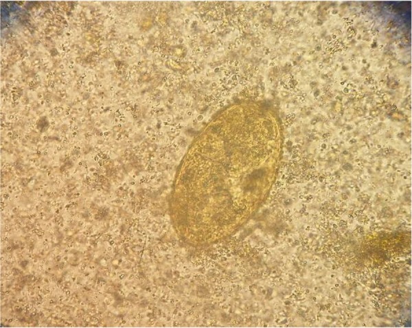
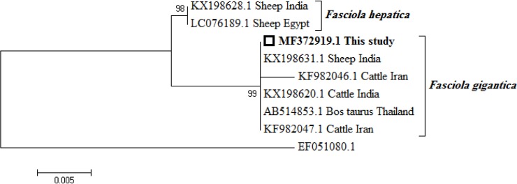
Fascioliasis is a foodborne zoonotic disease caused by the two parasite species Fasciola hepatica and F. gigantica. In spite of the presence of both species of Fasciola in the livestock, to our knowledge, to date, no cases of human F. gigantica infection have been reported in Iran officially. Here, we report such a case in a 25 yr old woman referred to The Department of Medical Parasitology and Mycology, School of Public Health, Tehran University of Medical Sciences, Tehran, Iran in 2016. CT imaging and MRCP revealed an ill-defined lesion of segments of liver. Specific ELISA produced a positive result besides detecting egg of the parasite via stool exam. The identification of parasite species was performed by the DNA extracted from the eggs and sequencing ITS-1, in addition to comparison to GenBank retrieved sequences, using the BLAST search tool. The sample showed 100% identity with F. gigantica. She was treated for fasciolosis with a single dose of Egaten® 10 mg/kg with positive response. This is the first case of human fasciolosis due to F. gigantica reported in Iran.
Keywords: Diagnosis; ELISA; Fasciola gigantica; Iran; PCR.
Conflict of interest statement
Conflict of interest We have no conflict of interest related to this work.
Figures
Similar articles
-
Morphological and molecular characterization of Fasciola isolates from livestock in Golestan province, northern Iran.Vet Med Sci. 2023 Jul;9(4):1824-1832. doi: 10.1002/vms3.1189. Epub 2023 Jun 15. Vet Med Sci. 2023. PMID: 37317979 Free PMC article.
-
Distribution of Fasciola hepatica and F. gigantica in the endemic area of Guilan, Iran: Relationships between zonal overlap and phenotypic traits.Infect Genet Evol. 2015 Apr;31:95-109. doi: 10.1016/j.meegid.2015.01.009. Epub 2015 Jan 17. Infect Genet Evol. 2015. PMID: 25602718
-
Which species is in the faeces at a time of global livestock movements: single nucleotide polymorphism genotyping assays for the differentiation of Fasciola spp.Int J Parasitol. 2020 Feb;50(2):91-101. doi: 10.1016/j.ijpara.2019.12.002. Epub 2020 Feb 22. Int J Parasitol. 2020. PMID: 32006549
-
Differential expression of microRNAs and tRNA fragments mediate the adaptation of the liver fluke Fasciola gigantica to its intermediate snail and definitive mammalian hosts.Int J Parasitol. 2021 Apr;51(5):405-414. doi: 10.1016/j.ijpara.2020.10.009. Epub 2021 Jan 26. Int J Parasitol. 2021. PMID: 33513403 Review.
-
Chapter 2. Fasciola, lymnaeids and human fascioliasis, with a global overview on disease transmission, epidemiology, evolutionary genetics, molecular epidemiology and control.Adv Parasitol. 2009;69:41-146. doi: 10.1016/S0065-308X(09)69002-3. Adv Parasitol. 2009. PMID: 19622408 Review.
Cited by
-
High-Resolution Melting Analysis as an Appropriate Method to Differentiate between Fasciola hepatica and F. gigantica.Iran J Public Health. 2019 Mar;48(3):501-507. Iran J Public Health. 2019. PMID: 31223578 Free PMC article.
-
Assessment of genetic markers for multilocus sequence typing (MLST) of Fasciola isolates from Iran.Vet Med Sci. 2023 Mar;9(2):924-933. doi: 10.1002/vms3.995. Epub 2022 Nov 7. Vet Med Sci. 2023. PMID: 36343016 Free PMC article.
-
Serological Study of Fascioliasis Using Indirect ELISA in Gorgan City, Golestan Province, Northern Iran.Iran J Parasitol. 2020 Jul-Sep;15(3):418-424. doi: 10.18502/ijpa.v15i3.4207. Iran J Parasitol. 2020. PMID: 33082807 Free PMC article.
-
Molecular phylogenetic and genetic variability of Fasciola gigantica in Kermanshah province, western Iran with an overview to understand haplotypes distribution in Asia and Africa.Vet Res Forum. 2020 Summer;11(3):265-271. doi: 10.30466/vrf.2019.98547.2350. Epub 2020 Sep 15. Vet Res Forum. 2020. PMID: 33133464 Free PMC article.
-
Evaluation of Semi-Nested PCR Compared with Indirect-ELISA to Diagnose Human Fasciolosis.Iran J Public Health. 2022 Mar;51(3):686-694. doi: 10.18502/ijph.v51i3.8947. Iran J Public Health. 2022. PMID: 35865068 Free PMC article.
References
-
- Rokni MB. Helminth-Trematode: Fasciola hepatica and F. gigantica. In: Motarjemi Y, editor. Encyclopedia of Food Safety. 1st ed Amsterdam: Elsevier/Academic Press; 2015. p. 140–5.
-
- Mas-Coma S, Valero MA, Bargues MD. (2009). Fasciola, lymnaeids and human fascioliasis, with a global overview on disease transmission, epidemiology, evolutionary genetics, molecular epidemiology and control. Adv Parasitol, 69:41–146. - PubMed
LinkOut - more resources
Full Text Sources
Research Materials