

Advances in the computational and molecular understanding of the prostate cancer cell nucleus
- PMID: 29923622
- PMCID: PMC6150831
- DOI: 10.1002/jcb.27156
Advances in the computational and molecular understanding of the prostate cancer cell nucleus
Abstract
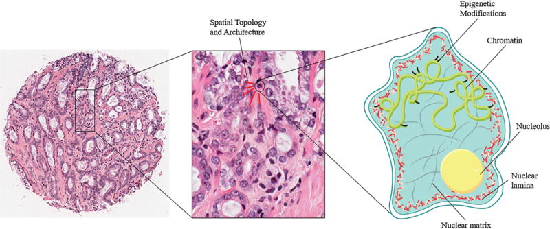
Nuclear alterations are a hallmark of many types of cancers, including prostate cancer (PCa). Recent evidence shows that subvisual changes, ones that may not be visually perceptible to a pathologist, to the nucleus and its ultrastructural components can precede visual histopathological recognition of cancer. Alterations to nuclear features, such as nuclear size and shape, texture, and spatial architecture, reflect the complex molecular-level changes that occur during oncogenesis. Quantitative nuclear morphometry, a field that uses computational approaches to identify and quantify malignancy-induced nuclear changes, can enable a detailed and objective analysis of the PCa cell nucleus. Recent advances in machine learning-based approaches can now automatically mine data related to these changes to aid in the diagnosis, decision making, and prediction of PCa prognoses. In this review, we use PCa as a case study to connect the molecular-level mechanisms that underlie these nuclear changes to the machine learning computational approaches, bridging the gap between the clinical and computational understanding of PCa. First, we will discuss recent developments to our understanding of the molecular events that drive nuclear alterations in the context of PCa: the role of the nuclear matrix and lamina in size and shape changes, the role of 3-dimensional chromatin organization and epigenetic modifications in textural changes, and the role of the tumor microenvironment in altering nuclear spatial topology. We will then discuss the advances in the applications of machine learning algorithms to automatically segment nuclei in prostate histopathological images, extract nuclear features to aid in diagnostic decision making, and predict potential outcomes, such as biochemical recurrence and survival. Finally, we will discuss the challenges and opportunities associated with translation of the quantitative nuclear morphometry methodology into the clinical space. Ultimately, accurate identification and quantification of nuclear alterations can contribute to the field of nucleomics and has applications for computationally driven precision oncologic patient care.
Keywords: machine learning in medicine; molecular-level nuclear changes; nuclear architecture; prostate cancer; quantitative nuclear morphometry.
© 2018 Wiley Periodicals, Inc.
Conflict of interest statement
The authors declare no conflicts of interest with respect to this work.
Figures

References
-
- Ali S, Veltri R, Epstein JA, Christudass C, Madabhushi A. Medical Imaging 2013: Digital Pathology. Vol. 8676. International Society for Optics and Photonics; 2013. Mar, Cell cluster graph for prediction of biochemical recurrence in prostate cancer patients from tissue microarrays; p. 86760H.
-
- Ali S, Veltri R, Epstein JI, Christudass C, Madabhushi A. International Conference on Medical Image Computing and Computer-Assisted Intervention. Springer; Berlin, Heidelberg: 2011. Sep, Adaptive energy selective active contour with shape priors for nuclear segmentation and gleason grading of prostate cancer; pp. 661–669. - PubMed
Publication types
MeSH terms
Substances
Grants and funding
LinkOut - more resources
Full Text Sources
Other Literature Sources
Medical
Miscellaneous
