. 2018 Jul 5;140(26):8096-8099.
doi: 10.1021/jacs.8b04833. Epub 2018 Jun 25.

Monitoring 15N Chemical Shifts During Protein Folding by Pressure-Jump NMR

Affiliations

Monitoring 15N Chemical Shifts During Protein Folding by Pressure-Jump NMR

Cyril Charlier et al. J Am Chem Soc. .

Abstract

Pressure-jump hardware permits direct observation of protein NMR spectra during a cyclically repeated protein folding process. For a two-state folding protein, the change in resonance frequency will occur nearly instantaneously when the protein clears the transition state barrier, resulting in a monoexponential change of the ensemble-averaged chemical shift. However, protein folding pathways can be more complex and contain metastable intermediates. With a pseudo-3D NMR experiment that utilizes stroboscopic observation, we measure the ensemble-averaged chemical shifts, including those of exchange-broadened intermediates, during the folding process. Such measurements for a pressure-sensitized mutant of ubiquitin show an on-pathway kinetic intermediate whose 15N chemical shifts differ most from the natively folded protein for strands β5, its preceding turn, and the two strands that pair with β5 in the native structure.


Figures

Figure 1.
Figure 1.
Pressure-jump pseudo-3D NMR pulse scheme used for stroboscopic measurement of 15N chemical shifts during protein folding. For details, see Figure S2.
Figure 2.
Figure 2.
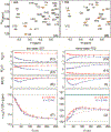
Measurement of 15N chemical shifts during VA2-ubiquitin folding. (A, B) Small region of the HSQC spectrum, recorded with the scheme of Figure 1 for τ = 10 ms and κ = 2 ms, modulated by (A) cos(ωNκ) and (B) sin(ωNκ). Extension of the 50-ms t1 time domain to 75 ms by the NUS reconstruction program SMILE was used to slightly increase both resolution and sensitivity. Negative intensity is shown in brown. For full spectra, see Figure S3. (C) Mx(τ) and (D) My(τ) magnetization of residue S57 after evolution for κ = 0.5 ms (red) and κ = 2 ms (blue), and (E) the corresponding magnitude |M(τ)|, with these values normalized to |M(τ)| = 1 at τ = 248 ms. Solid colored lines correspond to the values predicted for a two-state folding model with ρ = 14.3 s−1 and known chemical shifts of the unfolded (F, dashed line) and folded (F, dotted) states at 1 bar. (F) Apparent average chemical shift <ωN/2π> at time τ after the pressure drop. (G-J) Analogous to (C-F) but for residue R72 with solid lines corresponding to a three-state model with kui = 13.2 s−1; kuf = 6.5 s−1; and kif = 12.0 s−1, and colored dashed lines for the two-state model. The band at 126.6 ppm in (J) depicts the fitted 15N shift of the intermediate state. The full set of residues is shown in Figure S4. The error bars in (C-E) and (G-I) indicate the RMS noise in the Mx and My spectra, scaled by the normalization factor of |M|. Error bars in (F) and (J) indicate the uncertainty in the average chemical shift derived from the RMS noise in the Mx and My spectra
Figure 3.
Figure 3.
15N chemical shift differences, |Δδ(15N)| = |δI – δF|, between the meta-stable intermediate and natively folded VA2-ubiquitin. (A) Color coded on a backbone ribbon structure. Residues for which Δδ(15N) could not be determined are shown in grey. (B) Δδ(15N) as a function of residue number, with negative values depicted as open bars.

Similar articles

Cited by

References

    1. Hoeltzli SD; Frieden C, Stopped-flow NMR spectroscopy - Real-time unfolding studies of 6-F19-tryptophan-labeled E. Coli dihydrofolate reductase. Proc. Natl. Acad. Sci. U. S. A 1995, 92, 9318–9322. - PMC - PubMed
    1. Koide S; Dyson HJ; Wright PE, Characterization of a folding intermediate of apoplastocyanin trapped by proline isomerization. Biochemistry 1993, 32, 12299–12310. - PubMed
    1. Balbach J; Forge V; Vannuland NAJ; Winder SL; Hore PJ; Dobson CM, Following protein folding in real-time using NMR spectroscopy. Nat. Struct. Biol 1995, 2, 865–870. - PubMed
    1. Roche J; Dellarole M; Caro JA; Norberto DR; Garcia AE; Garcia-Moreno B; Roumestand C; Royer CA, Effect of Internal Cavities on Folding Rates and Routes Revealed by Real-Time Pressure-Jump NMR Spectroscopy. J. Am. Chem. Soc 2013, 135, 14610–14618. - PubMed
    1. Schlepckow K; Wirmer J; Bachmann A; Kiefhaber T; Schwalbe H, Conserved folding pathways of alpha-lactalbumin and lysozyme revealed by kinetic CD, fluorescence, NMR, and interrupted refolding experiments. J. Mol. Biol 2008, 378, 686–698. - PubMed

Publication types