. 2018 Nov 1;315(5):F1370-F1384.
doi: 10.1152/ajprenal.00566.2017. Epub 2018 Jun 20.

Novel hemodynamic structures in the human glomerulus

Affiliations

Novel hemodynamic structures in the human glomerulus

Christopher R Neal et al. Am J Physiol Renal Physiol. .

Abstract

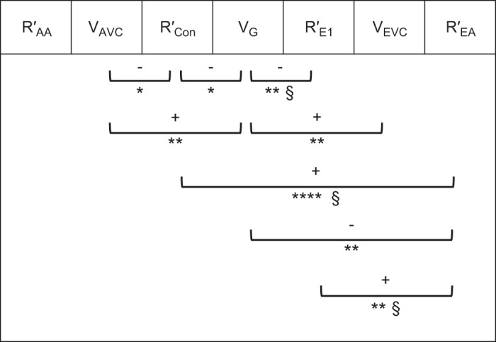
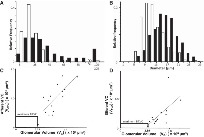
To investigate human glomerular structure under conditions of physiological perfusion, we have analyzed fresh and perfusion-fixed normal human glomeruli at physiological hydrostatic and oncotic pressures using serial resin section reconstruction, confocal, multiphoton, and electron microscope imaging. Afferent and efferent arterioles (21.5 ± 1.2 µm and 15.9 ± 1.2 µm diameter), recognized from vascular origins, lead into previously undescribed wider regions (43.2 ± 2.8 µm and 38.4 ± 4.9 µm diameter) we have termed vascular chambers (VCs) embedded in the mesangium of the vascular pole. Afferent VC (AVC) volume was 1.6-fold greater than efferent VC (EVC) volume. From the AVC, long nonbranching high-capacity conduit vessels ( n = 7) (Con; 15.9 ± 0.7 µm diameter) led to the glomerular edge, where branching was more frequent. Conduit vessels have fewer podocytes than filtration capillaries. VCs were confirmed in fixed and unfixed specimens with a layer of banded collagen identified in AVC walls by multiphoton and electron microscopy. Thirteen highly branched efferent first-order vessels (E1; 9.9 ± 0.4 µm diameter) converge on the EVC, draining into the efferent arteriole (15.9 ± 1.2 µm diameter). Banded collagen was scarce around EVCs. This previously undescribed branching topology does not conform to the branching of minimum energy expenditure (Murray's law), suggesting that even distribution of pressure/flow to the filtration capillaries is more important than maintaining the minimum work required for blood flow. We propose that AVCs act as plenum manifolds possibly aided by vortical flow in distributing and balancing blood flow/pressure to conduit vessels supplying glomerular lobules. These major adaptations to glomerular capillary structure could regulate hemodynamic pressure and flow in human glomerular capillaries.

Keywords: conduit vessels; glomerular microcirculation; hemodynamics; mesangial collagen; vascular chambers.


Figures

Fig. 1.
Fig. 1.
Afferent arteriole and glomerulus connectivity. Selected light micrographs from a 1 µm serial section stack to show the connectivity of an afferent arteriole (25 µm diameter) with a small artery (110 µm diameter interlobular or feed artery). Identifying the root/route of the vessels entering the glomerulus allows identification of afferent and efferent arterioles. Note that the afferent arteriole goes through a right angle as it enters the glomerulus. AA, afferent arteriole; GC, glomerular capillary. Serial section number at bottom right.
Fig. 2.
Fig. 2.
Vascular resistance and capacity relationships. Significant correlationships (8 out of 21) between 7 variables measured in human glomerular initial vasculature. Correlates of vascular resistance for afferent arterioles (R′AA), conduit vessels (R′Con), 1st-order efferent vessels (R′E1), and efferent arterioles (R′EA) were compared with each other and with afferent vascular chamber (AVC) volume (VAVC), glomerular volume (VG), and efferent vascular chamber (EVC) volume (VEVC). +, Positive correlation; –, negative correlation. *P < 0.05; **P ≤ 0.01; ****P ≤ 0.0001; §higher significance with outlier removed.
Fig. 3.
Fig. 3.
A and B: serial resin sections through a glomerulus. Selected light micrographs from 2 complete 1 µm serial section series to show the route that blood takes from an afferent arteriole (AA) into an afferent vascular chamber (AVC) leading into conduit vessels (Con) of high capacity and few branches. At the other end of the microcirculation, many branching efferent 1st-order vessels (E1) drain into a smaller efferent vascular chamber (EVC) leading to an efferent arteriole (EA). Serial section numbers at bottom left. Scale bar, 100 µm in micrograph of section 254 or 198 (see Supplemental Videos S2A and S2B for glomerular image stacks of A and B, respectively, and Supplemental Videos S2C and S2D for a reconstruction of afferent and efferent parts of B).
Fig. 4.
Fig. 4.
Scale diagram of glomerular vasculature; the smallest vascular chambers (VCs). A: scale diagram of the afferent (light gray) and efferent (white) ends of the glomerular vasculature. Diagram shows size and branch relationships between arterioles, VCs, and 1st-order vessels (mesangium close to vascular pole, dark gray) (diameters from Tables 1 and 2). To illustrate VC volume in relation to attached vessels, the length of attached vessels accommodating VC volume is shown; afferent VC volume (VAVC) would distribute along 112 µm length (delimited by hoops x, y) of afferent arteriole (AA) or distribute along 31 µm length (delimited by hoops x, y′) of 7 conduit vessels (Con; 3 of 7 shown). The efferent VC volume (EAVC) would fill 138 µm length of efferent arteriole (EA; hoops p, q) or 28 µm length of 13 1st-order vessels (E1; hoops p, q′, 4 of 13 shown). Scale bar, 100 µm. A2 and E2, 2nd-order vessel examples; Mes.Con, conduit vessel embedded in mesangium; GFB.Con, conduit vessel with glomerular filtration barrier (GFB) surface and minor mesangial attachment. B: minimal vascular chambers. Top: VCs shown as in our reconstructions, but both VAVC and VEVC decrease as glomerular volume (VG) decreases (Fig. 5, C and D). VC shrinkage in the radial direction would reduce the diameter and VC volume until it was a continuation of the attached arteriole.
Fig. 5.
Fig. 5.
Conduit branching and diameter; vascular chamber (VC) volume scales with glomerular volume (VG). A: histogram of branch separation between 2nd-order branches (A2 or E2) emerging from 1st-order vessels (Con or E1). Branch intervals were assessed in 9 glomeruli. Conduit vessels (Con, closed bars) are longer and less branched than 1st-order efferent vessels (E1, open bars) (Mann-Whitney U-test medians (32.8, 15 µm), P < 0.0001). B: histogram of 1st-order vessel diameter coming off vascular chambers. Conduit vessels (closed bars) are significantly wider than efferent 1st-order vessels (open bars). Efferent distribution is skewed towards lower values [15.3 (12.8–18.9) vs. 9.0 (7.0–11.1); median (interquartile range); Mann-Whitney U-test, P < 0.0001]. C and D: afferent VC volume (VAVC; C) and efferent VC volume (VEVC; D) scale with glomerular volume to a highly significant level (R2 = 0.517 P = 0.004; R2 = 0.419 P = 0.012, respectively). A minimum possible VAVC and VEVC (see Fig. 4B) is also plotted to show VG where VCs are a continuation of the attached arteriole (i.e., no VC widening).
Fig. 6.
Fig. 6.
Conduit diameter changes with mesangium; conduit podocyte attachment; resistance vs. capacity examples. A: conduit diameter changes relative to mesangial cover. Conduit vessel diameters adjacent to the afferent vascular chamber (VC) with mesangial cover of 80–100% [glomerular filtration barrier (GFB) coverage 0–20%] were compared with diameters of low mesangial covered (distal) regions of the same vessel. The fold change in diameter shows a significant diameter increase of 7.4% (*) when mesangial cover is minimal (0–14%, i.e., GFB 86–100%). P = 0.04, paired t-tests and Wilcoxon matched pair test. B: histogram of podocyte cell body (PCB) area coverage of the filtration barrier of conduit vessels (closed bars) and small filtration capillaries (open bars). Conduits have significantly less PCB coverage of the GFB than filtration capillaries (P < 0.0001, t-test). C: conduit resistance vs. afferent VC volume (VAVC). A significant negative correlation exists between a correlate of conduit resistance (R′Con) and VAVC (R2 = 0.327, P = 0.033). D: efferent arteriole resistance per unit length (R′EA) reduces in line with increasing glomerular volume (VG; R2 = 0.47, P = 0.007).
Fig. 7.
Fig. 7.
Vascular widenings in single sections. Murray constant from vascular radii. A: observed occurrence of glomerular vascular widening in single sections. The frequency with which widening [implying vascular chamber (VC) presence] was observed at vascular poles in immersion and perfusion-fixed glomeruli. SC, subcapsular glomeruli; JM, juxta-medullary glomeruli; JMSC, JM and SC glomeruli combined (n, no. of kidneys). B: in 14 glomeruli, a Murray constant (K = r3 nV, where r is radius and nV is vessel number; see text) was calculated for the afferent and efferent arteriolar tree leading through the VCs and thence into the 1st-order vessels (Con and E1). In 2 glomeruli, K was calculated for 2nd-order vessels. The Murray relationship of equal K at each vessel level is absent in the afferent VC (AVC), efferent VC (EVC), and conduit (Con) vessels. AA, afferent arteriole; EA, efferent arteriole.
Fig. 8.
Fig. 8.
Multiphoton imaging of glomeruli. Images were obtained by combining two-photon fluorescence (TPF) signal images with second harmonic generation (SHG) images of an unfixed human glomerulus. The capillary walls emit a TPF signal (green), with most of the smaller filtration capillaries showing collapse. A banded collagen signal (SHG blue) is located adjacent to a vascular chamber (VC) wall (intense Bowman’s capsule collagen has been blanked). Each optical section 1 µm thick. Section s1 is close to the tissues’ physical surface; A, arteriole. Section s31 shows a wide incomplete region of banded collagen around an uncollapsed region (VC) connected with A in s1. The banded collagen region has disappeared in section s37, but offshoots in attached vessels appear in sections s37 (right of VC) and s52 (left of VC position). Diameter of field, 200 µm. (See Supplemental Video S3 for full section series.)
Fig. 9.
Fig. 9.
Transmission electron micrographs of vascular chamber walls. Vascular chambers (VC) were imaged using a Tecnai 12 electron microscope. A: low power showing vascular pole, an afferent VC (AVC), conduit vessels (Con), and urinary space (US). B: montage of micrographs to show the disposition of the banded collagen fibers around the VC walls. White dotted lines show the extent of the mesangial matrix where banded collagen fibers were evident. C: area “C” from montage in B with matrix rich in banded collagen (BCM) and where collagen is absent (M). D: area “D” from montage in B with banded collagen fibers.

References

    1. Arendshorst WJ, Gottschalk CW. Glomerular ultrafiltration dynamics: historical perspective. Am J Physiol Renal Physiol 248: F163–F174, 1985. - PubMed
    1. Arkill KP, Moger J, Winlove CP. The structure and mechanical properties of collecting lymphatic vessels: an investigation using multimodal nonlinear microscopy. J Anat 216: 547–555, 2010. doi:10.1111/j.1469-7580.2010.01215.x. - DOI - PMC - PubMed
    1. Batchelor GK. An Introduction to Fluid Dynamics. Cambridge, UK: Cambridge Univ. Press, 2000. doi:10.1017/CBO9780511800955. - DOI
    1. Bohle A, Aeikens B, Eenboom A, Fronholt L, Plate WR, Xiao JC, Greschniok A, Wehrmann M. Human glomerular structure under normal conditions and in isolated glomerular disease. Kidney Int Suppl 67: S186–S188, 1998. doi:10.1046/j.1523-1755.1998.06742.x. - DOI - PubMed
    1. Bowman W. On the structure and use of the malpighian bodies of the kidney, with observations on the circulation through that gland. Philos Trans R Soc Lond B Biol Sci 132: 57–80, 1842. doi:10.1098/rstl.1842.0005. - DOI

Publication types