. 2018 Oct 1;315(4):F1042-F1057.
doi: 10.1152/ajprenal.00072.2018. Epub 2018 Jun 20.

Physiological functions of ferroportin in the regulation of renal iron recycling and ischemic acute kidney injury

Affiliations

Physiological functions of ferroportin in the regulation of renal iron recycling and ischemic acute kidney injury

Xueqiao Wang et al. Am J Physiol Renal Physiol. .

Abstract

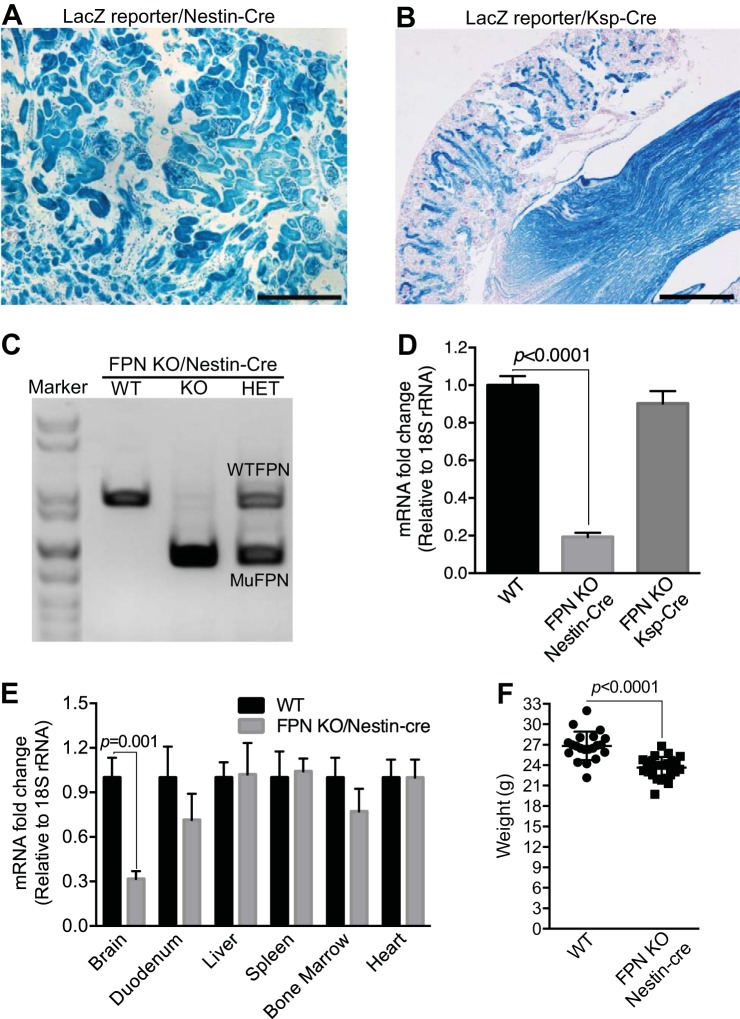
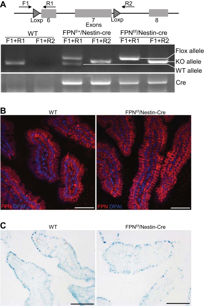
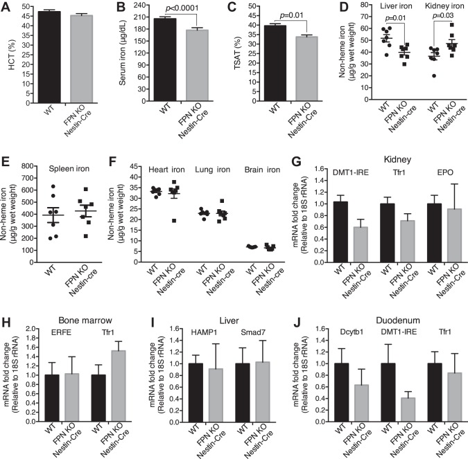
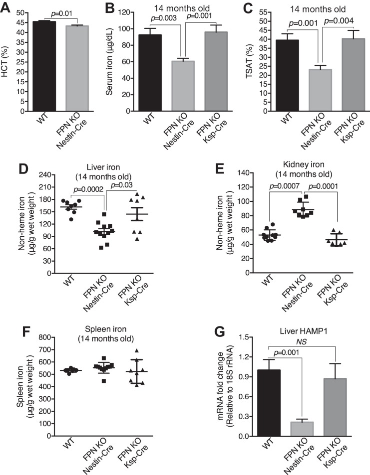
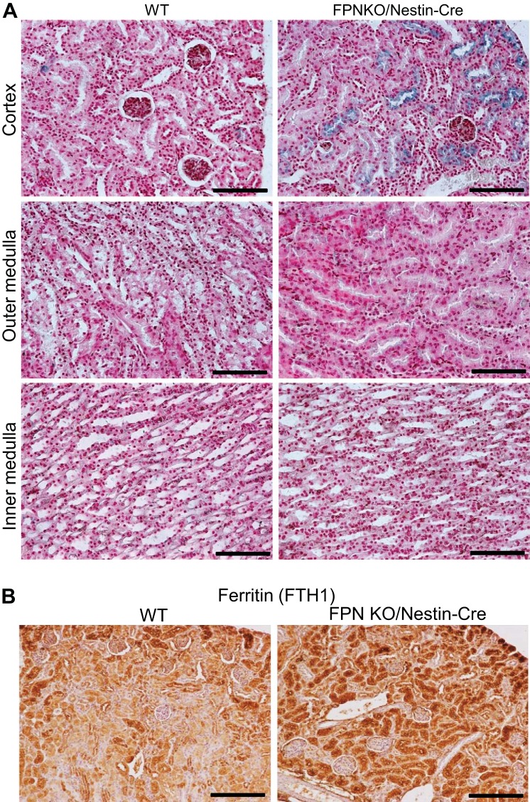
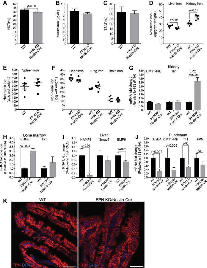
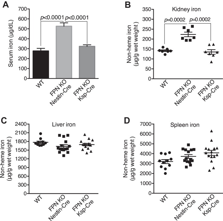
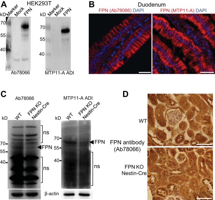
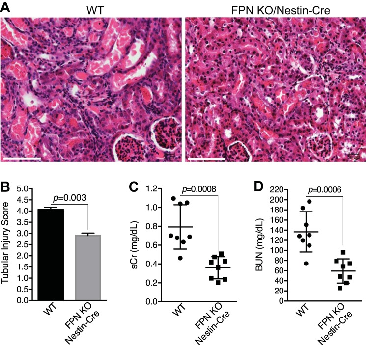
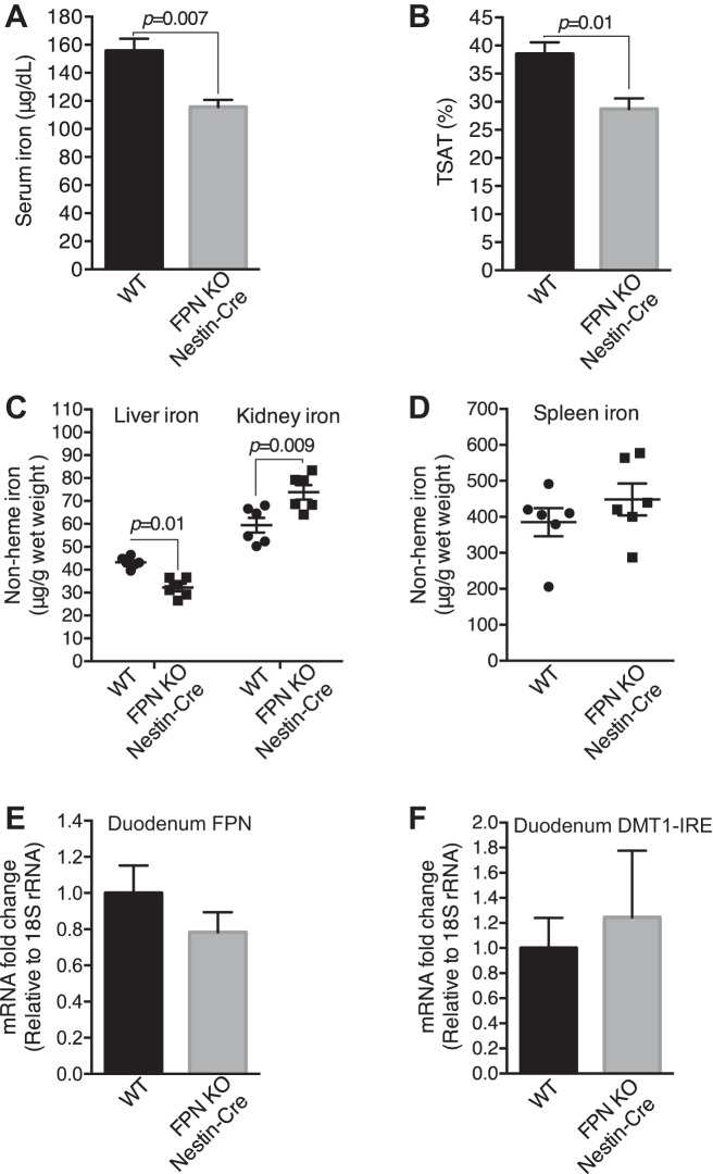
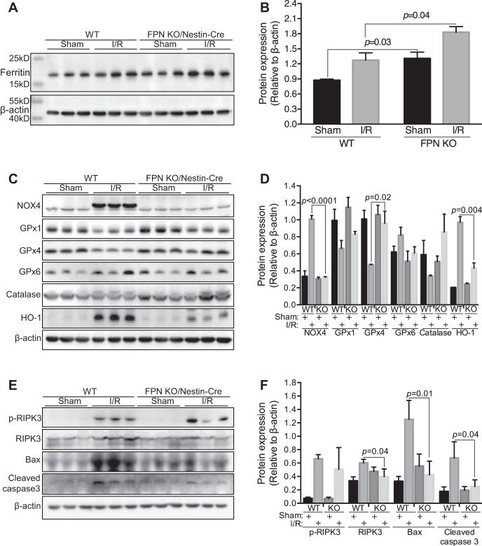
Renal iron recycling preserves filtered iron from urinary excretion. However, it remains debated whether ferroportin (FPN), the only known iron exporter, is functionally involved in renal iron recycling and whether renal iron recycling is required for systemic iron homeostasis. We deleted FPN in whole nephrons by use of a Nestin-Cre and in the distal nephrons and collecting ducts, using a Ksp-Cre, and investigated its impacts on renal iron recycling and systemic iron homeostasis. FPN deletion by Nestin-Cre, but not by Ksp-Cre, caused excess iron retention and increased ferritin heavy chain (FTH1) specifically in the proximal tubules and resulted in the reduction of serum and hepatic iron. The systemic iron redistribution was aggravated, resulting in anemia and the marked downregulation of hepatic hepcidin in elderly FPN knockout (KO)/Nestin-Cre mice. Similarly, in iron-deficient FPN KO/Nestin-Cre mice, the renal iron retention worsened anemia with the activation of the erythropoietin-erythroferrone-hepcidin pathway and the downregulation of hepatic hepcidin. Hence, FPN likely located at the basolateral membrane of the proximal tubules to export iron into the circulation and was required for renal iron recycling and systemic iron homeostasis particularly in elderly and iron-deficient mice. Moreover, FPN deletion in the proximal tubules alleviated ischemic acute kidney injury, possibly by upregulating FTH1 to limit catalytic iron and by priming antioxidant mechanisms, indicating that FPN could be deleterious in the pathophysiology of ischemic acute kidney injury (AKI) and thus may be a potential target for the prevention and mitigation of ischemic AKI.

Keywords: acute kidney injury; ferroportin; iron reabsorption; ischemia/reperfusion.


Figures

Fig. 1.
Fig. 1.
Ferroportin (FPN) was efficiently deleted in the kidney by Nestin-Cre but not by Ksp-Cre. Nestin-Cre (A) and Ksp-Cre (B) were crossed with Rosa26R-LacZ reporter mice, and β-galactosidase activity was stained to show gene deletion in the kidneys. RT-PCR (C) and qPCR (D) showed FPN knockout (KO) efficiency in the kidneys of floxed FPN (FPNf/f)/Nestin-Cre and FPNf/f/Ksp-Cre vs. wild-type (WT) mice (n = 8; male: 3; female: 5), respectively. E: qPCR showed the marked reduction of FPN mRNA in brain but not in duodenum, liver, spleen, bone marrow, and heart of FPNf/f/Nestin-Cre vs. WT mice (n = 8; male: 3; female: 5). F: body weight was slightly reduced in FPNf/f/Nestin-Cre vs. WT mice (3 mo; n = 21–26; male: 10–13; female: 10–13). Data are presented as means ± SE. D was analyzed by one-way ANOVA followed by Dunnett’s test; E and F were analyzed using Student’s t-test. Scale bars: A, 200 μm; B, 500 μm.
Fig. 2.
Fig. 2.
Nestin-Cre did not cause ferroportin (FPN) deletion in duodenal epithelia of floxed FPN (FPNf/f)/Nestin-Cre mice. A: PCR genotyping showed that the FPN gene was deleted in some duodenal cells of FPNf/+/Nestin-Cre and FPNf/f/Nestin-Cre mice. B: immunofluorescence showed that expression of FPN protein in duodenal epithelia of FPNf/f/Nestin-Cre mice was intact and comparable to WT counterparts. C: Perl’s staining showed comparable iron accumulation in duodenal epithelia of WT and FPNf/f/Nestin-Cre mice. Scale bars, 50 μm.
Fig. 3.
Fig. 3.
Ferroportin (FPN) knockout (KO) by Nestin-Cre resulted in the increase of renal iron and the decrease of serum and liver iron. A: hematocrit was comparable in floxed FPN (FPNf/f)/Nestin-Cre and WT mice (3 mo old; n = 37–40, male: 17–20, female: 17–20). Serum iron (B) and transferrin saturation (TSAT; C) were significantly decreased in FPNf/f/Nestin-Cre vs. WT mice (3 mo old). D: liver iron was decreased, whereas kidney iron was increased in FPNf/f/Nestin-Cre vs. WT mice (3 mo old). Iron contents in spleen (E) and heart, lung, and brain (F) were comparable in FPNf/f/Nestin-Cre and WT mice (3 mo old). G–J: qPCR showed that the expressions of divalent metal transporter 1(iron-responsive element) [DMT1(IRE)], transferrin receptor 1 (Tfr1), and erythropoietin (EPO) in kidney (G), erythroferrone (ERFE) and Tfr1 in bone marrow (H), hepcidin (HAMP1) and Smad7 in liver (I), and duodenal cytochrome B1 (Dcytb1), DMT-1(IRE), and Tfr1 in duodenum (J) were comparable in FPNf/f/Nestin-Cre and WT mice. Data are presented as means ± SE and were analyzed using Student’s t-test. B–J: n = 7 (male: 3, female: 4).
Fig. 4.
Fig. 4.
Ferroportin (FPN) recycled renal iron in the proximal tubules. A: hematocrit was significantly decreased in floxed FPN (FPNf/f)/Nestin-Cre vs. WT mice (14 mo; n = 20–21, male: 10–11, female: 10–11. Serum iron (B) and transferrin saturation (TSAT; C) were decreased in FPNf/f/Nestin-Cre vs. WT and FPNf/f/Ksp-Cre mice (14 mo old). Liver iron was decreased (D) and kidney iron was markedly increased (E) in FPNf/f/Nestin-Cre vs. WT and FPNf/f/Ksp-Cre mice. F: iron content in the spleen was comparable in WT, FPNf/f/Nestin-Cre, and FPNf/f/Ksp-Cre mice. G: hepcidin (HAMP1) was markedly downregulated in livers of FPNf/f/Nestin-Cre vs. WT and FPNf/f/Ksp-Cre mice. Data are presented as means ± SE. A was analyzed using Student’s t-test; B–G (n = 8–11; male: 5, female: 6) were analyzed using one-way ANOVA followed by Dunnett’s test.
Fig. 5.
Fig. 5.
Ferroportin (FPN) knockout (KO) by Nestin-Cre caused excess iron deposition and increased FTH1 expression specifically in the proximal tubules. A: Perl’s staining showed excess iron deposition specifically in the proximal tubules but not in the loop of Henle, distal tubules, and collecting ducts in floxed FPN (FPNf/f)/Nestin-Cre mice. Scale bars: 100 μm. B: immunochemistry showed increased FTH1 specifically in proximal tubules. Scale bars, 200 μm.
Fig. 6.
Fig. 6.
Ferroportin (FPN) knockout (KO) by Nestin-Cre resulted in increased renal iron and decreased liver iron under systemic iron deficiency. A: hematocrit was significantly decreased in iron-deficient floxed FPN (FPNf/f)/Nestin-Cre vs. WT mice (3 mo). Serum iron (B) and transferrin saturation (TSAT; C) were leveled in iron-deficient FPNf/f/Nestin-Cre and WT mice (3 mo), respectively. D: liver iron was significantly decreased and kidney iron increased in iron-deficient FPNf/f/Nestin-Cre vs. WT mice. Iron contents in spleen (E) and heart, lung, and brain (F) were comparable in iron-deficient FPNf/f/Nestin-Cre and WT mice. G: erythropoietin (EPO) but not divalent metal transporter 1(iron-responsive element) [DMT1(IRE)] and transferrin receptor 1 (Tfr1) mRNA was markedly upregulated in kidneys of iron-deficient FPNf/f/Nestin-Cre mice. H: erythroferrone (ERFE) but not Tfr1 mRNA was markedly upregulated in bone marrows of iron-deficient FPNf/f/Nestin-Cre mice. I: hepcidin (HAMP1) and bone morphogenetic protein-6 (BMP6) mRNA were markedly downregulated in livers of iron-deficient FPNf/f/Nestin-Cre mice. J: duodenal cytochrome B1 (Dcytb1) and DMT-1(IRE) mRNA were significantly decreased in duodenums of iron-deficient FPNf/f/Nestin-Cre mice. K: immunofluorescence showed lower expression levels of FPN protein in duodenal epithelia of FPNf/f/Nestin-Cre than in WT mice. Data are presented as means ± SE (n = 6; male: 3, female: 3) and were analyzed using Student’s t-test. Scale bars, 50 μm.
Fig. 7.
Fig. 7.
Ferroportin (FPN) knockout (KO) by Nestin-Cre resulted in increased renal iron under systemic iron overload. A: serum iron was increased in iron overload floxed FPN (FPNf/f)/Nestin-Cre vs. WT and FPNf/f/KSP-Cre mice. Kidney iron was markedly increased in iron overload FPNf/f/Nestin-Cre vs. WT and FPNf/f/KSP-Cre mice (B) despite liver iron being leveled (C). D: spleen iron was comparable in iron overload FPNf/f/Nestin-Cre, FPNf/f/KSP-Cre and WT mice. Data are presented as means ± SE (n = 7–17; male: 3–8, female: 4–10) and were analyzed using one-way ANOVA followed by Dunnett’s test.
Fig. 8.
Fig. 8.
Evaluation of ferroportin (FPN) antibodies for subcellular localization of FPN in the kidney. A: FPN antibodies Ab78066 from Abcam and MTP11-A from ADI recognized mouse FPN protein overexpressed in HEK-293T cells. B: both Ab78066 and MTP11-A recognized FPN protein at the basolateral membrane of duodenal epithelial cells. Scale bars, 25 µm. C: Western blot using Ab78066 and MTP11-A showed decreased FPN protein in kidneys of FPNf/f/Nestin-Cre vs. WT mice but also detected multiple abundant nonspecific (ns) proteins. D: immunochemistry using Ab78066 showed nonspecific intracellular staining in both WT and FPNf/f/Nestin-Cre kidneys. Scale bars, 50 µm.
Fig. 9.
Fig. 9.
Ferroportin (FPN) knockout (KO) by Nestin-Cre protected from ischemic acute kidney injury (AKI). Representative hematoxylin and eosin staining (A) and tubular injury score (B) showed less severe damage in ischemic kidneys of floxed FPN (FPNf/f)/Nestin-Cre vs. WT mice (ischemia, 20 min; reperfusion, 48 h). Scale bar, 100 µm. Both serum creatinine (sCr; C) and blood urea nitrogen (BUN; D) were lower in FPNf/f/Nestin-Cre than in WT mice with ischemic AKI. Data are presented as means ± SE (3 mo; n = 8, male) and were analyzed using Student’s t-test.
Fig. 10.
Fig. 10.
Ferroportin (FPN) knockout (KO) by Nestin-Cre resulted in decreased serum and liver iron and increased renal iron in ischemic acute kidney injury (AKI) mice. Serum iron (A), transferrin saturation (TSAT; B), and liver iron (C) were decreased, and kidney iron was increased (C) in floxed FPN (FPNf/f)/Nestin-Cre vs. WT ischemic AKI mice. D: spleen iron was comparable in FPNf/f/Nestin-Cre and WT ischemic AKI mice. E and F: FPN and divalent metal transporter 1(iron-responsive element) [DMT1(IRE)] mRNA were comparable in duodenums of FPNf/f/Nestin-Cre and WT ischemic AKI mice, respectively. Data are presented as means ± SE (3 mo; n = 6, male) and were analyzed using Student’s t-test.
Fig. 11.
Fig. 11.
Ferroportin (FPN) knockout (KO) by Nestin-Cre increased FTH1 and glutathione peroxidase-4 (GPx4) and decreased NADPH oxidase-4 (NOX4) and apoptosis in ischemia/reperfusion (I/R) kidneys. Western blot (A) and densitometric analysis (B) showed increased ferritin (FTH1) in both sham and I/R floxed FPN (FPNf/f)/Nestin-Cre vs. WT kidneys. C and D: NOX4 and heme oxygenase-1 (HO-1) proteins were decreased and GPx4 was increased in I/R kidneys of FPNf/f/Nestin-Cre vs. WT mice, and GPx1, GPx6, and catalase were comparable, respectively. E and F: phosphorylated receptor-interacting Ser/Thr kinase-3 (p-RIPK3) was comparable, and Bax and cleaved caspase-3 were decreased in I/R kidneys of FPNf/f/Nestin-Cre vs. WT mice. Data are presented as means ± SE (3 mo; n = 3, male) and were analyzed using two-way ANOVA followed by post hoc Tukey’s HSD test.

References

    1. Abboud S, Haile DJ. A novel mammalian iron-regulated protein involved in intracellular iron metabolism. J Biol Chem 275: 19906–19912, 2000. doi:10.1074/jbc.M000713200. - DOI - PubMed
    1. Aragno M, Cutrin JC, Mastrocola R, Perrelli MG, Restivo F, Poli G, Danni O, Boccuzzi G. Oxidative stress and kidney dysfunction due to ischemia/reperfusion in rat: attenuation by dehydroepiandrosterone. Kidney Int 64: 836–843, 2003. doi:10.1046/j.1523-1755.2003.00152.x. - DOI - PubMed
    1. Baliga R, Ueda N, Shah SV. Increase in bleomycin-detectable iron in ischaemia/reperfusion injury to rat kidneys. Biochem J 291: 901–905, 1993. doi:10.1042/bj2910901. - DOI - PMC - PubMed
    1. Baliga R, Zhang Z, Baliga M, Shah SV. Evidence for cytochrome P-450 as a source of catalytic iron in myoglobinuric acute renal failure. Kidney Int 49: 362–369, 1996. doi:10.1038/ki.1996.53. - DOI - PubMed
    1. Baliga R, Zhang Z, Baliga M, Ueda N, Shah SV. Role of cytochrome P-450 as a source of catalytic iron in cisplatin-induced nephrotoxicity. Kidney Int 54: 1562–1569, 1998. doi:10.1046/j.1523-1755.1998.00161.x. - DOI - PubMed

Publication types