Case Reports
. 2018 Jun;97(25):e11044.
doi: 10.1097/MD.0000000000011044.

Catheter radiofrequency ablation for arrhythmias under the guidance of the Carto 3 three-dimensional mapping system in an operating room without digital subtraction angiography

Affiliations
Case Reports

Catheter radiofrequency ablation for arrhythmias under the guidance of the Carto 3 three-dimensional mapping system in an operating room without digital subtraction angiography

Xingfu Huang et al. Medicine (Baltimore). 2018 Jun.

Abstract

Several studies have reported the efficacy of a zero-fluoroscopy approach for catheter radiofrequency ablation of arrhythmias in a digital subtraction angiography (DSA) room. However, no reports are available on the ablation of arrhythmias in the absence of DSA in the operating room. To investigate the efficacy and safety of catheter radiofrequency ablation for arrhythmias under the guidance of a Carto 3 three-dimensional (3D) mapping system in an operating room without DSA. Patients were enrolled according to the type of arrhythmia. The Carto 3 mapping system was used to reconstruct heart models and guide the electrophysiologic examination, mapping, and ablation. The total procedure, reconstruction, electrophysiologic examination, and mapping times were recorded. Furthermore, immediate success rates and complications were also recorded. A total of 20 patients were enrolled, including 12 males. The average age was 51.3 ± 17.2 (19-76) years. Nine cases of atrioventricular nodal re-entrant tachycardia, 7 cases of frequent ventricular premature contractions, 3 cases of Wolff-Parkinson-White syndrome, and 1 case of typical atrial flutter were included. All arrhythmias were successfully ablated. The procedure time was 127.0 ± 21.0 (99-177) minutes, the reconstruction time was 6.5 ± 2.9 (3-14) minutes, the electrophysiologic study time was 10.4 ± 3.4 (6-20) minutes, and the mapping time was 11.7 ± 8.3 (3-36) minutes. No complications occurred. Radiofrequency ablation of arrhythmias without DSA is effective and feasible under the guidance of the Carto 3 mapping system. However, the electrophysiology physician must have sufficient experience, and related emergency measures must be present to ensure safety.


Conflict of interest statement

The authors have no funding and conflicts of interest to disclose.

Figures

Figure 1
Figure 1
The setup of the operating room.
Figure 2
Figure 2
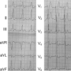
(A) Electrocardiogram (ECG) indicating type B pre-excitation syndrome in case 1. (B) The ablation catheter mapped the potential preceding the delta wave by 35 milliseconds. The yellow spot denotes the location of His, the blue dots denote the earliest area, and the red spots denote the ablation points. The pink points indicate the coronary sinus, and the green line indicates the tricuspid valve annulus. (C) The ablation catheter mapped the potential preceding the delta wave by 21 milliseconds. The yellow point indicates the left His location, the white spots indicate the earliest areas, and the red spots indicate the ablation points. (D) 3D image after merging. (E) The postablation ECG of case 1. LM, left main; LCC, left coronary cusp; RCC, right coronary cusp; NCC, noncoronary cusp.
Figure 2 (Continued)
(A) Electrocardiogram (ECG) indicating type B pre-excitation syndrome in case 1. (B) The ablation catheter mapped the potential preceding the delta wave by 35 milliseconds. The yellow spot denotes the location of His, the blue dots denote the earliest area, and the red spots denote the ablation points. The pink points indicate the coronary sinus, and the green line indicates the tricuspid valve annulus. (C) The ablation catheter mapped the potential preceding the delta wave by 21 milliseconds. The yellow point indicates the left His location, the white spots indicate the earliest areas, and the red spots indicate the ablation points. (D) 3D image after merging. (E) The postablation ECG of case 1. LM, left main; LCC, left coronary cusp; RCC, right coronary cusp; NCC, noncoronary cusp.
Figure 3
Figure 3
(A) Electrocardiogram (ECG) suggesting frequent ventricular premature contractions (VPCs). (B) The ablation catheter mapped the potential preceding the delta wave by 55 milliseconds. (C) The pacing ECG was consistent with spontaneous VPCs. (D) 3D mapping shows the LL and LAO 45° positions: the red spot indicates the earliest excitation spot, and the white spot indicates the LCC position. (E) The postablation ECG of case 2.
Figure 3 (Continued)
(A) Electrocardiogram (ECG) suggesting frequent ventricular premature contractions (VPCs). (B) The ablation catheter mapped the potential preceding the delta wave by 55 milliseconds. (C) The pacing ECG was consistent with spontaneous VPCs. (D) 3D mapping shows the LL and LAO 45° positions: the red spot indicates the earliest excitation spot, and the white spot indicates the LCC position. (E) The postablation ECG of case 2.

References

    1. Casella M, Dello Russo A, Pelargonio G, et al. Near zerO fluoroscopic exPosure during catheter ablAtion of supRavenTricular arrhYthmias: the NO-PARTY multicentre randomized trial. Europace 2016;18:1565–72. - PMC - PubMed
    1. Wang Y, Chen GZ, Yao Y, et al. Ablation of idiopathic ventricular arrhythmia using zero-fluoroscopy approach with equivalent efficacy and less fatigue: a multicenter comparative study. Medicine (Baltimore) 2017;96:e6080. - PMC - PubMed
    1. Zhu TY, Liu SR, Chen YY, et al. Zero-fluoroscopy catheter ablation for idiopathic premature ventricular contractions from the aortic sinus cusp [in Chinese]. Nan Fang Yi Ke Da Xue Xue Bao 2016;36:1105–9. - PubMed
    1. Giaccardi M, Del Rosso A, Guarnaccia V, et al. Near-zero x-ray in arrhythmia ablation using a 3-dimensional electroanatomic mapping system: a multicenter experience. Heart Rhythm 2016;13:150–6. - PubMed
    1. Kerst G, Parade U, Weig HJ, et al. A novel technique for zero-fluoroscopy catheter ablation used to manage Wolff-Parkinson-White syndrome with a left-sided accessory pathway. Pediatr Cardiol 2012;33:820–3. - PubMed