Review
. 2018 Jul 21;143(14):3249-3283.
doi: 10.1039/c8an00731d. Epub 2018 Jun 20.

Optical assays based on colloidal inorganic nanoparticles

Affiliations
Review

Optical assays based on colloidal inorganic nanoparticles

Amir Ghasemi et al. Analyst. .

Abstract

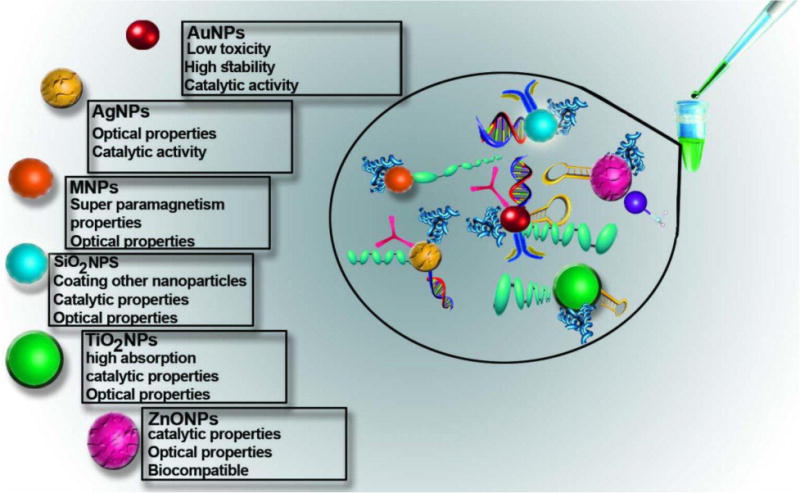
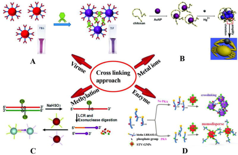
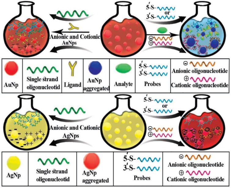
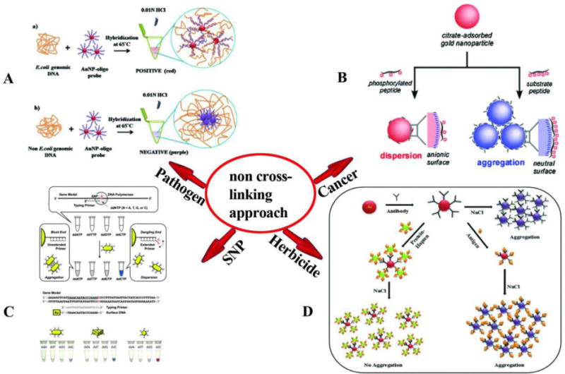
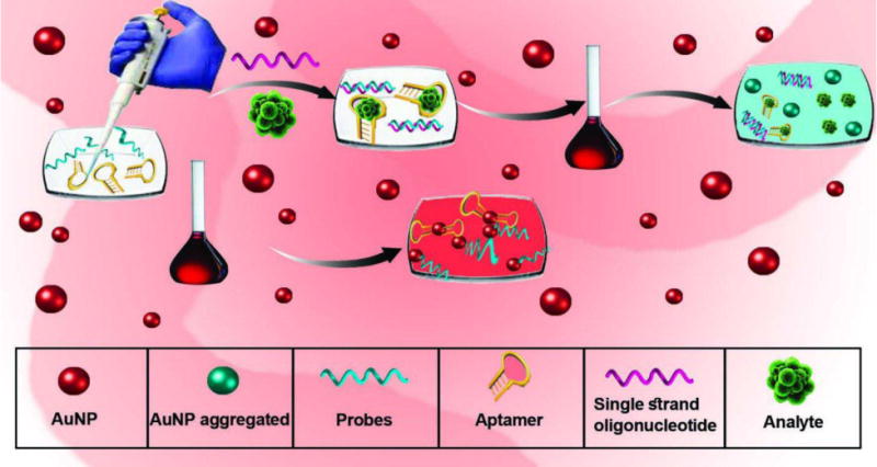
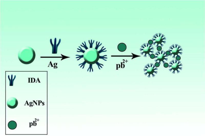
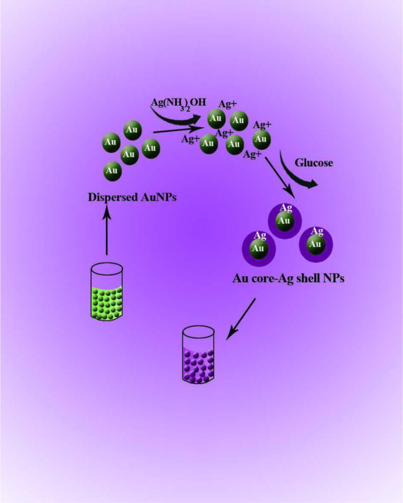
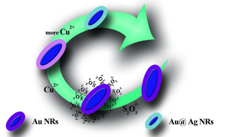
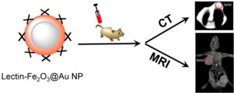
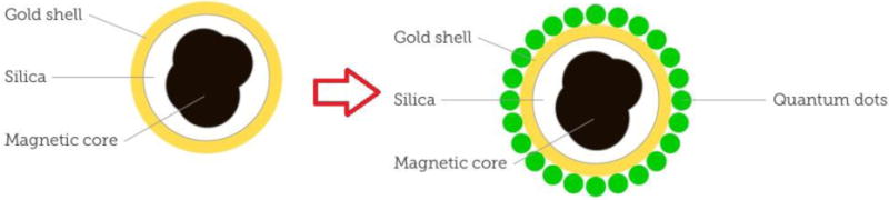
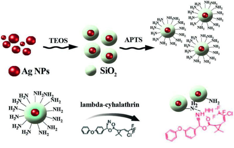
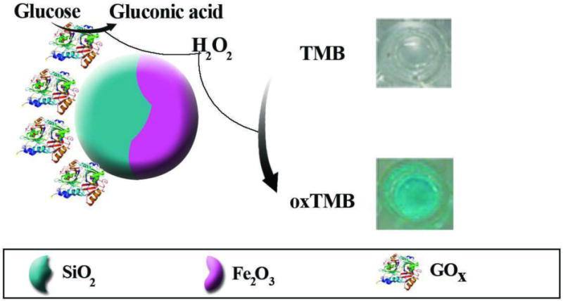
Colloidal inorganic nanoparticles have wide applications in the detection of analytes and in biological assays. A large number of these assays rely on the ability of gold nanoparticles (AuNPs, in the 20 nm diameter size range) to undergo a color change from red to blue upon aggregation. AuNP assays can be based on cross-linking, non-cross linking or unmodified charge-based aggregation. Nucleic acid-based probes, monoclonal antibodies, and molecular-affinity agents can be attached by covalent or non-covalent means. Surface plasmon resonance and SERS techniques can be utilized. Silver NPs also have attractive optical properties (higher extinction coefficient). Combinations of AuNPs and AgNPs in nanocomposites can have additional advantages. Magnetic NPs and ZnO, TiO2 and ZnS as well as insulator NPs including SiO2 can be employed in colorimetric assays, and some can act as peroxidase mimics in catalytic applications. This review covers the synthesis and stabilization of inorganic NPs and their diverse applications in colorimetric and optical assays for analytes related to environmental contamination (metal ions and pesticides), and for early diagnosis and monitoring of diseases, using medically important biomarkers.


Conflict of interest statement

Transparency Declaration. The authors have declared that no competing interests exist.

Figures

Fig. 1
Fig. 1
Properties and functionalization of inorganic nanoparticles in colorimetric assay.
Fig. 2
Fig. 2
Schematic illustration of crosslinking based colorimetric assays. (A) The detection of bacteriophage T7 using AuNPs modified with covalently bonded anti-T7 antibodies and color change based on antibody antigen interaction which causes them to aggregate [111]. (B) Colorimetric detection of Hg2+ based on chelation reaction between Hg2+ and chitosan and observed color changes in AuNPs [112]. (C) LCR amplification and colorimetric assay of CpG methylation in DNA: In the presence of methylated genomic DNA a red to purple color change can be detected [113]. (D) Colorimetric assay for detection of protein kinase activities based on hybridization between STV-AuNPs and biotinylated peptide (biotin-LRRASLG), and the PKA catalyzed phosphorylation of biotin-peptide prevented AuNPs crosslinking and the monodisperse AuNPs remained red [81]. Reprinted with Permission
Fig. 3
Fig. 3
A schematic illustration of non-crosslinking AuNP and AgNP aggregation-based colorimetric detection
Fig. 4
Fig. 4
Schematic illustration of the non-crosslinking based colorimetric assay. (A) colorimetric detection of E. coli genomic DNA based on AuNPs probes; in presence of genomic DNA, AuNPs probes do not aggregate and the color remains red; in contrast, in the absence of genomic DNA, AuNPs probes lose stability and tend to aggregate [131]. (B) Non-crosslinking aggregation-based colorimetric detection of cancers. This assay is based on the non-crosslinking aggregation using a cationic PKC-specific peptide substrate [133]. (C) Colorimetric detection of SNP based on DNA-functionalized anisotropic AuNPs and showing a mixture of AuNR AuNT and the AuNS probe after mixing with the primer solution and color changes of NPs [134]. (D) Colorimetric detection of phenylurea herbicide, in the presence of antigen, color change is visible upon aggregation of Ab-AuNPs [136]. Reprinted with permission
Fig. 5
Fig. 5
Schematic of colorimetric assays based on unmodified AuNPs
Fig. 6
Fig. 6
Aggregation and color change of modified AgNPs in the presence of lead ions [231]. Reprinted with permission
Fig. 7
Fig. 7
Color change induced by Ag shell formation on AuNP core. The shell thickens and the resultant color change is proportional to the glucose concentration [271]. Reprinted with permission
Fig. 8
Fig. 8
Shell etching according to the concentration of copper ions [275]. Reprinted with permission
Fig. 9
Fig. 9
(A) Silane molecular structures. (B) Schematic of the silane condensation reaction [290]. Reprinted with permission
Fig. 10
Fig. 10
Immobilization of Chiral Ru Catalyst on MNPs. Reprinted (adapted) with permission from [293] . Copyright (2005) American Chemical Society.
Fig. 11
Fig. 11
Zeta-potential of Fe3O4 NPs (a), Fe3O4/SiO2 NPs (b) and native silica (c) [283]. Reprinted with permission
Fig. 12
Fig. 12
MNPs catalyze the oxidation of a colorimetric substrate, TMB, to generate a color reaction in the presence of AChE and CHO producing hydrogen peroxide after incubation in acetylcholine chloride solution. [302]. Reprinted with permission
Fig. 13
Fig. 13
Lectin conjugated to core-shell IONPs-GNPs. Reprinted (adapted) with permission from [306]. Copyright (2014) American Chemical Society
Fig. 14
Fig. 14
Magnetic-fluorescent composite particles consisting of a magnetic core, silica spacer, and fluorescent quantum dots covalently bonded to the silica surface.
Fig. 15
Fig. 15
Surface modified Fe3O4 with mercaptopropionic acid, followed by conjugation of nitrilotriacetic acid (NTA) and subsequently chelating Ni2+ chelation. Reprinted (adapted) with permission from [309] Copyright (2010) American Chemical Society
Fig. 16
Fig. 16
Determination of dopamine in an artificial sample of cerebrospinal fluid and in mouse striatum brain tissue using a Fe3O4/Ag nanocomposite. Reprinted (adapted) with permission from [313] Copyright (2014) American Chemical Society
Fig. 17
Fig. 17
Schematic illustration of Ag@SiO2-NH2 NPs detection of lambda-cyhalothrin (LC) [317]. Reprinted with permission
Fig. 18
Fig. 18
Schematic illustration of colorimetric detection of H2O2 and glucose by glucose oxidase and peroxidase-like activity of JFSNs. In presence of glucose, GOx catalyzes oxidation of glucose. Subsequently, glucoronic acid and H2O2 are produced. H2O2 decomposition is catalyzed by JFSNs, and if TMB is present a blue color is formed. Reprinted (adapted) with permission from [319] Copyright (2015) American Chemical Society.
Fig. 19
Fig. 19
Schematic illustration of colorimetric detection of H2O2 by catalytic activity of ZnS-MMT nanocomposites. ZnS-MMT catalyzes the decomposition of H2O2 to OH radicals to oxidize TMB to form blue-colored oxTMB [329]. Reprinted with permission

Similar articles

Cited by

References

    1. Cho-Chung YS. Autoantibody biomarkers in the detection of cancer. Biochimica et Biophysica Acta (BBA) - Molecular Basis of Disease. 2006;1762(6):587–591. - PubMed
    1. Zaenker P, Ziman MR. Serologic autoantibodies as diagnostic cancer biomarkers–a review. Cancer Epidemiol Biomarkers Prev. 2013;22(12):2161–81. - PubMed
    1. Suo B, et al. Development of an oligonucleotide-based microarray to detect multiple foodborne pathogens. Mol Cell Probes. 2010;24(2):77–86. - PubMed
    1. Steinberg TH. Protein gel staining methods: an introduction and overview. Methods Enzymol. 2009;463:541–63. - PubMed
    1. Mandal P, et al. Methods for Rapid Detection of Foodborne Pathogens: An Overview. 2011;6