. 2018 Oct 1;28(10):786-801.
doi: 10.1093/glycob/cwy057.

Sialylated keratan sulfate proteoglycans are Siglec-8 ligands in human airways

Affiliations

Sialylated keratan sulfate proteoglycans are Siglec-8 ligands in human airways

Anabel Gonzalez-Gil et al. Glycobiology. .

Abstract

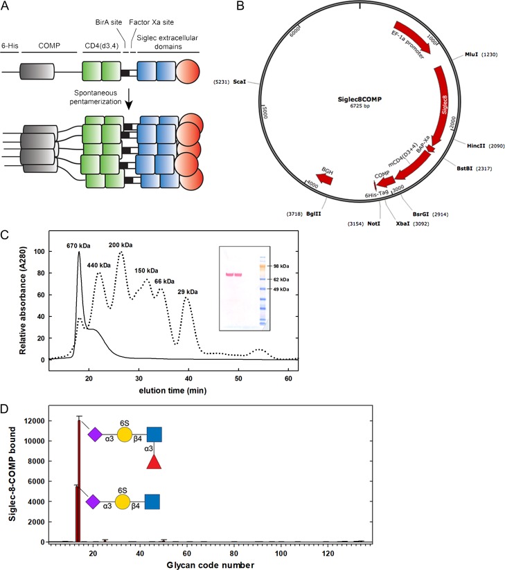
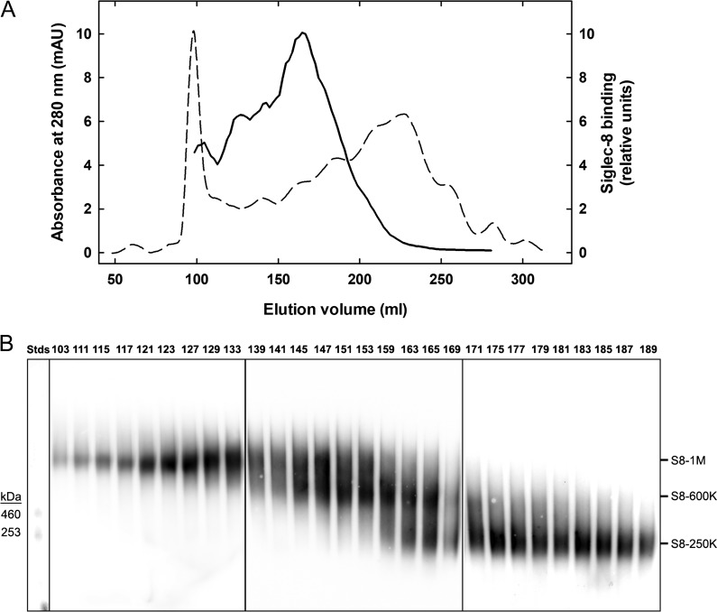
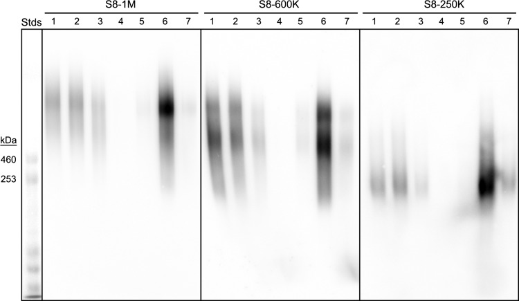
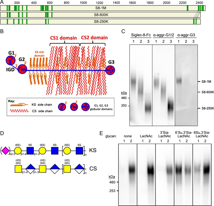
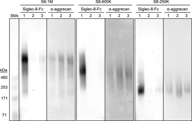
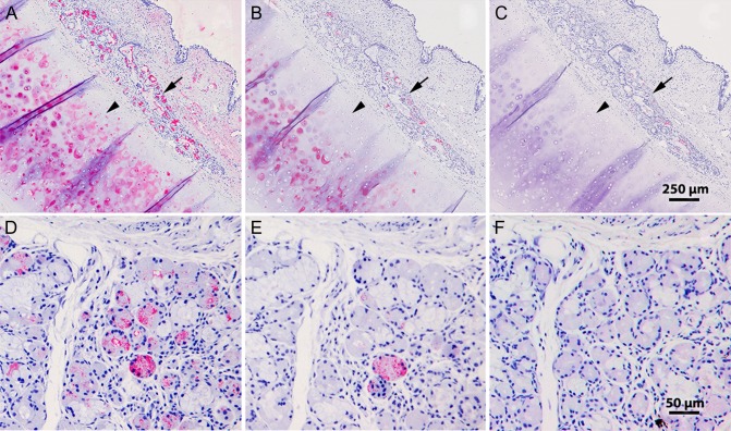
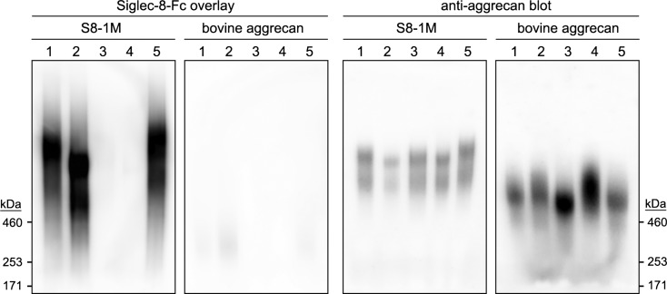
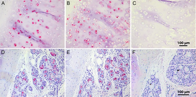
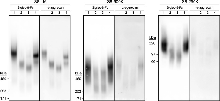
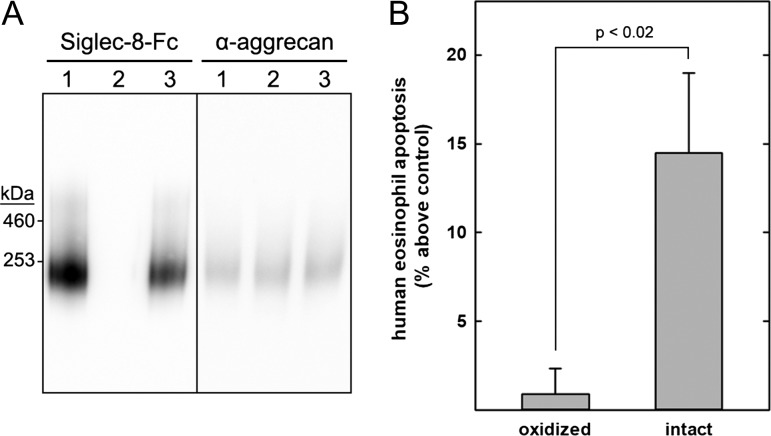
Human siglecs are a family of 14 sialic acid-binding proteins, most of which are expressed on subsets of immune cells where they regulate immune responses. Siglec-8 is expressed selectively on human allergic inflammatory cells-primarily eosinophils and mast cells-where engagement causes eosinophil apoptosis and inhibits mast cell mediator release. Evidence supports a model in which human eosinophils and mast cells bind to Siglec-8 sialoglycan ligands on inflammatory target tissues to resolve allergic inflammation and limit tissue damage. To identify Siglec-8-binding sialoglycans from human airways, proteins extracted from postmortem human trachea were resolved by size-exclusion chromatography and composite agarose-acrylamide gel electrophoresis, blotted and probed by Siglec-8-Fc blot overlay. Three size classes of Siglec-8 ligands were identified: 250 kDa, 600 kDa and 1 MDa, each of which was purified by affinity chromatography using a recombinant pentameric form of Siglec-8. Proteomic mass spectrometry identified all size classes as the proteoglycan aggrecan, a finding validated by immunoblotting. Glycan array studies demonstrated Siglec-8 binding to synthetic glycans with a terminal Neu5Acα2-3(6-sulfo)-Gal determinant, a quantitatively minor terminus on keratan sulfate (KS) chains of aggrecan. Treating human tracheal extracts with sialidase or keratanase eliminated Siglec-8 binding, indicating sialylated KS chains as Siglec-8-binding determinants. Treating human tracheal histological sections with keratanase also completely eliminated the binding of Siglec-8-Fc. Finally, Siglec-8 ligand purified from human trachea extracts induced increased apoptosis of freshly isolated human eosinophils in vitro. We conclude that sialylated KS proteoglycans are endogenous human airway ligands that bind Siglec-8 and may regulate allergic inflammation.


Figures

Fig. 1.
Fig. 1.
Pentavalent Siglec-8-COMP. (A) Chimeric Siglec-8-COMP schematic design includes the extracellular domain of Siglec-8, two spacer domains from CD4, spontaneously pentamerizing COMP and a 6-His tag. (B) Plasmid design for Siglec-8-COMP. (C) Superdex-200 size-exclusion chromatography of expressed Siglec-8-COMP with protein size standards indicated. Denaturing SDS-PAGE gel (insert) immunostained for the 6-His tag indicates a monomer molecular mass of ~80 kDa. (D) Glycan array screening of Siglec-8-COMP binding. A custom glycan array containing 10 neutral glycans, 69 α2,3-linked sialoglycans and 56 α2,6-linked sialoglycans (Supplementary data, Table I) was probed with Siglec-8-COMP and binding detected with anti-6-His antibody. The mean and standard error for binding to six spots of each glycan are presented. Glycans that supported Siglec-8-COMP binding are shown using symbol nomenclature (Varki et al. 2015).
Fig. 2.
Fig. 2.
Chromatographic resolution of Siglec-8 ligands extracted from human airway. (A) Sephacryl S-500 column chromatography of Siglec-8 ligands extracted from human trachea. Elution of proteins (A280, dashed line) and Siglec-8 binding activity (solid line) determined by semiquantitative dot blot Siglec-8-Fc overlay are plotted against the elution volume. (B) Composite gel electrophoresis of active binding fractions. Equal aliquots of fractions with Siglec-8-Fc-binding activity detected by semiquantitative dot blot were resolved by 1.5% acrylamide, 2% agarose composite gel electrophoresis, blotted to PVDF membranes and probed for Siglec-8-Fc binding. Images of three replicate gels (boxed) used to accommodate fractions across the active elution fractions were stitched together by lining up common molecular weight standards (Stds, left). Fractions were pooled to represent three size classes of Siglec-8 ligands (designated by estimated molecular weight in daltons): S8-1M, S8-600K and S8-250K.
Fig. 3.
Fig. 3.
Siglec-8-COMP affinity purification of Siglec-8 ligands from human trachea. Siglec-8 ligands extracted from human trachea were resolved by Sephacryl S-500 size-exclusion chromatography (Figure 2) and fractions corresponding to S8-1M, S8-600K and S8-250K separately combined for affinity purification. Pooled fractions of each molecular size were loaded separately onto 1-mL nickel Sepharose affinity columns carrying 6-His-tagged pentameric Siglec-8-COMP. After loading and washing each column, bound Siglec-8 ligands were eluted with high salt. Equal aliquots of washes and eluates were subjected to 1.5% acrylamide, 2% agarose composite gel electrophoresis, blotted to PVDF membranes and probed with Siglec-8-Fc. Lanes: (1) pooled Sephacryl S-500 fraction; (2) affinity column flow-through; (3) wash 1; (4) wash 2; (5) elution 1; (6) elution 2; (7) elution 3. HiMark-prestained molecular weight standards are shown at the left (Stds).
Fig. 4.
Fig. 4.
Aggrecan carries Siglec-8-binding glycans. (A) Proteins extracted from human trachea were purified by sequential size-exclusion and Siglec-8 affinity chromatography, proteolyzed, and peptides identified by mass spectrometry. Maps of aggrecan protein sequences with identified peptides from three size classes of affinity-purified Siglec-8 ligands are shown. Green bars are peptides (see Table I) that exceed strict false discovery rates for each of the Siglec-8 ligand size classes as indicated. The smaller size classes (S8-600 K and S8-250K) are mapped on aggrecan Uniprot reference sequence P16112 (2415 amino acids); whereas the largest size class (S8-1M) is mapped on C-terminal alternatively spliced aggrecan sequence H0YM81 (2492 amino acids). (B) Schematic map of aggrecan. IGD, interglobular domain; CS1, CS2, chondroitin sulfate (CS)-rich domains. Modified from Caterson and Melrose (2018), with permission. (C) Co-migration of purified human Siglec-8 ligands and aggrecan immunoreactivity. Three purified size classes of Siglec-8 ligands from human trachea were resolved by electrophoresis on replicate 1.5% acrylamide, 2% agarose composite gels, blotted to PVDF membranes and probed with Siglec-8-Fc, anti-aggrecan antibody 7D4 (α-aggr-G1/2) or anti-aggrecan antibody PA1-1745 (α-aggr-G3). Migration positions of HiMark-prestained standards are indicated at the left. Lanes: (1) S81M; (2) S8-600K and (3) S8-250K. (D) Generalized schematic structures of KS and CS chains depicted using symbol nomenclature (Varki et al. 2015). Sialic acid and sulfates that are variable are shown in parentheses. (E) Elution of S8-1M from Siglec-8-COMP affinity chromatography with soluble glycans. Pooled size-exclusion fractions containing S8-1M (Figure 2) were captured on Siglec-8-COMP magnetic beads. The beads were thoroughly washed prior to eluting with β-azidoethylglycosides (lane 1) followed by 500 mM imidazole to elute the bound Siglec-8-COMP with any remaining ligand attached (lane 2). Eluates were resolved by composite gel electrophoresis, blotted and probed with Siglec-8-Fc. Elution was tested with the following β-azidoethylglycosides: none, N-acetyllactosamine (LacNAc, Galβ1-4GlcNAc), 3′-sialyl LacNac (3′Sia-LacNAc, Neu5Acα2-3Galβ1-4GlcNAc), 6′-sulfo-3′-sialyl-LacNAc (6′Su,3′Sia-LacNAc, Neu5Acα2-3[6 S]Galβ1-4GlcNAc) and 6-sulfo-3′-sialyl-LacNAc (6 Su,3′Sia-LacNAc, Neu5Acα2-3Galβ1-4[6S]GlcNAc).
Fig. 5.
Fig. 5.
Keratanase and sialidase pretreatments diminish Siglec-8 binding to purified Siglec-8 ligands. Siglec-8 ligands were extracted from human trachea, purified by sequential size-exclusion and Siglec-8 affinity chromatography and subjected to keratanase or sialidase treatments. Samples were resolved on 1.5% acrylamide, 2% agarose composite gels, blotted to PVDF membranes and probed with precomplexed Siglec-8-Fc or anti-aggrecan antibody (7D4) as indicated. Lanes: (1) incubation without enzyme; (2) sialidase (67 mU/mL, 90 min); (3) keratanase II (6 mU/mL, 16 h). HiMark molecular weight standards are shown at the left (Stds).
Fig. 6.
Fig. 6.
Keratanase treatment of human trachea tissue sections diminishes Siglec-8-Fc binding. Cross sections of human trachea were incubated with Siglec-8-Fc precomplexed with AP-conjugated anti-human-Fc (AC) or Siglec-8-Fc followed with the same secondary antibody (DF) as described in the text. Lectin binding was detected using Vector Red stain and sections counterstained using hematoxylin QS. (A–C) Low-power images of serial sections of human trachea preincubated for 24 h at 37°C with buffer alone (A), 0.2 mU/mL keratanase I (B), or 5 mU/mL keratanase II (C) prior to Siglec-8-Fc overlay. Arrows, submucosal glands; arrowheads, cartilage. (D–F) Higher power images of human tracheal submucosal glands from sections preincubated for 25 h at 37°C with buffer alone (D), 21 mU/mL keratanase I (E), or 10 mU/mL keratanase II (F).
Fig. 7.
Fig. 7.
Chondroitinase ABC (ChABC) and keratanase treatments of a human tracheal Siglec-8 ligand reveal a chondroitin sulfate (CS) proteoglycan with Siglec-8-binding keratan sulfate (KS) chains. Siglec-8 ligands were extracted from human trachea and S8-1M purified by sequential size-exclusion and Siglec-8 affinity chromatography. Equal aliquots containing the isolated ligand (S8-1M) or commercial bovine articular cartilage aggrecan (bovine aggrecan) were treated with buffer alone (no enzyme), ChABC, keratanase I or both enzymes for 20 h at 37°C. Samples were denatured and resolved on 1.5% acrylamide, 2% agarose composite gels, blotted to PVDF membranes and probed with precomplexed Siglec-8-Fc overlay or anti-aggrecan antibody. Lanes (all enzyme treatments 20 h at 37°C): (1) no incubation; (2) ChABC (500 mU/mL); (3) keratanase I (21 mU/mL); (4) ChABC (500 mU/mL) plus keratanase I (21 mU/mL); (5) incubation without enzymes. Migration positions of HiMark molecular weight markers are shown.
Fig. 8.
Fig. 8.
Chondroitinase ABC (ChABC) treatment of human trachea tissue sections enhances Siglec-8-Fc binding. Cross sections of human trachea were incubated with Siglec-8-Fc precomplexed with AP-conjugated anti-human-Fc. Lectin binding was detected using Vector Red stain and sections counterstained using hematoxylin QS. (A–C) Human tracheal cartilage from sections treated: (A) for 44 h with buffer alone; (B) for 18 h with 150 mU/mL ChABC followed by 25 h with buffer alone, or (C) for 18 h with 150 mU/mL ChABC followed by a 1-h wash and further incubation for 25 h with 10 mU/mL keratanase II. (D–F) Human trachea submucosal glands from the same experiment: (D), buffer incubation; (E) ChABC followed by buffer incubation; (F) ChABC followed by keratanase II.
Fig. 9.
Fig. 9.
ADAMTS-4 (aggrecanase-1) treatment of human airway Siglec-8 ligands shifts their electrophoretic migration. Siglec-8 ligands were extracted from human trachea, purified by sequential size exclusion followed by Siglec-8 affinity chromatography and subjected to ChABC, aggrecanase or both enzymes. Samples were resolved on 1.5% acrylamide, 2% agarose composite gels, blotted to PVDF membranes and probed with precomplexed Siglec-8-Fc or anti-aggrecan antibody (7D4) as indicated. Lanes (all enzyme treatments 16 h at 37°C): (1) incubation without enzyme; (2) aggrecanase (0.3 mU/mL); (3) aggrecanase (0.3 mU/mL) plus ChABC (0.5 U/mL); (4) ChABC (0.5 U/mL). S8-1M and S8-600K were resolved on 1.5% acrylamide, 2% agarose composite gels with migration positions of HiMark molecular weight markers shown; S8-250K was resolved on 3% acrylamide, 2% agarose composite gels with migration positions of SeeBlue Plus2 molecular weight markers shown.
Fig. 10.
Fig. 10.
Purified human tracheal Siglec-8 ligand induces human eosinophil apoptosis. Siglec-8 ligands were extracted from human trachea and purified by size-exclusion chromatography followed by Siglec-8 affinity chromatography. A portion of purified S8-250K was treated with cold periodate to selectively oxidize the glycerol sidearm of its sialic acid followed by sodium borohydride reduction (negative control). An equal portion was incubated without periodate and treated with sodium borohydride (intact ligand). Oxidized and intact S8-250K were dialyzed against RPMI medium and added at equal concentrations to freshly isolated primary human eosinophils. After 24 h in culture, eosinophil apoptosis was quantified by flow cytometry. (A) Oxidized and intact S8-250 K were electrophoretically resolved on a composite agarose–acrylamide gel, blotted to PVDF membranes. Replicate blots were probed with Siglec-8-Fc to reveal Siglec-8 ligands or anti-aggrecan antibody (7D4). Lanes: (1) untreated S8-250K; (2) periodate oxidized and reduced S8-250K; (3) reduced S8-250K (intact ligand). (B) After isolation and overnight interleukin 5 (IL-5) priming, human eosinophils were incubated with equal portions of oxidized and intact S8-250K and apoptosis was assessed 18–24 h later. Results are expressed relative to untreated eosinophils, which had average apoptosis of 42 ± 5% (SEM). Data are displayed as mean and SEM of six replicates performed on three separate primary human eosinophil preparations.

References

    1. Akhtar S, Kerr BC, Hayes AJ, Hughes CE, Meek KM, Caterson B. 2008. Immunochemical localization of keratan sulfate proteoglycans in cornea, sclera, and limbus using a keratanase-generated neoepitope monoclonal antibody. Invest Ophthalmol Vis Sci. 49:2424–2431. - PubMed
    1. Angata T, Margulies EH, Green ED, Varki A. 2004. Large-scale sequencing of the CD33-related Siglec gene cluster in five mammalian species reveals rapid evolution by multiple mechanisms. Proc Natl Acad Sci USA. 101:13251–13256. - PMC - PubMed
    1. Barnig C, Frossard N, Levy BD. 2018. Towards targeting resolution pathways of airway inflammation in asthma. Pharmacol Ther. 186:98–113. - PubMed
    1. Bochner BS. 2016. "Siglec"ting the allergic response for therapeutic targeting. Glycobiology. 26:546–552. - PMC - PubMed
    1. Bochner BS, Alvarez RA, Mehta P, Bovin NV, Blixt O, White JR, Schnaar RL. 2005. Glycan array screening reveals a candidate ligand for siglec-8. J Biol Chem. 280:4307–4312. - PubMed

Publication types

MeSH terms