. 2018 Aug 7;57(31):4741-4746.
doi: 10.1021/acs.biochem.8b00257. Epub 2018 Jul 5.

Dynamic Nuclear Polarization Nuclear Magnetic Resonance in Human Cells Using Fluorescent Polarizing Agents

Affiliations

Dynamic Nuclear Polarization Nuclear Magnetic Resonance in Human Cells Using Fluorescent Polarizing Agents

Brice J Albert et al. Biochemistry. .

Abstract

Solid state nuclear magnetic resonance (NMR) enables atomic-resolution characterization of the molecular structure and dynamics within complex heterogeneous samples, but it is typically insensitive. Dynamic nuclear polarization (DNP) increases the NMR signal intensity by orders of magnitude and can be performed in combination with magic angle spinning (MAS) for sensitive, high-resolution spectroscopy. Here we report MAS DNP experiments, for the first time, within intact human cells with >40-fold DNP enhancement and a sample temperature of <6 K. In addition to cryogenic MAS results at <6 K, we also show in-cell DNP enhancements of 57-fold at 90 K. In-cell DNP is demonstrated using biradicals and sterically shielded monoradicals as polarizing agents. A novel trimodal polarizing agent is introduced for DNP, which contains a nitroxide biradical, a targeting peptide for cell penetration, and a fluorophore for subcellular localization with confocal microscopy. The fluorescent polarizing agent provides in-cell DNP enhancements of 63-fold at a concentration of 2.7 mM. These experiments pave the way for structural characterization of biomolecules in an endogenous cellular context.


Figures

Figure 1.
Figure 1.
Characterization of nitroxide radicals in HEK293F cells. DNP CPMAS NMR spectra below 6 K using (A) 20 mM AMUPol, (B) 40 mM sterically-shielded nitroxide monoradical, and (C) 2.7 mM TotaFAM. Black spectra represent no microwave irradiation while red spectra are recorded with microwave irradiation. Asterisks (*) denote spinning side bands. The 13C resonances in the 50–100 ppm chemical shift range are attributed to sugars (Supplementary Figure S9). Intensity of integrated EPR spectra versus time of cellular samples prepared with (D) AMUPol, (E) sterically-shielded nitroxide monoradical, and (F) TotaFAM. Polarizing agents (G) AMUPol, (H) sterically-shielded nitroxide monoradical, and (I) trimodal polarizing agent, TotaFAM. The three moieties of TotaFAM include a TOTAPOL nitroxide radical (red), 11 residues of the HIV-1 Tat protein (blue), and a 6-FAM fluorophore (green).
Figure 2.
Figure 2.
DNP CPMAS NMR of HEK293F cells at 90K. 13C DNP spectra recorded at 90 K of cells washed with phosphate buffered saline (PBS) and suspended in PBS with 20 mM AMUPol. Black spectra represent no microwave irradiation while red spectra are recorded with microwave irradiation.
Figure 3.
Figure 3.
Confocal microscopy of a subset of cellular culture which was used for DNP NMR experiments. Images confirm cellular uptake of TotaFAM through comparison of DIC image (A) and fluorescent image (B). Scale bar = 25 μm.
Figure 4.
Figure 4.
TotaFAM localizes to nucleoli of HEK293F cells. Confocal microscopy shows (A) DIC image, (B) staining of nuclei with DAPI, (C) uptake of trimodal polarizing agent, TotaFAM, and (D) overlay of (A), (B), and (C). White arrows indicate nucleoli. Scale bar = 25 μm.
Figure 5.
Figure 5.
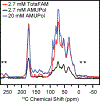
A comparison between DNP-enhanced spectra of HEK293F cells below 6 K with TotaFAM at 2.7 mM (red), AMUPol at 2.7 mM (black), and AMUPol at 20 mM (blue). Experimental details can be found in Supplementary Table S1. Asterisks (*) denote spinning side bands.

References

    1. Hoop CL, Lin H-K, Kar K, Magyarfalvi G, Lamley JM, Boatz JC, Mandal A, Lewandowski JR, Wetzel R, and van der Wel PCA (2016) Huntingtin exon 1 fibrils feature an interdigitated β-hairpin–based polyglutamine core. Proc. Natl. Acad. Sci 113, 1546–1551. - PMC - PubMed
    1. Tuttle MD, Comellas G, Nieuwkoop AJ, Covell DJ, Berthold DA, Kloepper KD, Courtney JM, Kim JK, Barclay AM, Kendall A, Wan W, Stubbs G, Schwieters CD, Lee VMY, George JM, and Rienstra CM (2016) Solid-state NMR structure of a pathogenic fibril of full-length human α-synuclein. Nat. Struct. Mol. Biol 23, 1–9. - PMC - PubMed
    1. Zech SG, Wand AJ, and McDermott AE (2005) Protein Structure Determination by High-Resolution Solid-State NMR Spectroscopy: Application to Microcrystalline Ubiquitin. J. Am. Chem. Soc 127, 8618–8626. - PubMed
    1. Thongsomboon W, Serra DO, Possling A, Hadjineophytou C, Hengge R, and Cegelski L (2018) Phosphoethanolamine cellulose: A naturally produced chemically modified cellulose. Science 359, 334–338. - PubMed
    1. Petkova AT, Ishii Y, Balbach JJ, Antzutkin ON, Leapman RD, Delaglio F, and Tycko R (2002) A structural model for Alzheimer’s β-amyloid fibrils based on experimental constraints from solid state NMR. Proc. Natl. Acad. Sci. U. S. A 99, 16742–7. - PMC - PubMed

Publication types

MeSH terms

Substances