. 2018 Oct 1;120(4):1680-1694.
doi: 10.1152/jn.00715.2017. Epub 2018 Jun 20.

Abnormally reduced primary motor cortex output is related to impaired hand function in chronic stroke

Affiliations

Abnormally reduced primary motor cortex output is related to impaired hand function in chronic stroke

C M Buetefisch et al. J Neurophysiol. .

Abstract

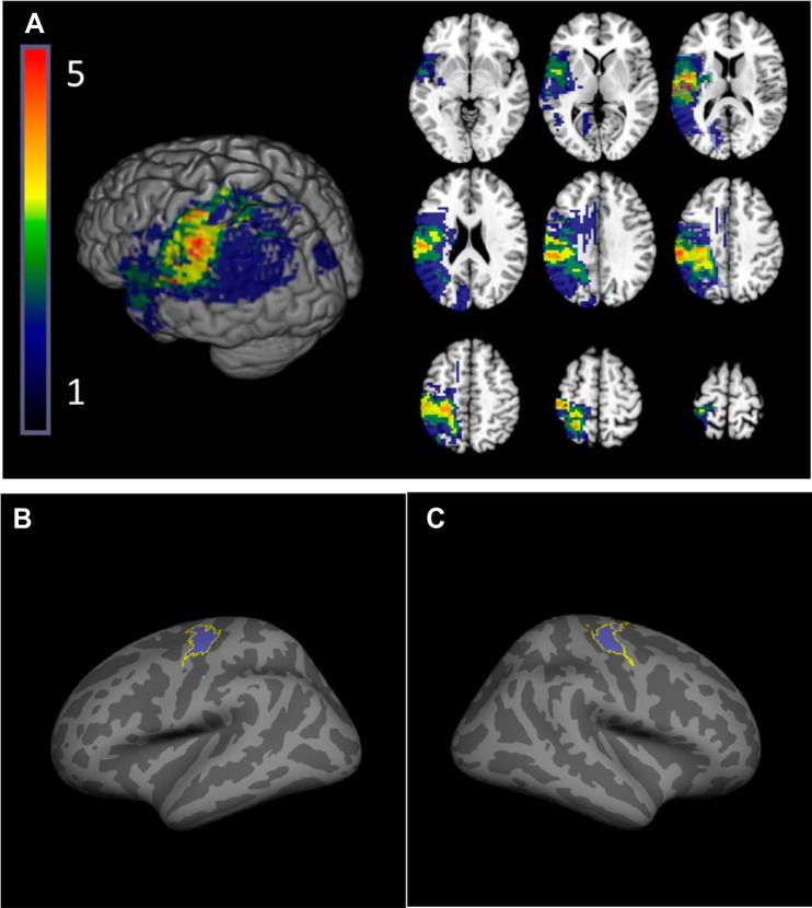
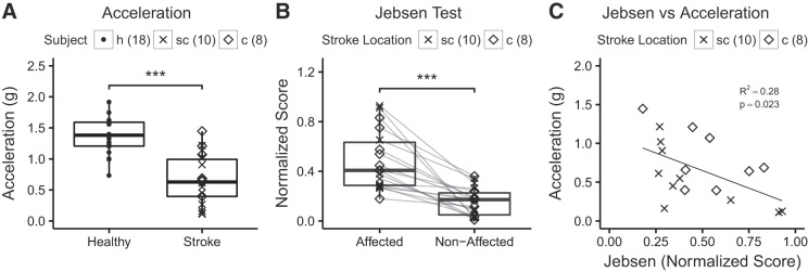
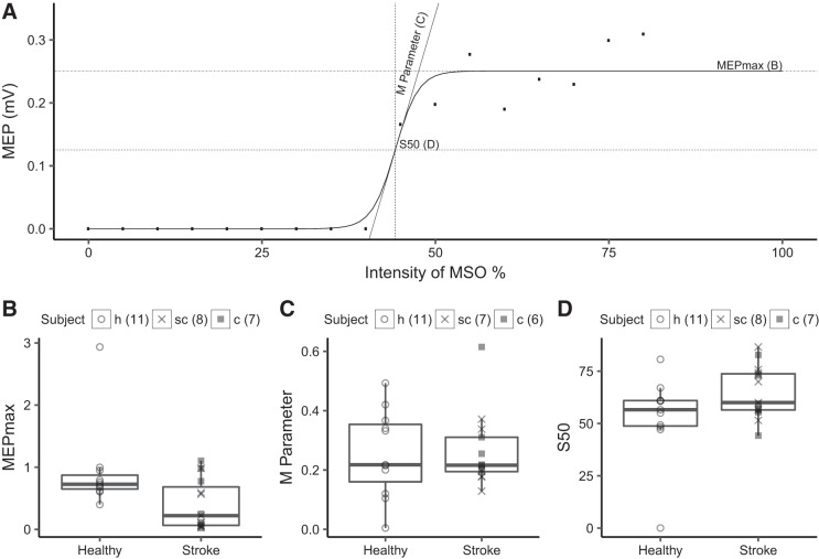
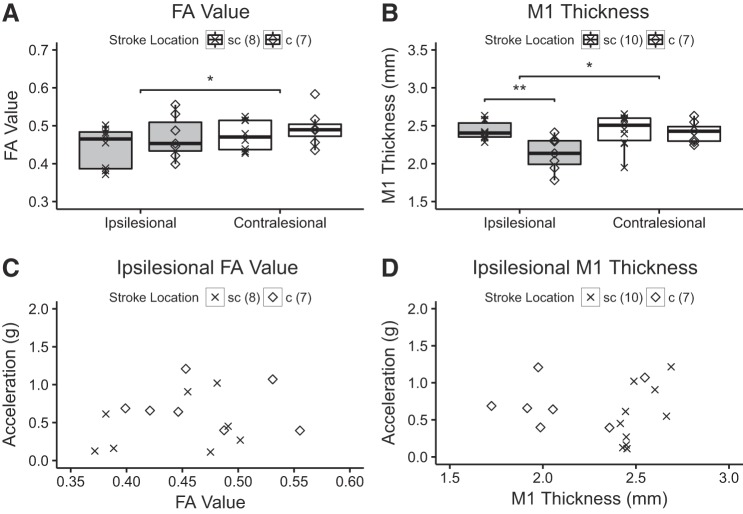
Stroke often involves primary motor cortex (M1) and its corticospinal projections (CST). As hand function is critically dependent on these structures, its recovery is often incomplete. The neuronal substrate supporting affected hand function is not well understood but likely involves reorganized M1 and CST of the lesioned hemisphere (M1IL and CSTIL). We hypothesized that affected hand function in chronic stroke is related to structural and functional reorganization of M1IL and CSTIL. We tested 18 patients with chronic ischemic stroke involving M1 or CST. Their hand function was compared with 18 age-matched healthy subjects. M1IL thickness and CSTIL fractional anisotropy (FA) were determined with MRI and compared with measures of the other hemisphere. Transcranial magnetic stimulation (TMS) was applied to M1IL to determine its input-output function [stimulus response curve (SRC)]. The plateau of the SRC (MEPmax), inflection point, and slope parameters of the curve were extracted. Results were compared with measures in 12 age-matched healthy controls. MEPmax of M1IL was significantly smaller ( P = 0.02) in the patients, indicating reduced CSTIL motor output, and was correlated with impaired hand function ( P = 0.02). M1IL thickness ( P < 0.01) and CSTIL-FA ( P < 0.01) were reduced but did not correlate with hand function. The results indicate that employed M1IL or CSTIL structural measures do not explain the extent of impairment in hand function once M1 and CST are sufficiently functional for TMS to evoke a motor potential. Instead, impairment of hand function is best explained by the abnormally low output from M1IL. NEW & NOTEWORTHY Hand function often remains impaired after stroke. While the critical role of the primary motor cortex (M1) and its corticospinal output (CST) for hand function has been described in the nonhuman primate stroke model, their structure and function have not been systematically evaluated for patients after stroke. We report that in chronic stroke patients with injury to M1 and/or CST an abnormally reduced M1 output is related to impaired hand function.

Keywords: human; rehabilitation; stroke.


Figures

Fig. 1.
Fig. 1.
Stroke lesion overlap. A: participants’ lesions were normalized to standard space and flipped if necessary to the left hemisphere for display purposes, shown overlaid on the ch2.better.nii brain distributed with MRIcron. Color indicates the number of participants with lesions in each voxel. B and C: for association between primary motor cortex of the lesioned hemisphere (M1IL) structure and hand function, we restricted the extraction of cortical thickness values to the hand area of M1IL using the intersection of anatomically defined BA4 ex vivo label supplied by the FreeSurfer toolkit (Fischl et al. 2008) and a hand task-based functional MRI mask.
Fig. 2.
Fig. 2.
Measures of hand motor function. A: peak acceleration of wrist extension movements was significantly lower in stroke patients [displayed separately for subcortical (sc) and cortical (c) location of stroke] when compared with healthy subjects (h) (***P < 0.01, unpaired one-tailed t-tests). B: normalized Jebsen-Taylor Test score data for the affected hand was significantly higher when compared with the nonaffected hand (***P < 0.01, unpaired one-tailed t-tests). Note that this is a time based test where higher scores indicate worse performance. C: relationship between peak acceleration and normalized Jebsen-Taylor Test score. For the correlation analysis, the R2 and P value are given. The number of subjects is indicated. Location of stroke is indicated but statistical testing was done across all stroke patients and healthy subjects.
Fig. 3.
Fig. 3.
Transcranial magnetic stimulation (TMS) measures of primary motor cortex of the lesioned hemisphere (M1IL). A: the stimulus response curve (SRC) evoked by TMS of extensor carpi ulnaris hot spot at 35–80% maximum stimulator output (MSO) intensity for a single stroke patient. We extracted 3 SRC parameters: the plateau of the curve, which is the maximum motor-evoked potential (MEPmax; B), the M parameter (C), and the inflection point of the curve (S50; D) for 15 of the 18 stroke and 11 of the 12 healthy subjects. The number of subjects for each parameter is indicated. B: MEPmax was statistically significant lower in stroke than in healthy subjects (P = 0.02) indicating that maximum corticospinal projection output of surviving neurons was reduced. C and D: there was no statistically significant difference in M parameter and S50 between stroke and healthy subjects (see Table 3 for details of the statistical testing).
Fig. 4.
Fig. 4.
A: short-interval intracortical inhibition (SICI) was weaker in primary motor cortex of the lesioned hemisphere (M1IL) than healthy M1 when tested at a conditioning stimulus (CS) of 80% (P = 0.051) but not at CS of 60% (P = 0.08). B and C: in these scatterplots of healthy controls (B) and stroke patients (C) SICI is plotted against the CS intensity expressed as %maximum stimulator output (MSO). For each subject, the 2 measures of SICI are shown corresponding the CS of 60 and 80% motor threshold (MT). Note that in stroke patients higher CS intensities result in less inhibition consistent with the notion that after stroke, the relationship between MT and CS intensity that produces maximum inhibition is weak and that CS intensity of 80% MT may be too high to capture the effects of lower threshold inhibitory interneurons (Bütefisch et al. 2003). The number of subjects is indicated. Location of stroke is indicated, but statistical testing was done across all stroke patients and healthy subjects. MEP, motor-evoked potential; c, cortical stroke location; sc, subcortical location; h, healthy.
Fig. 5.
Fig. 5.
Measures of primary motor cortex (M1) and corticospinal projection (CST) structure and hand function. A: the fractional anisotropy (FA) value of stroke patients is significantly lower when compared with healthy subjects (*P < 0.01, one-tailed t-test). The location of stroke is indicated, but comparison was done across the entire group. There was no difference for FA value between cortical and subcortical location of the stroke. B: the M1 of the affected hemisphere was significantly thinner when compared with M1 of the nonaffected hemisphere (**P < 0.04, one-tailed t-test). For the cortical location of the stroke, M1 was significantly thinner when compared with M1 of patients with subcortical stroke healthy subjects (*P < 0.01, one-tailed t-test). C: ipsilesional FA value. D: ipsilesional M1 thickness. c, Cortical stroke location, sc, subcortical location.

References

    1. Allred RP, Jones TA. Maladaptive effects of learning with the less-affected forelimb after focal cortical infarcts in rats. Exp Neurol 210: 172–181, 2008. doi:10.1016/j.expneurol.2007.10.010. - DOI - PMC - PubMed
    1. Amassian VE, Cracco RQ. Human cerebral cortical responses to contralateral transcranial stimulation. Neurosurgery 20: 148–155, 1987. doi:10.1097/00006123-198701000-00031. - DOI - PubMed
    1. Basser PJ. Inferring microstructural features and the physiological state of tissues from diffusion-weighted images. NMR Biomed 8: 333–344, 1995. doi:10.1002/nbm.1940080707. - DOI - PubMed
    1. Bennett KM, Lemon RN. Corticomotoneuronal contribution to the fractionation of muscle activity during precision grip in the monkey. J Neurophysiol 75: 1826–1842, 1996. doi:10.1152/jn.1996.75.5.1826. - DOI - PubMed
    1. Bestmann S, Swayne O, Blankenburg F, Ruff CC, Teo J, Weiskopf N, Driver J, Rothwell JC, Ward NS. The role of contralesional dorsal premotor cortex after stroke as studied with concurrent TMS-fMRI. J Neurosci 30: 11926–11937, 2010. doi:10.1523/JNEUROSCI.5642-09.2010. - DOI - PMC - PubMed

Publication types