. 2018 Jun 20;13(6):e0198013.
doi: 10.1371/journal.pone.0198013. eCollection 2018.

Nephrin is necessary for podocyte recovery following injury in an adult mature glomerulus

Affiliations

Nephrin is necessary for podocyte recovery following injury in an adult mature glomerulus

Rakesh Verma et al. PLoS One. .

Abstract

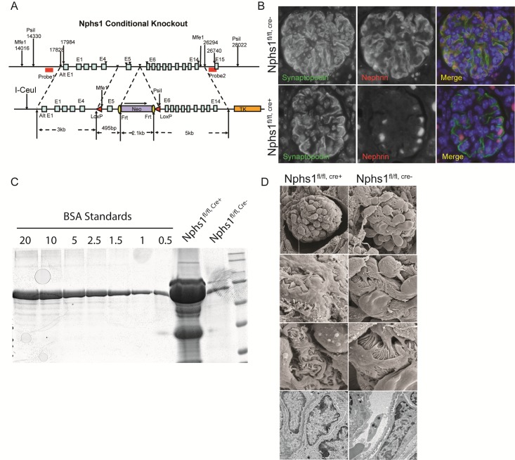
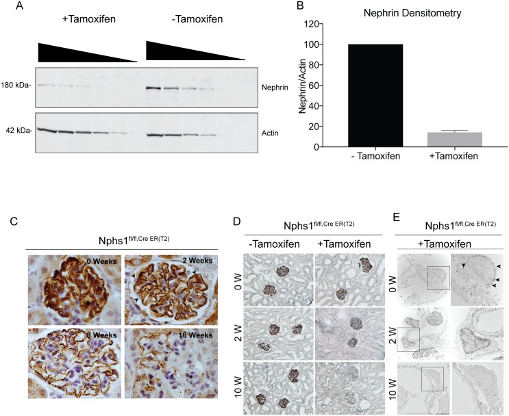
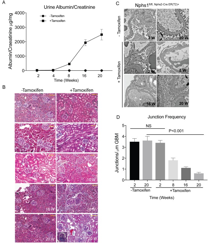
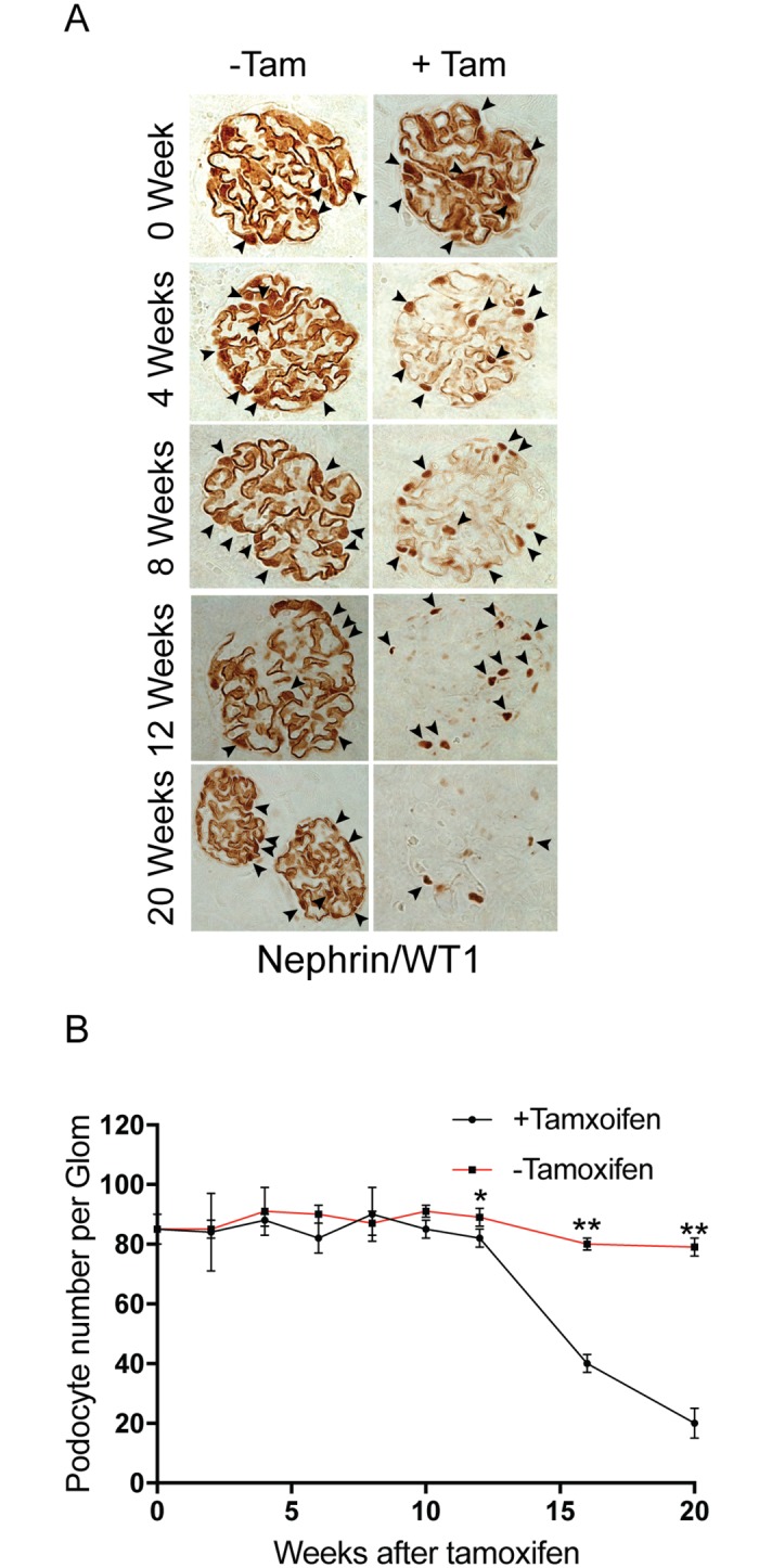
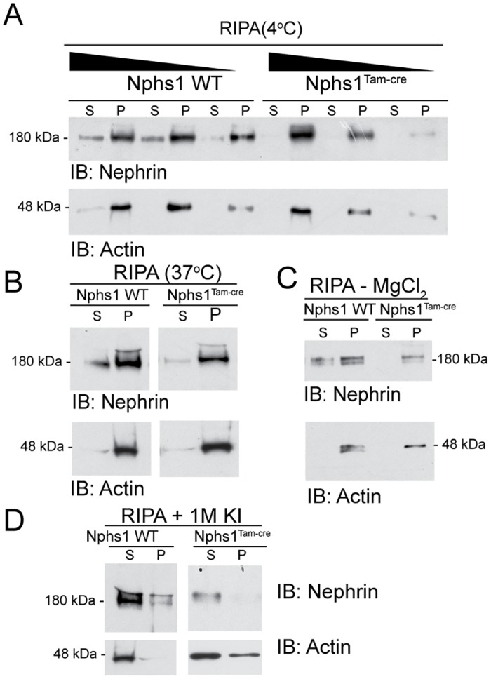
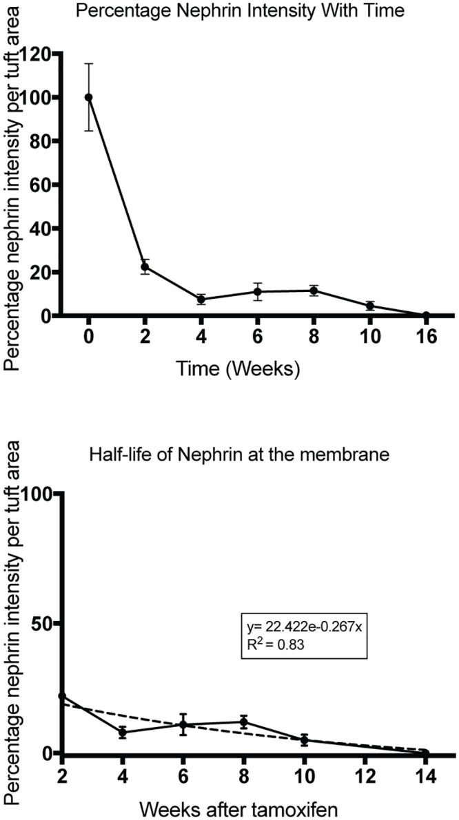
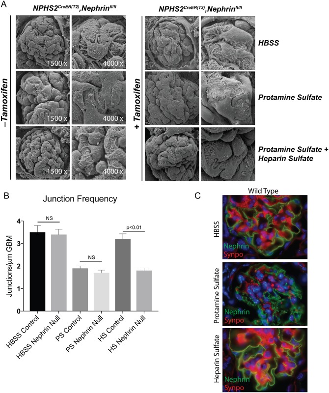
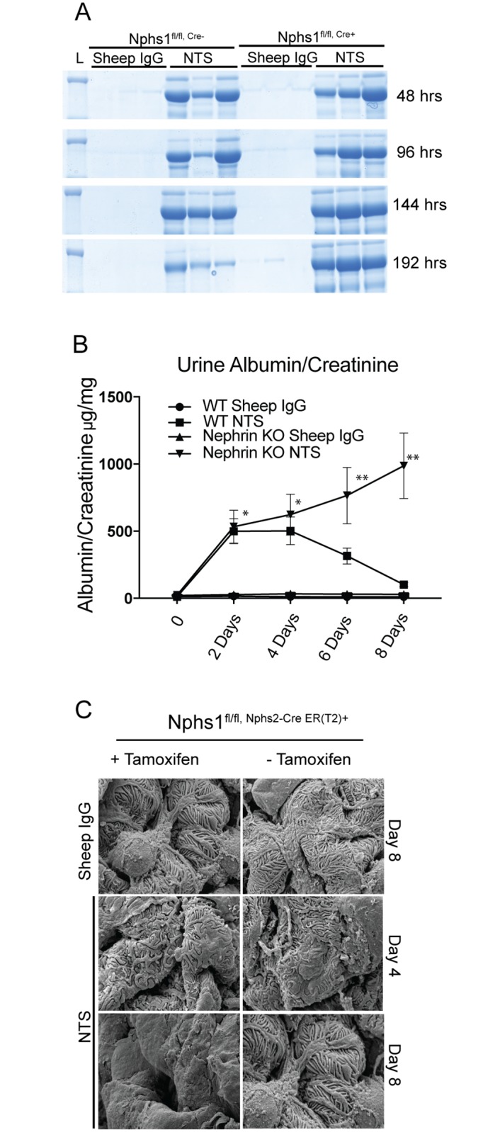
Nephrin (Nphs1) is an adhesion protein that is expressed at the podocyte intercellular junction in the glomerulus. Nphs1 mutations in humans or deletion in animal genetic models results in a developmental failure of foot process formation. A number of studies have shown decrease in expression of nephrin in various proteinuric kidney diseases as well as in animal models of glomerular disease. Decrease in nephrin expression has been suggested to precede podocyte loss and linked to the progression of kidney disease. Whether the decrease in expression of nephrin is related to loss of podocytes or lead to podocyte detachment is unclear. To answer this central question we generated an inducible model of nephrin deletion (Nphs1Tam-Cre) in order to lower nephrin expression in healthy adult mice. Following tamoxifen-induction there was a 75% decrease in nephrin expression by 14 days. The Nphs1Tam-Cre mice had normal foot process ultrastructure and intact filtration barriers up to 4-6 weeks post-induction. Despite the loss of nephrin expression, the podocyte number and density remained unchanged during the initial period. Unexpectedly, nephrin expression, albeit at low levels persisted at the slit diaphragm up to 16-20 weeks post-tamoxifen induction. The mice became progressively proteinuric with glomerular hypertrophy and scarring reminiscent of focal and segmental glomerulosclerosis at 20 weeks. Four week-old Nphs1 knockout mice subjected to protamine sulfate model of podocyte injury demonstrated failure to recover from foot process effacement following heparin sulfate. Similarly, Nphs1 knockout mice failed to recover following nephrotoxic serum (NTS) with persistence of proteinuria and foot process effacement. Our results suggest that as in development, nephrin is necessary for maintenance of a healthy glomerular filter. In contrast to the developmental phenotype, lowering nephrin expression in a mature glomerulus resulted in a slowly progressive disease that histologically resembles FSGS a disease linked closely with podocyte depletion. Podocytes with low levels of nephrin expression are both susceptible and unable to recover following perturbation. Our results suggest that decreased nephrin expression independent of podocyte loss occurring as an early event in proteinuric kidney diseases might play a role in disease progression.


Conflict of interest statement

The authors have declared that no competing interests exist.

Figures

Fig 1
Fig 1. Conditional deletion of nephrin in podocytes.
(A) Schematic of the targeting vector containing a neomycin cassette, Frt sites and loxP sites were engineered to flank exon 5 of mouse nephrin. Successful recombination was verified using Southern blot with probes 1 and 2 flanking the nephrin gene. (B) Immunofluorescence images showing absence of nephrin in paraffin embedded mouse kidney section. Synaptopodin staining was used to identify podocytes. C) Coomassie blue stained SDS-PAGE gel showing severe albuminuria/proteinuria in the Nphs1fl/fl,Cre+ animals at 2 weeks following birth. BSA standards (0.5–20 μg/dl). (D) Transmission and Scanning electron microscopy images showing podocyte foot process developmental abnormalities following nephrin deletion.
Fig 2
Fig 2. Induced model of nephrin deletion in podocytes.
(A) Western blot showing decrease in nephrin in glomerular lysates from Nphs1fl/fl,iCre(ER)T2 mice kidneys 14 weeks post-induction with tamoxifen. Decreasing amounts of lysate volume were loaded in each well to avoid reaching the threshold of the x-ray film. Actin is used as loading control. (B) Densitometry showing quantification of the decrease in total nephrin following deletion. Results are representative of 4 separate experiments. (C) Immunoperoxidase staining for nephrin at various time points following tamoxifen-induction. Arrowheads show loss of cytoplasmic nephrin staining in podocytes at 2 weeks. (D) Light microscopy images of kidney tissue sections following silver enhancement of immunogold particles. (E) Immunogold electron microscopy of mouse kidney sections at 2 weeks and 10 weeks showing gold particles (arrowheads) at the slit diaphragm and within the cell body. Gold particles are seen primarily at the slit diaphragm in Nphs1fl/fl,Cre+ at 10 weeks. Right panel shows enlarged images of the boxed areas in the left panel. Images were taken at X12,200 and cropped to emphasize the immunogold particles.
Fig 3
Fig 3. Deletion of nephrin in adult mature glomerulus.
(A) Immunofluorescence images showing nephrin and synaptopodin staining at 2 weeks following Cre-induction with tamoxifen. There is abrogation of peri-nuclear nephrin staining following induction (arrowheads). (B) Immunofluorescence images showing nephrin and synaptopodin staining at various time points following tamoxifen. Scale bars, 20 μm.
Fig 4
Fig 4. Nephrin deletion in adult mature glomerulus results in focal segmental glomerulosclerosis and proteinuria.
(A) Mice with tamoxifen-induced nephrin deletion develop proteinuria at 6–8 weeks following tamoxifen. (B) Masson Trichrome Verhoeff staining of mouse kidney sections at different time points following tamoxifen-induction showing development of focal and segmental glomerular scarring at 16 weeks following tamoxifen. Scale bars, 20μm. (C) Transmission EM images show normal foot process structure at 4 weeks following tamoxifen (Images were acquired at 10,400X and cropped for each panel). There is widespread foot process spreading at 8 weeks following tamoxifen. (D) Assessment of junction frequency shows decrease in the number of podocyte intercellular junction frequency at 8 weeks following tamoxifen. All results are representative of 4 sets of experiments with 5 animals in each group. Data are Mean ± SEM.
Fig 5
Fig 5. Podocyte numbers remain unchanged for 10–12 weeks following nephrin gene deletion.
(A) Immunohistochemistry using nephrin and WT1 antibodies simultaneously shows presence of nuclear WTI (arrowheads) staining despite loss of nephrin in kidney sections at 12 weeks after tamoxifen-induced nephrin deletion. (B) Podocyte counts per glomerulus at various time points in controls and tamoxifen-induced nephrin deletion. All results are representative of 4 sets of experiments with 5 animals in each group. Data are Mean ± SEM.
Fig 6
Fig 6. Residual nephrin at the membrane is associated with actin.
(A) Isolated glomeruli from wild type and Nphs1Tam-Cre mice kidneys were lysed in RIPA buffer containing 1% Triton X-100 at 4°C. Lysates were resolved with SDS-PAGE gel under reducing conditions and probed with nephrin and actin antibodies. Triton X1000 soluble fraction (S) and insoluble pellet (P) were separated by centrifugation. Nephrin is seen primarily in the insoluble fraction or pellet. (B) Glomerular lysates in RIPA buffer at 37°C showing no improvement in nephrin solubulity. (C) Glomerular lysates in RIPA buffer without 1.5 M MgCl2 shows slight improvement in nephrin solubility with prominence of the high molecular weight band in the Nphs1Tam-Cre mice. (D) Glomerular lysates in RIPA buffer containing 1M potassium iodide (KI) show dramatic improvement in nephrin solubility. IB: Immunoblot.
Fig 7
Fig 7. Nephrin has a half-life of 2.59 weeks at the membrane.
(A) Mouse kidney sections stained for nephrin using immunohistochemistry were analyzed for nephrin staining intensity at various time points following tamoxifen-induction using MetaMorph image analysis software. (B) Nephrin intensity at time = 0 weeks was plotted as 100%. There is rapid decline in nephrin staining in the initial 2 weeks followed by a slow decline over the next 14–16 weeks. The initial decline represents loss of new-nephrin production and represents nephrin that is primarily within the podocyte cell body. Nephrin staining after 2 weeks is primarily at the membrane (see S4 Fig). (C) Half-life analysis of nephrin at the membrane after the 2 weeks time point. Non-linear regression analysis (dotted line) shows a nephrin half-life 2.59 weeks at the membrane (R2 = 0.83). The data is representative of 70–100 glomeruli per kidney from 3–4 animals in each group. Data are Mean ± SD.
Fig 8
Fig 8. Nephrin-deleted mature podocyte fail to recover following protamine sulfate injury.
(A) SEM images of kidneys from 8 weeks old Nphs1fl/fl,iCre(ER)T2 mice following protamine sulfate injury. Nphs1fl/fl,iCre(ER)T2 mice that received tamoxifen show failure of foot process recovery in response to heparin sulfate following injury with protamine sulfate. (B) Analysis of junction frequency in TEM images show failure of recovery following heparin sulfate in Nphs1fl/fl,iCre(ER)T2 mice that received tamoxifen. (C) Immunofluorescence images of wild type animals (Nphs1fl/fl,iCre(ER)T2, tamoxifen-) show change in nephrin distribution from the membrane to punctate structures following protamine sulfate injury (middle panel). Nephrin staining is reversed back to the membrane on perfusion with heparin sulfate (lower panel). Synaptopodin (synpo) was used to for double staining the podocytes. All mice used in these experiments were 8–10 weeks old prior to receiving tamoxifen.
Fig 9
Fig 9. Nphs1fl/fl,iCre(ER)T2 mice continue to be proteinuric following NTS injury.
(A) Coomassie blue stained SDS-PAGE gel show persistence of proteinuria following NTS single dose administration in Nphs1fl/fl,iCre(ER)T2 mice that received tamoxifen. (B) Urine albumin/creatinine ratio from Nphs1fl/fl,iCre(ER)T2 mice that were fed tamoxifen and normal chow following NTS injury. Most nephrin deleted mice did not survive beyond 8–10 days following NTS. (C) SEM images from Nphs1fl/fl,iCre(ER)T2 mice kidneys with and without tamoxifen induction following NTS injury show failure of recovery of nephrin-deleted podocytes at day 8 compared to control. The data is representative of 4 sets of experiments with 3 animals in each group. Sheep IgG was used as control. Data are Mean ± SEM. *P = Not significant, ** P <0.01, when compared between WT and Nephrin knock out in the NTS group.

References

    1. Ruotsalainen V, Ljungberg P, Wartiovaara J, Lenkkeri U, Kestila M, Jalanko H, et al. (1999) Nephrin is specifically located at the slit diaphragm of glomerular podocytes. Proc Natl Acad Sci U S A 96: 7962–7967. - PMC - PubMed
    1. Kestila M, Lenkkeri U, Mannikko M, Lamerdin J, McCready P, Putaala H, et al. (1998) Positionally cloned gene for a novel glomerular protein—nephrin—is mutated in congenital nephrotic syndrome. Mol Cell 1: 575–582. - PubMed
    1. Putaala H, Soininen R, Kilpelainen P, Wartiovaara J, Tryggvason K (2001) The murine nephrin gene is specifically expressed in kidney, brain and pancreas: inactivation of the gene leads to massive proteinuria and neonatal death. Hum Mol Genet 10: 1–8. - PubMed
    1. Wang SX, Rastaldi MP, Patari A, Ahola H, Heikkila E, Holthofer H (2002) Patterns of nephrin and a new proteinuria-associated protein expression in human renal diseases. Kidney Int 61: 141–147. doi: 10.1046/j.1523-1755.2002.00114.x - DOI - PubMed
    1. Luimula P, Ahola H, Wang SX, Solin ML, Aaltonen P, Tikkanen I, et al. (2000) Nephrin in experimental glomerular disease. Kidney Int 58: 1461–1468. doi: 10.1046/j.1523-1755.2000.00308.x - DOI - PubMed

Publication types

LinkOut - more resources