. 2018 Jun 20;13(6):e0199517.
doi: 10.1371/journal.pone.0199517. eCollection 2018.

ARQ-197, a small-molecule inhibitor of c-Met, reduces tumour burden and prevents myeloma-induced bone disease in vivo

Affiliations

ARQ-197, a small-molecule inhibitor of c-Met, reduces tumour burden and prevents myeloma-induced bone disease in vivo

Darren L Lath et al. PLoS One. .

Abstract

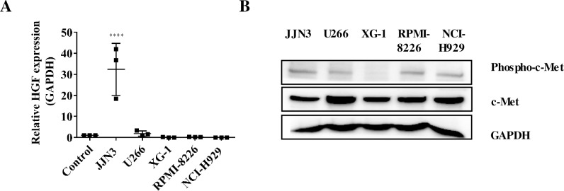
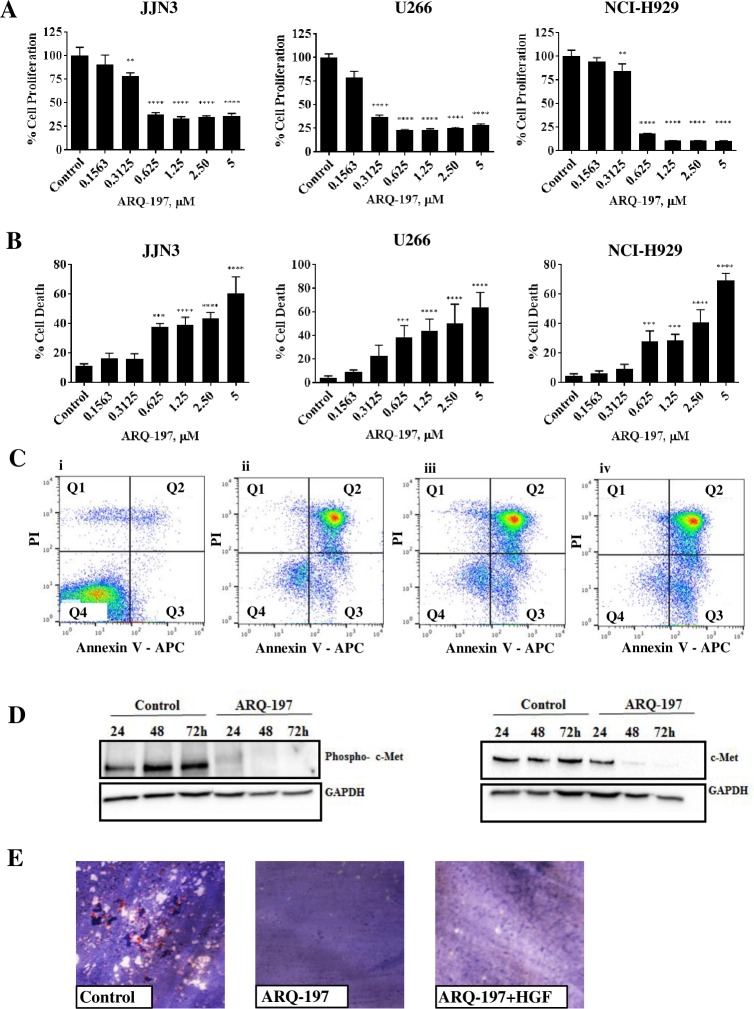
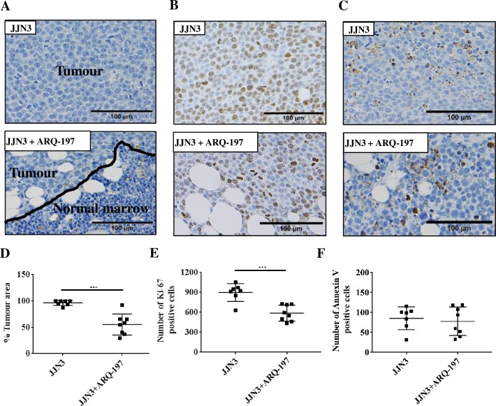
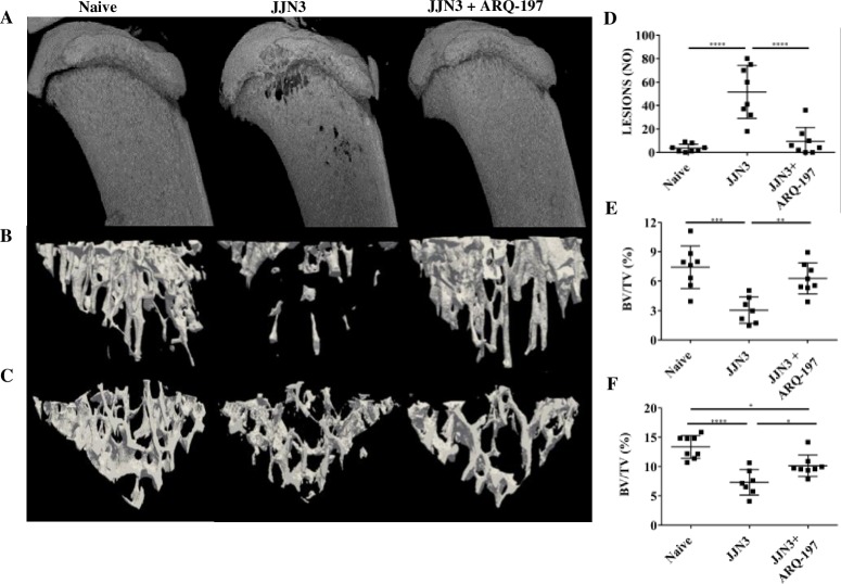
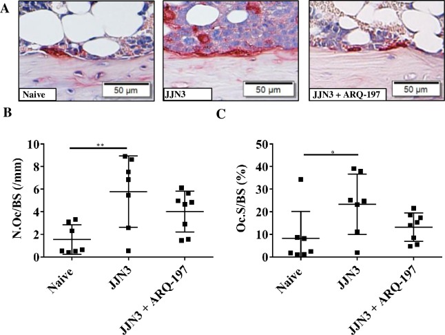
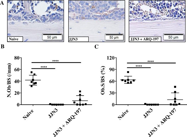
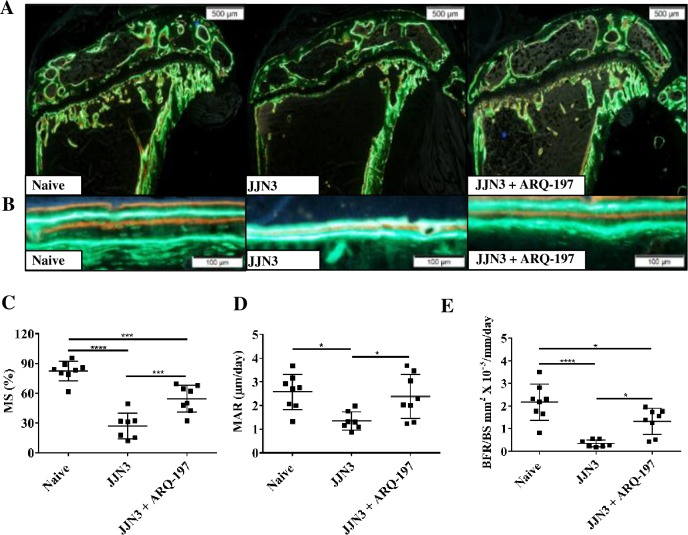
The receptor tyrosine kinase c-Met, its ligand HGF, and components of the downstream signalling pathway, have all been implicated in the pathogenesis of myeloma, both as modulators of plasma cell proliferation and as agents driving osteoclast differentiation and osteoblast inhibition thus, all these contribute substantially to the bone destruction typically caused by myeloma. Patients with elevated levels of HGF have a poor prognosis, therefore, targeting these entities in such patients may be of substantial benefit. We hypothesized that ARQ-197 (Tivantinib), a small molecule c-Met inhibitor, would reduce myeloma cell growth and prevent myeloma-associated bone disease in a murine model. In vitro we assessed the effects of ARQ-197 on myeloma cell proliferation, cytotoxicity and c-Met protein expression in human myeloma cell lines. In vivo we injected NOD/SCID-γ mice with PBS (non-tumour bearing) or JJN3 cells and treated them with either ARQ-197 or vehicle. In vitro exposure of JJN3, U266 or NCI-H929 cells to ARQ-197 resulted in a significant inhibition of cell proliferation and an induction of cell death by necrosis, probably caused by significantly reduced levels of phosphorylated c-Met. In vivo ARQ-197 treatment of JJN3 tumour-bearing mice resulted in a significant reduction in tumour burden, tumour cell proliferation, bone lesion number, trabecular bone loss and prevented significant decreases in the bone formation rate on the cortico-endosteal bone surface compared to the vehicle group. However, no significant differences on bone parameters were observed in non-tumour mice treated with ARQ-197 compared to vehicle, implying that in tumour-bearing mice the effects of ARQ-197 on bone cells was indirect. In summary, these res ults suggest that ARQ-197 could be a promising therapeutic in myeloma patients, leading to both a reduction in tumour burden and an inhibition of myeloma-induced bone disease.


Conflict of interest statement

The authors have declared no competing interests exist.

Figures

Fig 1
Fig 1. JJN3 human myeloma cells exhibited higher HGF gene expression compared to other human myeloma cell lines and healthy plasma cells and a higher phospho-c-Met protein level than other human myeloma cell lines.
(A) Relative HGF gene expression of human myeloma cell lines JJN3, NCIH-H929, U226, XG-1 and RPMI-8226 and healthy human plasma cells (control). All data displayed as mean ± SD and analysed using a normal one-way ANOVA, where significance is indicated by ****P<0.0001. (B) Phospho-c-Met and total c-Met protein levels in human myeloma cells.
Fig 2
Fig 2. ARQ-197 inhibits cell proliferation, induces cell death by necrosis and reduces protein expression of phospho-c-Met and c-Met.
(A) JJN3, U266 and NCI-H929 cells were incubated with control media containing DMSO or ARQ-197 at 0.1563, 0.3125, 0.625, 1.25, 2.50 or 5 μM for 48 h. Cell proliferation was measured compared to DMSO control. (B) JJN3, U266 and NCI-H929 cells were incubated with control media containing DMSO or ARQ-197 at 0.1563, 0.3125, 0.625, 1.25, 2.50 or 5 μM for 48 h and counted using trypan blue exclusion and the percentage cell death calculated. All data displayed as mean ± SD and analysed using a normal one-way ANOVA, where significance is indicated by **P<0.01, ***P<0.001 or ****P<0.0001. (C) JJN3 cells were treated with control media containing DMSO (i) or ARQ-197 at 0.3125 (ii), 1.25 (iii) or 5 μM (iv) and then stained with annexin V/PI before flow cytometric analysis. In the flow cytrometry plots, Q1 relates to dead cells, Q2 necrotic, Q3 apoptotic and Q4 viable cells. (D) JJN3 cells were treated with control media containing DMSO or 1 μM ARQ-197 for 24 h, 48 h and 72 h and immunoblotted with an anti-phospho-c-Met antibody and an anti-c-Met antibody. (E) Representative images of osteoclasts treated with either control media containing DMSO, ARQ-197 1 μM or ARQ-197 1 μM with HGF 50 ng.
Fig 3
Fig 3. ARQ-197 reduces tumour burden and tumour cell proliferation but does not induce cell death by necrosis in JJN3 tumour-bearing mice.
(A) Representative images of histological sections stained by Haematoxylin for tumour burden, (B) immunohistological staining by anti-Ki-67 or (C) anti-Annexin V. All scale bars = 100 μm. Histomorphometric analysis of (D) tumour burden, (E) Ki-67 and (F) Annexin V on tibiae from JJN3 tumour-bearing mice treated with either vehicle (JJN3) or ARQ-197 (JJN3+ARQ-197). All data displayed as mean ± SD and analysed using an unpaired t-test, where significance is indicated by ***P<0.001.
Fig 4
Fig 4. ARQ-197 reduces the number of osteolytic lesions and prevents the loss of trabecular bone in JJN3 tumour-bearing mice.
(A) Representative μCT 3D and (B) cross-sectional images of tibiae or (C) vertebrae (L3) from naïve mice (Naïve) and tumour-bearing mice treated with vehicle (JJN3) or ARQ-197 (JJN3+ARQ-197). (D) Lesion counts (Lesions NO) and (E) trabecular bone fraction (BV/TV %) analysed by μCT of the tibiae or (F) vertebrae (L3) from naïve mice (Naïve) and tumour-bearing mice treated with vehicle (JJN3) or ARQ-197 (JJN3+ARQ-197). All data displayed as mean ± SD and analysed using a normal one-way ANOVA, where significance is indicated by *P<0.05, **P<0.01, ***P<0.001 or ****P<0.0001.
Fig 5
Fig 5. ARQ-197 reduces osteoclastic bone resorption on cortico-endosteal surfaces of the tibiae from JJN3 tumour-bearing mice.
(A) Representative images of TRAP positive osteoclasts (stained red) on the cortico-endosteal surface of tibiae from naïve mice (Naïve) and tumour-bearing mice treated with vehicle (JJN3) or ARQ-197 (JJN3+ARQ-197) (scale bar = 50 μm). (B) Histomorphometric analysis of the number of TRAP positive osteoclasts per mm cortico-endosteal bone (N.Oc/BS,mm) and (C) the percentage coverage of TRAP positive osteoclasts on the cortico-endosteal bone (Oc.S/BS (%) from naive mice (Naïve) and tumour-bearing mice treated with vehicle (JJN3) or ARQ-197 (JJN3+ARQ-197). All data displayed as mean ± SD and analysed using a normal one-way ANOVA, where significance is indicated by *P<0.05 or **P<0.01.
Fig 6
Fig 6. ARQ-197 inhibits osteoblast loss on the cortico-endosteal surfaces of the tibiae from JJN3 tumour-bearing mice.
(A) Representative images of osteoblasts on the cortico-endosteal surface of tibiae from naïve mice (Naïve) and tumour-bearing mice treated with vehicle (JJN3) or ARQ-197 (JJN3+ARQ-197) (scale bar = 50 μm). (B) Histomorphometric analysis of the number of osteoblasts per mm cortico-endosteal bone (N.Ob/BS,mm) and (C) of the percentage coverage of osteoblasts on the cortico-endosteal bone (Ob.S/BS (%) from naive mice (Naïve) and tumour-bearing mice treated with vehicle (JJN3) or ARQ-197 (JJN3+ARQ-197). All data displayed as mean ± SD and analysed using a normal one-way ANOVA, where significance is indicated ****P<0.0001.
Fig 7
Fig 7. ARQ-197 inhibits bone formation loss on the cortico-endosteal surface of the tibiae from JJN3 tumour-bearing mice.
Representative images of bone formation on the, cortico-endosteal surfaces, (A) magnification x4 (scale bar = 500 μm) and (B) magnification x20 (scale bar = 100 μm) of tibiae from naïve mice (Naïve) and tumour-bearing mice treated with vehicle (JJN3) or ARQ-197 (JJN3+ARQ-197). Histomorphometric analysis of (C) the mineralising surface (MS, %) (D) the mineral apposition rate (MAR, μm/day) and (E) the bone formation rate (BFR/BS, mm2 X 10−3/mm/day) on the cortico-endosteal bone surface of tibiae from naive mice (Naïve) and tumour-bearing mice treated with vehicle (JJN3) or ARQ-197 (JJN3+ARQ-197). All data displayed as mean ± SD and analysed using a normal one-way ANOVA, where significance is indicated by *P<0.05, ***P<0.001 or ****P<0.0001.

References

    1. Palumbo A, Giaccone L, Bertola A, Pregno P, Bringhen S, Rus C, et al. Low-dose thalidomide plus dexamethasone is an effective salvage therapy for advanced myeloma. Haematologica. 2001;4:399–403. - PubMed
    1. Dimopoulos MA, Zervas K, Kouvatseas G, Galani E, Grigoraki V, Kiamouris C, et al. Thalidomide and dexamethasone combination for refractory multiple myeloma. Ann Oncol. 2001;12:991–5. - PubMed
    1. Richardson PG, Blood E, Mitsiades CS, Jagannath S, Zeldenrust SR, Alsina M, et al. A randomized phase 2 study of lenalidomide therapy for patients with relapsed or relapsed and refractory multiple myeloma. Blood. 2006;108:3458–64. doi: 10.1182/blood-2006-04-015909 - DOI - PMC - PubMed
    1. Stadtmauer EA, Weber DM, Niesvizky R, Belch A, Prince MH, San Miguel JF, et al. Lenalidomide in combination with dexamethasone at first relapse in comparison with its use as later salvage therapy in relapsed or refractory multiple myeloma. Eur J Haematol. 2009;82:426–32. doi: 10.1111/j.1600-0609.2009.01257.x - DOI - PMC - PubMed
    1. Lacy MQ, Hayman SR, Gertz MA, Short KD, Dispenziere A, Kumar S, et al. Pomalidomide (CC4047) plus low dose dexamethasone (Pom/dex) is active and well tolerated in lenalidomide refractory multiple myeloma (MM). Leukemia. 2010;24:1934–9. doi: 10.1038/leu.2010.190 - DOI - PMC - PubMed

Publication types

MeSH terms