. 2018 Jul;592(14):2466-2475.
doi: 10.1002/1873-3468.13168. Epub 2018 Jun 30.

Hansenula polymorpha Aat2p is targeted to peroxisomes via a novel Pex20p-dependent pathway

Affiliations

Hansenula polymorpha Aat2p is targeted to peroxisomes via a novel Pex20p-dependent pathway

Ann S Thomas et al. FEBS Lett. 2018 Jul.

Abstract

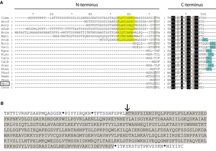
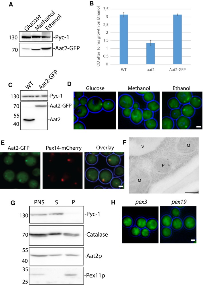
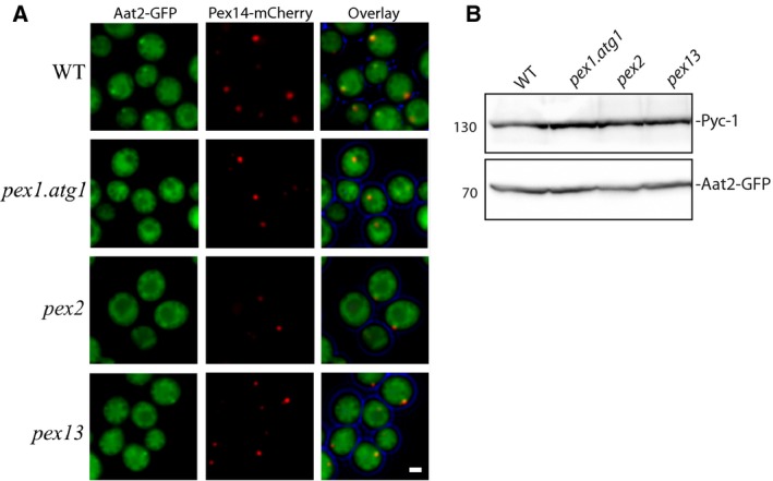
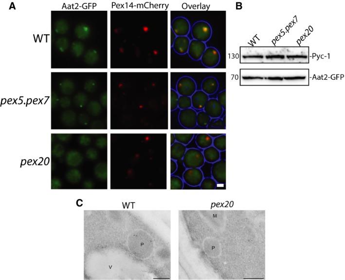
Saccharomyces cerevisiae Aat2p contains a peroxisomal targeting signal type-1 and localizes to peroxisomes in oleate-grown cells, but not in glucose-grown cells. Here, we have investigated Aat2p from the yeast Hansenula polymorpha, which lacks a recognizable peroxisomal targeting signal. Aat2p tagged with GFP at its C terminus displays a dual cytosol-peroxisome localization in ethanol-grown cells. The partial peroxisomal localization of Aat2p persisted in the absence of the classical cycling receptors Pex5p and Pex7p but Aat2p targeting to peroxisomes was reduced in cells deleted for the matrix protein import factors PEX1, PEX2 and PEX13. Furthermore, we demonstrate that Aat2p targeting to peroxisomes requires Pex20p. Together, our data identify a Pex20p-dependent pathway for targeting Aat2p to peroxisomes.

Keywords: Hansenula polymorpha; Aat2p; Peroxisome protein import; Pex20p; aspartate aminotransferase.


Figures

Figure 1
Figure 1
HpAat2p lacks a recognizable Peroxisomal Targeting Sequence. (A) Multiple sequence alignment of the N‐ and C‐terminal regions of Aat2p from different yeast and fungi. Black shading indicates identity. Similarity is indicated as white letters that are shaded dark grey when present in 19 of the 22 sequences and as black letters shaded light grey when present in 14 of the 22 sequences. Putative PTS1 sequences are shaded cyan, putative PTS2 sequences in yellow. (Fungi: Cimm, Coccidioides immitis; Anid, Aspergillus nidulans; Aory, Aspergillus oryzae; Afum, Aspergillus fumigatus; Ncra, Neurospora crassa; Mgri, Magnaporthe grisea; Prub, Penicillium rubens Wisconsin; Bcin, Botrytis cinerea; Sscl, Sclerotinia sclerotiorum. Yeasts: Egos, Eremothecium gossypii; Klac, Kluyveromyces lactis; Cgla, Candida glabrata; Calb, Candida albicans; Scer, Saccharomyces cerevisiae; Dhan, Debaryomyces hansenii; Pkud, Pichia kudriavzevii; Pmem, Pichia membranifaciens; Ppas, Pichia pastoris; Hpol (boxed), Hansenula polymorpha; Cara, Candida arabinofermentas). (B) Hansenula polymorpha Aat2 protein sequence (shaded in grey) showing potential upstream and downstream amino acid residues. The start codon of Aat2p is indicated with an arrow, while stop codons are indicated with an asterisk.
Figure 2
Figure 2
HpAat2p partially localizes to peroxisomes. (A) Cell lysates of WT cells grown on different carbon sources expressing Aat2p tagged with GFP at the C terminus were subjected to SDS/PAGE and immunoblotting using antibodies directed against GFP or Pyc‐1 (loading control). (B) Graph representing growth of WT, aat2 or a strain harbouring Aat2‐GFP on ethanol containing media. Growth of cells is indicated as a measure of optical density of the culture at an absorbance of 660 nm. (C) Western blot showing WT or a strain containing Aat2‐GFP, probed with antibodies directed against Aat2p. (D) Fluorescence microscopy analysis of WT cells expressing Aat2p tagged with GFP at the C terminus, grown on various carbon sources. Scale bar represents 1 μm. (E) Colocalization analysis of Aat2‐GFP with the peroxisomal marker Pex14‐mCherry. Scale bar represents 1 μm. (F) Immuno‐Electron microscopy analysis showing WT cells grown on ethanol. Aat2p was labelled with antibodies against Aat2p and detected with goat anti‐rabbit antibodies conjugated to 6‐nm gold particles. M‐Mitochondria, P‐Peroxisomes, V‐Vacuole. Scale bar represents 200 nm. (G) Cell fractionation analysis of WT cells, displaying the postnuclear supernatant (PNS), supernatant (S) and organelle pellet (P) fractions probed with SDS/PAGE, western blotting and antibodies against the cytosolic protein Pyc‐1, the peroxisomal matrix protein Catalase, the peroxisomal membrane protein Pex11p and Aat2p. (H) Fluorescence microscopy analysis of ethanol‐grown pex3 or pex19 cells expressing Aat2‐GFP. Scale bars represent 1 μm. Note that all GFP fluorescent images were processed differently, in order to visualize the GFP signal optimally.
Figure 3
Figure 3
Pex1p, Pex2p and Pex13p play a role in targeting HpAat2p to peroxisomes (A) Fluorescence microscopy images of WT, pex1.atg1, pex2 and pex13 cells grown for 16 h on ethanol containing media. Besides the peroxisomal marker Pex14‐mCherry, all cells produced the fusion protein Aat2‐GFP. Scale bar represents 1 μm. (B) SDS/PAGE and immunoblot analysis of lysates from the deletion strains presented in (A) probed with antibodies directed against GFP or Pyc‐1 (loading control).
Figure 4
Figure 4
HpAat2p targets to peroxisomes in a Pex20p‐dependent manner. (A) Fluorescence microscopy images of WT, pex5.pex7 and pex20 deletion strains grown for 16 h on ethanol containing media. Besides the peroxisomal marker Pex14‐mCherry, all cells produced the fusion protein Aat2‐GFP. Scale bar represents 1 μm. (B) SDS/PAGE and immunoblot analysis of lysates from the deletion strains presented in (A) probed with antibodies directed against GFP or Pyc‐1 (loading control). (C) Immuno‐Electron microscopy analysis showing WT and pex20 cells grown on ethanol for 16 h. Aat2p was labelled with anti‐Aat2p and detected with goat anti‐rabbit antibodies conjugated to 6‐nm gold particles. M‐Mitochondria, P‐Peroxisomes, V‐Vacuole. Scale bar represents 200 nm.

Similar articles

Cited by

References

    1. Kohlwein SD, Veenhuis M and van der Klei IJ (2013) Lipid droplets and peroxisomes: key players in cellular lipid homeostasis or a matter of fat–store ‘em up or burn ‘em down. Genetics 193, 1–50. - PMC - PubMed
    1. Smith JJ and Aitchison JD (2013) Peroxisomes take shape. Nat Rev Mol Cell Biol 14, 803–817. - PMC - PubMed
    1. Steinberg SJ, Dodt G, Raymond GV, Braverman NE, Moser AB and Moser HW (2006) Peroxisome biogenesis disorders. Biochim Biophys Acta 1763, 1733–1748. - PubMed
    1. Walton PA, Gould SJ, Feramisco JR and Subramani S (1992) Transport of microinjected proteins into peroxisomes of mammalian cells: inability of Zellweger cell lines to import proteins with the SKL tripeptide peroxisomal targeting signal. Mol Cell Biol 12, 531–541. - PMC - PubMed
    1. Gould SJ, Keller GA and Subramani S (1987) Identification of a peroxisomal targeting signal at the carboxy terminus of firefly luciferase. J Cell Biol 105, 2923–2931. - PMC - PubMed

Publication types

MeSH terms

Associated data

LinkOut - more resources