. 2018 Sep;62(18):e1800228.
doi: 10.1002/mnfr.201800228. Epub 2018 Jul 19.

Heterocyclic Analogs of Sulforaphane Trigger DNA Damage and Impede DNA Repair in Colon Cancer Cells: Interplay of HATs and HDACs

Affiliations

Heterocyclic Analogs of Sulforaphane Trigger DNA Damage and Impede DNA Repair in Colon Cancer Cells: Interplay of HATs and HDACs

Adaobi Okonkwo et al. Mol Nutr Food Res. 2018 Sep.

Abstract

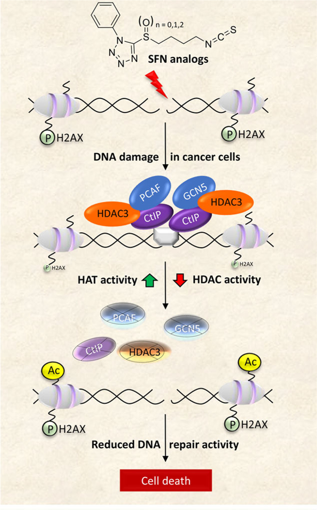
Scope: DNA repair inhibitors have broad clinical applications in tumor types with DNA repair defects, including colorectal cancer (CRC). Structural analogs of the anticancer agent sulforaphane (SFN) were investigated as modifiers of histone deacetylase (HDAC) and histone acetyltransferase (HAT) activity, and for effects on DNA damage/repair pertinent to human CRC.

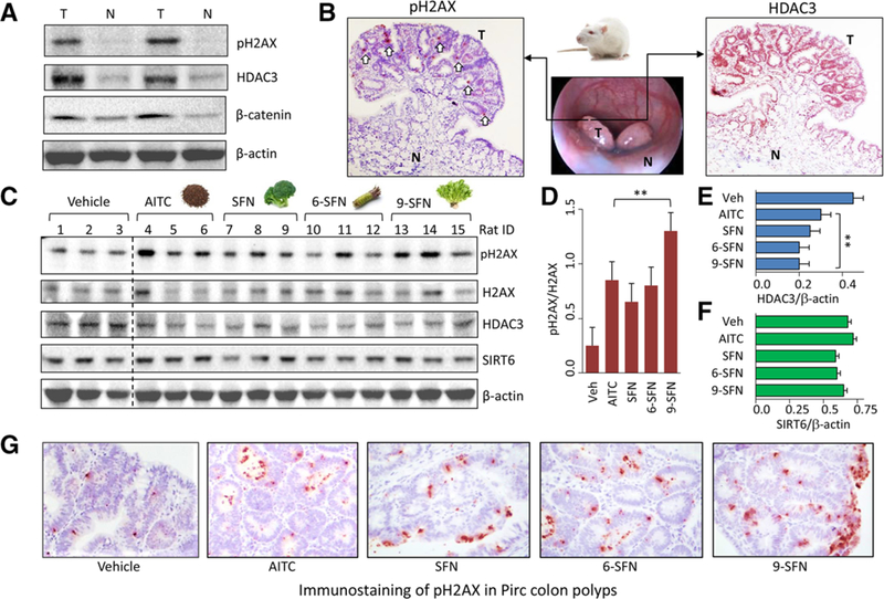
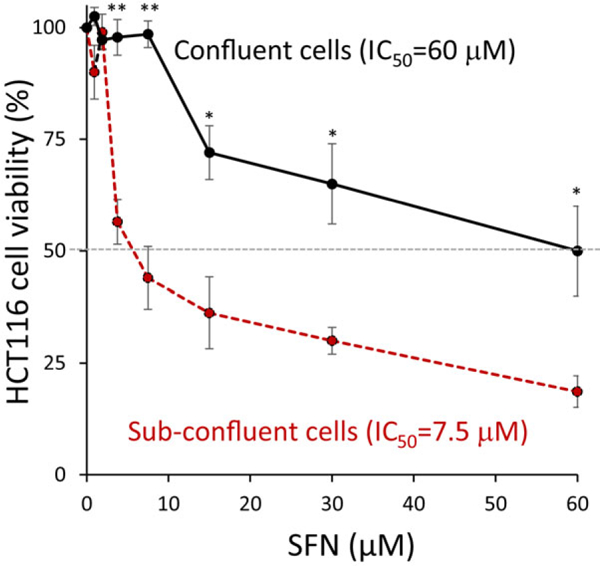
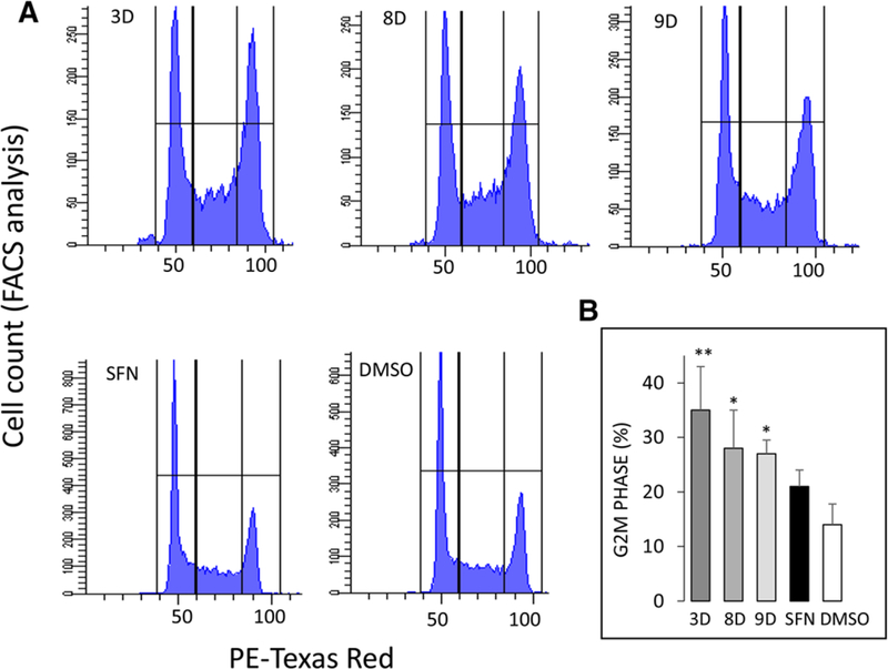
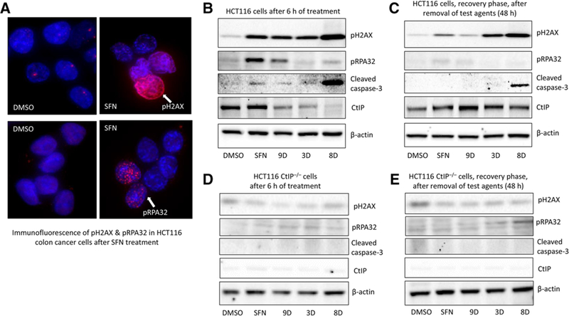
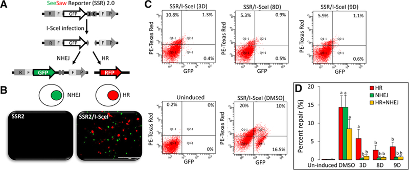
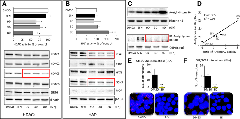
Methods and results: In the polyposis in rat colon (Pirc) model, single oral administration of SFN and structurally related long-chain isothiocyanates (ITCs) decreased histone deacetylase 3 (HDAC3) expression and increased pH2AX levels markedly in adenomatous colon polyps, extending prior observations on HDAC3 inhibition/turnover in cell-based assays. Colon cancer cells at a high initial plating density had diminished cytotoxicity from SFN, whereas novel tetrazole-containing heterocyclic analogs of SFN retained their efficacy. The potent SFN analogs triggered DNA damage, cell cycle arrest, apoptosis, and loss of a key DNA repair regulator, C-terminal binding protein (CtBP) interacting protein (CtIP). These SFN analogs also altered HAT/HDAC activities and histone acetylation status, lowered the expression of HDAC3, P300/CBP-associated factor (PCAF) and lysine acetyltransferase 2A (KAT2A/GCN5), and attenuated homologous recombination (HR)/non-homologous end joining (NHEJ) repair activities in colon cancer cells.

Conclusion: Novel tetrazole-containing heterocyclic analogs of SFN provide a new avenue for chemosensitization in colon cancer cells via modulation of HAT/HDAC activities and associated DNA damage/repair signaling pathways.

Keywords: C-terminal binding protein (CtBP) interacting protein; DNA damage; DNA repair; colon cancer; histone acetyltransferase; histone deacetylase; sulforaphane analogs.


Conflict of interest statement

Conflict of Interest

The authors declare no conflict of interest.

Figures

Figure 1.
Figure 1.
Decreased HDAC3 expression and increased pH2AX in the Pirc model. Tumor and adjacent normal colon from Pirc males (≈8 months old) were probed for HDAC3 and pH2AX by A) immunoblotting with β-actin as loading control and B) immunohistochemistry (IHC); T, tumor; N, normal; white arrows, pH2AX immunostaining. C) Pirc males received corn oil vehicle or ITC (60 mg kg–1) by single oral gavage and were sacrificed 6 h later. Tumor tissue lysates (n = 3) were immunoblotted for pH2AX, H2AX, HDAC3, and SIRT6, with β-actin as loading control. D–F) Immunoblots were quantified by densitometry and the data plotted as mean ± SEM (n = 3) by one-way ANOVA (**p < 0.01, compared to vehicle). G) Immunostaining of pH2AX in Pirc colon polyps after IHC analyses.
Figure 2.
Figure 2.
Cytotoxicity of SFN is dependent on plating density. HCT116 colon cancer cells were seeded at two different cell densities (30%, sub-confluent; 80%, confluent) and treated for 24 h with SFN in the dose range 0–60 μm. Cell viability was assessed using the CCK-8 assay (n = 4 experiments); *p < 0.05, < 0.01, confluent versus the corresponding sub-confluent cell density data point using Student’s t-test.
Figure 3.
Figure 3.
Cell cycle arrest by SFN and structural analogs. HCT116 cells were seeded overnight at confluent cell density and treated for 24 h with 15 μm SFN or selected structural analogs from Table 1. A) DNA content via flow cytometry and B) FACS data quantified (mean ± SEM), *p < 0.05, **p < 0.01 vs. DMSO vehicle control, by one-way ANOVA (n = 3 experiments).
Figure 4.
Figure 4.
DNA damage response by SFN and structural analogs. A) HCT116 cells were treated with DMSO or 15 μm SFN for 24 h and examined for pH2AX and pRPA32 immuno-fluorescence (arrows); DAPI stained nuclei (blue). Confluent HCT116 cells were treated with DMSO (vehicle), 15 μm SFN, or its analogs (3D, 8D, and 9D), and whole cell lysates were assessed by immunoblotting B) 6 h after treatment with compounds or C) 48 h after treatment, which includes 6 h of treatment followed by compound-free media for 42 h (recovery). D,E) Repeat of experiments in (B) and (C), using HCT116 CtIP knockout cells (HCT116 CtIP–/–), generated via CRISPR/Cas9 genome editing. Antibodies detected DNA damage signaling (pH2AX and pRPA32), apoptosis (cleaved caspase-3), and DNA repair (CtIP). β-actin, loading control. Data are representative of at least three independent experiments.
Figure 5.
Figure 5.
Inhibition of DNA repair by SFN analogs. A) A schematic representation of the See Saw reporter SSR 2.0, as published.[18] B) Images of HCT116 cells transfected with SSR 2.0 for 24 h (left panel), followed by transfection with I-SceI for 6 h (right panel) to induce DNA damage response, as indicated by HR-mediated (red) or NHEJ-mediated repair (green) fluorescence. C) SH-SY5Y cells were co-transfected with SSR 2.0/I-SceI and treated with 5 μm of SFN analogs for 6 h, as described in Experimental Section. Percent NHEJ (GFP) or HR (RFP) was measured via flow cytometry in negative controls without I-SceI (Uninduced), positive controls (SSR 2.0/I-SceI, DMSO), and test samples (SSR 2.0/I-SceI, treated with SFN analogs). D) Percentage NHEJ or HR quantified from three independent experiments, mean ± variance. a) p < 0.05 (DMSO vs. uninduced), b) p < 0.05 (DMSO vs. treatment), analyzed by one-way ANOVA.
Figure 6.
Figure 6.
HAT and HDAC changes produced by SFN analogs. HCT116 cells were treated with DMSO (vehicle) or 15 μm SFN or its analogs for 6 h and whole cell lysates were examined for A) HDAC activity (top panel) or HDAC protein expression, with β-actin as loading control in the immunoblots (bottom panel). B) Repeat of the experiments in (A), but for HATs. C) Histone H4 acetylation, normalized to histone H4 (top panel) assessed by immunoblotting, and CtIP acetylation changes normalized to CtIP input controls (bottom panel) by immunoprecipitation after Ac-Lys pull-down. D) The ratio of HAT:HDAC activity (from A,B) and the level of histone acetylation (densitometry from C) were plotted for each compound. Data are representative ofthree experiments (*p < 0.05 vs. DMSO). E,F) In HCT116 cells, proximity ligation assays (PLA) identified endogenous interactions ofCtIP:PCAF and CTIP:GCN5 proteins in situ (red dots). DAPI-stained nuclei (blue). Cells were imaged 6 h after treatment with DMSO (vehicle) or analog 8D, and the interactions per nuclei were quantified and averaged across five different microscopic fields. Data = mean ± SE, **p < 0.01, ***p < 0.001 for 8D versus vehicle controls using Student’s t-test. Data are representative of at least three independent experiments.
Figure 7.
Figure 7.
Working model of HDAC/HAT modulation by SFN analogs impacting DNA repair pathways, leading to cancer cell death. Ac, acetylation; P, phosphorylation. Through various mechanisms, not well characterized, SFN analogs trigger DSB in DNA that become flanked by increased pH2AX levels. A functional DNA damage response requires the formation of an efficient repair complex, typically comprising an MRN complex (gray box bridging the DNA break point) bound by the DNA resection and repair factor, CtIP. HATs and HDACs, such as PCAF, GCN5, and HDAC3, regulate the acetylation status of CtIP and maintain its activity/stability, leading to efficient DNA repair and reduced pH2AX levels. Treatment with SFN analogs causes an imbalance in HAT/HDAC activity and protein expression, leading to increased histone acetylation. This also adversely affects the stability of CtIP and its binding partners, including PCAF, GCN5, and HDAC3. As a crucial factor for DNA end resection, reduced levels of CtIP negatively influence homologous recombination (HR), concluding in impairment of DSB repair and eventual cell death. As described in the text, normal cells escape these outcomes, and are refractory to the mechanisms that trigger DNA damage and apoptosis in cancer cells.

Similar articles

Cited by

References

    1. American Cancer Society. Cancer Facts & Figures 2018. Atlanta, Ga: American Cancer Society, 2018. Retrieved from https://www.cancer.org/cancer/colon-rectal-cancer/about/key-statistics.html
    1. Wolpin BM, Mayer RJ, Gastroenterology, 2008, 134, 1296. - PMC - PubMed
    1. Huertas P, Nat. Struct. Mol. Biol. 2010, 17, 11. - PMC - PubMed
    1. Lord CJ, Ashworth A, Curr. Opin. Pharmacol. 2008, 8, 363. - PubMed
    1. Sinicrope FA, Nat. Rev. Clin. Oncol. 2010, 7, 174. - PMC - PubMed

Publication types

MeSH terms