. 2018 Jun 20;32(12):fj201800691R.
doi: 10.1096/fj.201800691R. Online ahead of print.

Peptidylarginine deiminase 4: a nuclear button triggering neutrophil extracellular traps in inflammatory diseases and aging

Affiliations

Peptidylarginine deiminase 4: a nuclear button triggering neutrophil extracellular traps in inflammatory diseases and aging

Siu Ling Wong et al. FASEB J. .

Abstract

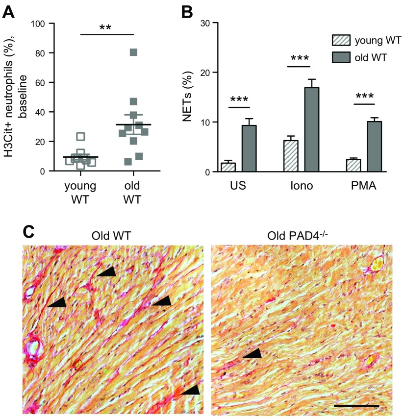
Peptidylarginine deiminase 4 (PAD4) is a nuclear citrullinating enzyme that is critically involved in the release of decondensed chromatin from neutrophils as neutrophil extracellular traps (NETs). NETs, together with fibrin, are implicated in host defense against pathogens; however, the formation of NETs (NETosis) has injurious effects that may outweigh their protective role. For example, PAD4 activity produces citrullinated neoantigens that promote autoimmune diseases, such as rheumatoid arthritis, to which PAD4 is genetically linked and where NETosis is prominent. NETs are also generated in basic sterile inflammatory responses that are induced by many inflammatory stimuli, including cytokines, hypoxia, and activated platelets. Mice that lack PAD4-deficient in NETosis-serve as an excellent tool with which to study the importance of NETs in disease models. In recent years, animal and human studies have demonstrated that NETs contribute to the etiology and propagation of many common noninfectious diseases, the focus of our review. We will discuss the role of NETs in thrombotic and cardiovascular disease, the induction of NETs by cancers and its implications for cancer progression and cancer-associated thrombosis, and elevated NETosis in diabetes and its negative impact on wound healing, and will propose a link between PAD4/NETs and age-related organ fibrosis. We identify unresolved issues and new research directions.-Wong, S. L., Wagner, D. D. Peptidylarginine deiminase 4: a nuclear button triggering neutrophil extracellular traps in inflammatory diseases and aging.

Keywords: NETs; PAD4; cancer; diabetes; thrombosis.


Conflict of interest statement

The authors thank Deya Cherpokova and Elise DeRoo for critical reading of the manuscript, Caleb Staudinger and Sarah Walker (all from Boston Children’s Hospital) for assistance in manuscript preparation. Some of the research described here was supported by the U.S. National Institutes of Health (NIH) National Heart, Lung, and Blood Institute Grant R35HL135765 (to D.D.W.). The authors declare no conflicts of interest.

Figures

Figure 1
Figure 1
Spontaneous NETosis in a neutrophil that was isolated from a patient with RA. Scanning electron micrograph shows a NETting neutrophil (pseudocolored in red). The NET (pseudocolored in green) appears as a large web-like structure emanating from a break in the plasma membrane. Scale bar, 5 μm. Image courtesy of Chanchal Sur Chowdhury (Center for Microscopy, University of Basel, Basel, Switzerland).
Figure 2
Figure 2
NETs formed in the myocardium after I/R injury contribute to myocardial infarction in mice. A) Immunofluorescence microscopy revealed that Gr-1–positive neutrophils (pink) are releasing H3Cit-positive NETs (yellow) in the infarcted myocardium of wild-type (WT) mice. Arrow indicates a NET. B) Triphenyltetrazolium chloride (TTC) staining of cross-sections of the left ventricle at 24 h after myocardial I/R injury (top). Infarcted areas appear as white tissue are outlined with dotted lines. Summarized data of left ventricle infarct size, expressed as the percentage of left ventricle (LV; bottom). The infarct size is remarkably smaller in PAD4−/− mice compared with WT mice. Systemic treatment with DNase 1 does not further reduce the infarct size of PAD4−/− mice, which indicates that the extracellular DNA in the damaged heart predominantly originates from NETs. From Savchenko et al. (57). Reprinted with permission from the American Society of Hematology.
Figure 3
Figure 3
Targeting NETs with DNase 1 reduces metastasis. Mice were treated with DNase 1–coated nanoparticles or control nanoparticles i.p. 2 h before intravenous injection of 4T1 breast cancer cells. Mice continued to receive nanoparticle treatment daily for 2 wk, and were euthanized when weight loss occurred. Hematoxylin and eosin–stained lung sections (left), and quantitative analysis shows that the metastatic burden was reduced by the DNase 1 treatment (right). Arrows indicate metastases. Scale bars, 4 mm. From Park et al. (86). Reprinted with permission from the American Association for the Advancement of Science.
Figure 4
Figure 4
Enhanced NETosis in neutrophils from patients with diabetes and deposition of NETs in mouse skin wounds. A) Both T1DM and T2DM prime human neutrophils to produce NETs. B) PAD4 protein expression is strikingly up-regulated in the neutrophils of patients with diabetes. T1DM, pink circle; T2DM, purple square. C) Hematoxylin and eosin staining revealed prominent extracellular DNA structures, appearing as blue streaks (arrows), in the scab of WT mice 3 d postinjury (top). Neutrophils remain intact (arrowheads) and NET structures are absent in the scab of PAD4−/− mice. Confocal immunofluorescence microscopy showed that H3Cit (green) colocalizes with externalized DNA (blue) in neutrophil (Ly6G) -rich area (red) in WT, but not PAD4−/− scab, which indicates the crucial role of PAD4 in NETosis in the mouse skin wounds (bottom). Scale bars, 50 μm. Adapted from Wong et al. (95).
Figure 5
Figure 5
PAD4 activity/NETosis increase with age, and PAD4/NETs are implicated in age-related organ fibrosis. A) More circulating neutrophils from old (age 14–17 mo) wild-type (WT) mice were H3Cit positive compared with those of young (age 1.5–3.5 mo) mice. B) Compared with young WT mice, a higher percentage of neutrophils from old WT mice produced NETs spontaneously, as well as in the presence of stimulants, after incubation ex vivo. C) Sirius red staining revealed more interstitial collagen deposition (red, arrowheads) in the heart of old WT mice than in age-matched PAD4−/− mice. Scale bar, 100 μm. Iono, ionomycin; PMA, phorbol 12-myristate 13-acetate; US, unstimulated. From Martinod et al. (113).

References

    1. Wang Y., Li M., Stadler S., Correll S., Li P., Wang D., Hayama R., Leonelli L., Han H., Grigoryev S. A., Allis C. D., Coonrod S. A. (2009) Histone hypercitrullination mediates chromatin decondensation and neutrophil extracellular trap formation. J. Cell Biol. 184, 205–213 10.1083/jcb.200806072 - DOI - PMC - PubMed
    1. Brinkmann V., Reichard U., Goosmann C., Fauler B., Uhlemann Y., Weiss D. S., Weinrauch Y., Zychlinsky A. (2004) Neutrophil extracellular traps kill bacteria. Science 303, 1532–1535 10.1126/science.1092385 - DOI - PubMed
    1. Brinkmann V., Zychlinsky A. (2012) Neutrophil extracellular traps: is immunity the second function of chromatin? J. Cell Biol. 198, 773–783 10.1083/jcb.201203170 - DOI - PMC - PubMed
    1. Gupta A. K., Hasler P., Holzgreve W., Gebhardt S., Hahn S. (2005) Induction of neutrophil extracellular DNA lattices by placental microparticles and IL-8 and their presence in preeclampsia. Hum. Immunol. 66, 1146–1154 10.1016/j.humimm.2005.11.003 - DOI - PubMed
    1. Zhong X. Y., Laivuori H., Livingston J. C., Ylikorkala O., Sibai B. M., Holzgreve W., Hahn S. (2001) Elevation of both maternal and fetal extracellular circulating deoxyribonucleic acid concentrations in the plasma of pregnant women with preeclampsia. Am. J. Obstet. Gynecol. 184, 414–419 10.1067/mob.2001.109594 - DOI - PubMed